Abstract
Analyses in mice and humans indicate that non-caloric artificial sweeteners may promote obesity-associated metabolic changes by changing the function of the bacteria that colonize the gut.
In many parts of the world, obesity is becoming increasingly prevalent. Weight control is important for reducing the risk of metabolic diseases such as type 2 diabetes, which is characterized by high blood-glucose levels and insulin resistance. Limiting calorie intake and replacing dietary fat and sugar with low- or non-caloric alternatives is a common weight-loss strategy1. Non-caloric artificial sweeteners (NAS) are often chosen to combat weight problems, because they do not contribute to overall calorie intake and are thought to subvert the rise in blood-glucose levels that occurs in response to food intake1 (Fig. 1). For unknown reasons, however, NAS are not always effective for weight loss. In a paper published on Nature’s website today, Suez et al.2 describe an unexpected effect of NAS that may shed some light on this issue.
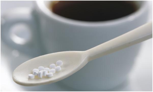
Figure 1.
Non-caloric artificial sweeteners are often used as an alternative to sugar.
Suez and colleagues added an NAS supplement (saccharin, sucralose or aspartame) to the diets of mice, and found that the sweeteners altered the animals’ metabolism, raising blood glucose to significantly higher levels than those of sugar-consuming mice. This was true both for mice fed a normal diet and for those on a high-fat diet — a model for a situation in which NAS supplements might be used to control weight. Because variations in diet have been shown to directly lead to changes in the populations of bacteria that occupy the gut, the authors examined whether these bacteria were responsible for the metabolic changes that they observed. And, indeed, when they used antibiotics to deplete the gut bacteria, they found that this eliminated NAS-induced glucose intolerance in mice fed either diet.
Next, the researchers transplanted faeces from NAS-fed or glucose-fed mice into germfree mice (those with no gut bacteria of their own) that had never consumed NAS. Transfer from NAS-fed mice induced elevated blood-glucose levels in the transplant recipients. Furthermore, the composition of the recipients’ gut bacterial community was different from that of mice receiving transplants from glucose-fed mice, suggesting that changes in this gut microbiota mediate glucose intolerance in NAS-fed mice. Genetic analysis revealed that this altered composition was accompanied by changes in bacterial function. In particular, Suez and co-workers detected an increase in carbohydrate-degradation pathways in the microbiota of NAS-fed mice. This connection parallels a previous report2 that the microbiota of obese mice has a higher carbohydrate-metabolizing capacity than the microbiota of normal-weight mice.
What is the relevance of these results for human disease? Suez et al. studied around 400 people, and found that bacterial populations in the guts of those who consumed NAS were significantly different from those who did not. Moreover, NAS consumption correlated with disease markers linked to obesity, such as elevated fasting blood-glucose levels and impaired glucose tolerance.
The authors placed seven volunteers who did not normally consume NAS on a seven-day regimen of controlled high NAS intake. After only four days, half the individuals had elevated blood-glucose levels and altered bacterial-community composition, mirroring the results seen in the mice. Transfer of faeces from NAS-fed human donors induced elevated blood-glucose levels in germ-free mouse recipients that had never consumed NAS. Taken together, Suez and colleagues’ data indicate that NAS consumption may contribute to, rather than alleviate, obesity-related metabolic conditions, by altering the composition and function of bacterial populations in the gut.
Studies examining genetic3,4 and diet-induced5 mouse models of obesity and obesity in humans6,7 have demonstrated that the disease is associated with changes in the composition of the gut microbiota. Most bacteria colonizing the gut come from two phyla, Bacteroidetes and Firmicutes. Obese mice and humans both have reduced bacterial diversity, with reduced proportions of Bacteroidetes and increased Firmicutes, when compared to lean littermates or twin controls4–7. Obesity-induced changes in the microbiota can be reversed by diet — obese mice or humans on fat- or carbohydrate-restricted diets have an increased abundance of Bacteroidetes5,6.
It is difficult to directly compare Suez and colleagues’ findings with earlier work, because the current report describes changes in a mix of bacteria (including Bacteroidetes and Firmicutes) after NAS treatment. Certain gut bacteria are well adapted to break down dietary components that the human body cannot. It could be that expansion of these populations in response to NAS increases extraction of energy — often stored as fat — from the diet, contributing to obesity2. Alternatively, NAS might exert their effect by suppressing the growth of particular bacterial taxa. In obese mice, the growth of certain bacterial species is suppressed, and there is an increased production of metabolites that can contribute to insulin resistance8. These two possibilities cannot be distinguished in the current report.
Bacterial communities in the gut have been linked to elevated lipid production, and increased storage of lipids and the carbohydrate glycogen9,10, correlating with an increase in adiposity and in cellular energy extraction from food. Furthermore, obesity-induced alterations to the composition of gut microbiota are associated with metabolic changes2,8, including enrichment of pathways related to bacterial growth. This suggests that obesity maintains alterations to the microbiota, allowing for the continued increase in production and storage of lipids and glycogen, further exacerbating the condition. Future work must determine whether the changes in the microbiota brought about by NAS consumption activate any of the same molecular pathways as are active in the obese microbiota.
Type 2 diabetes and impaired glucose tolerance have also been linked to alterations in gut microbiota composition11,12. Analysis of gut bacterial genomes shows that microbial-gene signatures differ between patients with and without diabetes, and people with impaired glucose tolerance11,12. Whether the bacterial populations or metabolic pathways altered by the consumption of NAS are similar to those described in people with or developing diabetes remains to be seen.
Many diseases associated with Western lifestyles have now been linked to environmentally induced alterations in the composition of the gut microbiota13. Questions remain regarding the precise mechanisms by which NAS disrupt the relationship between gut bacteria and their host. Studies to identify specific bacterial populations that promote resistance to weight gain or improve glucose tolerance may prove useful for devising therapies that modulate bacteria or their metabolites.
References
- 1.Gardner C, et al. Diabetes Care. 2012;35:1798–1808. doi: 10.2337/dc12-9002. [DOI] [PMC free article] [PubMed] [Google Scholar]
- 2.Suez J, et al. Nature. 2014 http://dx.doi.org/10.1038/nature13793.
- 3.Turnbaugh PJ, et al. Nature. 2006;444:1027–1031. doi: 10.1038/nature05414. [DOI] [PubMed] [Google Scholar]
- 4.Ley RE, et al. Proc. Natl Acad. Sci. USA. 2005;102:11070–11075. doi: 10.1073/pnas.0504978102. [DOI] [PMC free article] [PubMed] [Google Scholar]
- 5.Turnbaugh PJ, Bäckhed F, Fulton L, Gordon JI. Cell Host Microbe. 2008;3:213–223. doi: 10.1016/j.chom.2008.02.015. [DOI] [PMC free article] [PubMed] [Google Scholar]
- 6.Ley RE, Turnbaugh PJ, Klein S, Gordon JI. Nature. 2006;444:1022–1023. doi: 10.1038/4441022a. [DOI] [PubMed] [Google Scholar]
- 7.Turnbaugh PJ, et al. Nature. 2009;457:480–484. doi: 10.1038/nature07540. [DOI] [PMC free article] [PubMed] [Google Scholar]
- 8.Ridaura VK, et al. Science. 2013;341:1241214. doi: 10.1126/science.1241214. [DOI] [PMC free article] [PubMed] [Google Scholar]
- 9.Bäckhed F, et al. Proc. Natl Acad. Sci. USA. 2004;101:15718–15723. doi: 10.1073/pnas.0407076101. [DOI] [PMC free article] [PubMed] [Google Scholar]
- 10.Bäckhed F, Manchester JK, Semenkovich CF, Gordon JI. Proc. Natl Acad. Sci. USA. 2007;104:979–984. doi: 10.1073/pnas.0605374104. [DOI] [PMC free article] [PubMed] [Google Scholar]
- 11.Qin J, et al. Nature. 2012;490:55–60. doi: 10.1038/nature11450. [DOI] [PubMed] [Google Scholar]
- 12.Karlsson FH, et al. Nature. 2013;498:99–103. doi: 10.1038/nature12198. [DOI] [PubMed] [Google Scholar]
- 13.Cho I, Blaser MJ. Nature Rev. Genet. 2012;13:260–270. doi: 10.1038/nrg3182. [DOI] [PMC free article] [PubMed] [Google Scholar]


