Abstract
Background
The femoral approach has been the preferably used access in interventional cardiology as well for coronary diagnostics as for percutaneous coronary intervention, being perceived as easy and facilitating quick access with relatively low risk. Due to the results of the latest studies, however, the radial approach has become increasingly popular. The aim of this study was a safety analysis of cardiological interventional procedures (i.e., coronarography and PCI) according to the vessel approach.
Material/Methods
The 204 coronary interventions done in our Department of Interventional Cardiology were retrospectively analyzed. All the procedures were classified according to femoral or radial access. The incidence of local complications (e.g., major bleedings and hematomas) was assessed as well as the volume of contrast agent administered during the procedure and the radiation dose.
Results
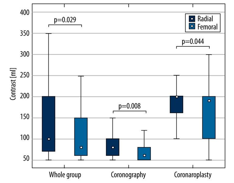
It has been shown that radial approach, which is obviously more comfortable for patients, reduces the risk of local complications (0 vs. 2.97% and 0 vs. 3.96%) and does not lead to increased radiation exposure (p=0.88). However, there could be a larger volume of contrast agent administered (p=0.029), which in some cases could increase the risk of contrast-induced nephropathy.
Conclusions
The radial approach should be recommended as a first choice because it is safer than the classical femoral approach, but one must be cautious in choosing radial approach patients with renal insufficiency.
Keywords: Cardiac Catheterization, Femoral Artery, Percutaneous Coronary Intervention, Radial Artery
Background
Until recently, the femoral approach was the preferably used method in interventional cardiology for diagnostics and therapy of coronary artery disease. It has been perceived as being easy and facilitating quick access with relatively low risk. Due to the results of latest studies, however, the radial approach has become more popular. The use of the radial approach not only reduced incidence of local and general operation-related complications, but also proved to be preferred by patients [1–3]. Despite the almost unequivocal results concerning the selection of the vessel approach, radial access continues to have its detractors.
The most common arguments brought up are the relatively large proportion of permanent radial artery occlusions following surgery and the need to learn a new method that is more difficult for the operator than the radial approach is. Thus, the largest opposition to this access is encountered in surgeons experienced in the femoral approach. This safety analysis was performed to experimentally assess the legitimacy of the radial approach. The analysis was performed in the Department during the transition period from the femoral to the radial approach.
The aim of this paper was a retrospective comparison of safety between the femoral and radial approaches during coronal arterial angiography procedures conducted in patients hospitalized between the years 2008 and 2009 at the Hemodynamics Laboratory in the Department of Invasive Cardiology in the Military Institute of Medicine.
Material and Methods
The 204 coronary interventions done in the Department of Interventional Cardiology were retrospectively analyzed. All the procedures were divided according to the femoral or radial access. The analysis covered diagnostic and therapeutic coronary artery procedures in year 2008 when the femoral approach was preferred (90 patients) and in the year 2009 when the radial approach was introduced (114 patients). In 2008, all patients were submitted to the femoral approach but in the year 2009, eleven (11) patients were subjected to the femoral approach whereas 103 to the radial approach. Time intervals (01–02.2008 and 04–05.2009) were selected accordingly to obtain comparable groups with respect to the number of patients with the femoral approach (FA) and the radial approach (RA). Two patients were excluded from the FA group because of a previous, ineffective radial approach procedure and as a result the access was changed to femoral. The opposite process (a shift from the radial to the femoral approach) was not observed. In the end, 204 patients were assessed (RA – 103 and FA – 101).
Statistical analysis
All analyses were prepared using Statistica 10.0 with the medical set, StatSoft Inc. Continuous variables are expressed as medians with 1st to 3rd percentile and qualitative variables as percentages. The normality of each continuous variable was at first tested with the Shapiro-Wilk W test. Because there were non-normal variables in further analyses, nonparametric, two-sided tests were used. Qualitative variables were analyzed with the Fisher exact test. P<0.05 was considered statistically significant.
Basic demographic data, the type of procedure, and indications to coronary intervention were assessed (Table 1) and no differences between FA and RA groups were observed.
Table 1.
Basic demographic and clinical data regarding to included patients.
| Radial catheterization | Femoral catheterization | p | |
|---|---|---|---|
| Demographic data | |||
| Age | 62.6±10.2 | 62.9±12.8 | ns. (0.88) |
| Sex | 38.8% (female) | 27.7% (female) | ns. (0.09) |
| Patients’ burdens | |||
| Arterial hypertension | 72.8% | 68.3% | ns. (0.48) |
| Diabetes | 27.2% | 30.7% | ns. (0.58) |
| Hyperlipidemia | 45.6% | 34.6% | ns. (0.11) |
| Cardiac infarction | 24.3% | 32.7% | ns. (0.18) |
| CABG | 5.8% | 5.9% | ns. (0.97) |
| Aortic valve disease | 3.9% | 4.0% | ns. (0.74) |
| Indications | |||
| STEMI | 6.8% | 9.9% | ns. (0.42) |
| NSTEMI | 14.6% | 15.8% | ns. (0.80) |
| UA | 1.94% | 6.93% | ns. (0.075) |
| Stable CAD | 75.7% | 68.3% | ns. (0.24) |
| Kind of intervention | |||
| Coronarography | 64.1% | 60.4% | ns. (0.59) |
| Coronarography and PCI | 35.9% | 39.6% | |
Groups of patients were also analyzed with respect to ionizing radiation exposure, the volume of administered contrast agent, and procedure-related complications, which were divided into hemorrhagic (major bleedings) and local (false aneurysm and large hematomas).
Results
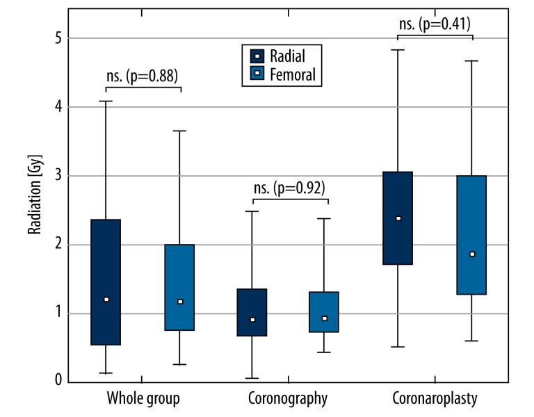
The volume of contrast agent used and radiation dose are presented in Table 1. The analysis shows that there are differences in the volumes of contrast used during between the FA and RA groups (Figure 1) but no differences recorded in ionizing radiation dose (Figure 2). Similar results were observed between the distinct subgroups of coronarographies and percutaneous coronary angioplasty. Use of contrast agent was greater during the radial approach than in the femoral approach in each studied group. There was no significant difference between groups according to the radiation dose.
Figure 1.
The comparison of the median (1st to 3rd quartile) of the volume of contrast agent used during coronary procedure in groups of patients.
Figure 2.
The comparison of the median (1st to 3rd quartile) of radiation dose in analyzed groups of patients.
Patients were also compared with regard to complications, but no statistically significant differences were observed between the groups (Table 3). There was, however, a tendency to an increased number of complications in the FA group, accompanied with a borderline value using the Fisher exact test (p=0.058). It is noteworthy that despite there being no statistical differences in the RA group, no complications were reported, whereas in the FA group they did occur (Table 3).
Table 3.
Complications of a percutaneous procedure.
| Radial | Femoral | p* | |
|---|---|---|---|
| Analyzed group | |||
| Catheterization | |||
| Haemorrhagic complications | 0 | 3 (2.97%) | ns. (0.12) |
| Local complications (aneurysm + hematoma) | 0 | 4 (3.96%) | ns. (0.058) |
| Coronarography | |||
| Haemorrhagic complications | 0 | 0 | ns. |
| Local complications (aneurysm + hematoma) | 0 | 2 (3.28%) | ns. (0.23) |
| Angioplasty | |||
| Haemorrhagic complications | 0 | 3 (7.5%) | ns. (0.24) |
| Local complications (aneurysm + hematoma) | 2 (5.0%) | ns. (0.49) | |
Due to no complications reported in the radial approach group the Fisher exact test was performed.
Discussion
The aim of this work was to demonstrate the benefits of the radial approach in comparison to the femoral approach for patients with coronary interventions (diagnostic or therapeutic).
We selected from among factors which can be easily assessed by retrospective analysis and contribute to patient benefit in terms of the volume of applied contrast agent, ionizing radiation dose, and incidence of complications. In short-term observations, the ionizing radiation dose was not connected with any complications. In contrast, in patients requiring multiple ionizations for diagnostic and therapeutic purposes, some skin and hematopoietic disturbances were reported even in a short observation period. Moreover, the risk of carcinogenic activity of ionizing radiation cannot be excluded, as was proven in patients submitted to CT diagnostics [4]. Between the compared groups, no statistically significant difference in absorbed ionizing radiation dose was found. In recently published studies, similar results were obtained by the STEMI-RADIAL team, who admittedly analyzed the time of fluoroscopy, not the radiation dose; nonetheless, were no differences between groups. However, there are studies which demonstrate a small but statistically significant difference in radiation dose, which was smaller in the femoral approach [3, 5].
Another aspect of the present comparison was used – the volume of contrast agent. Contrast agent is crucial for radio-diagnostics because quality of obtained pictures depends on volume and quality of contrast agent. However, it should be remembered that some complications such as CIN (contrast-induced nephropathy) and hypersensitive reactions, are connected with excessive volume of contrast agent. In previous papers, there were no differences in volume of contrast agent in terms of the vessel approach used [3] or the volume was greater in the radial approach [6]. Our results are particularly interesting, because contrary to previous reports, they indicate that the volume of contrast agent is statistically greater in the radial approach procedure. It corresponds, however, to the results of “the learning curve” in radial approach procedures [7,8]. Please note that the period of 2008–2009 was a time when the Department shifted from the femoral to the radial approach. As mentioned previously, in 2008 most of the procedures were performed using the femoral approach. In the year 2009, this relationship was reversed. Thus, in 2009, our surgeons passed through consecutive stages of “the learning curve”. Most published reports were derived from sites with a long-standing use of and experience in the femoral and radial approaches. This could explain the divergence between the aforementioned large studies and the presented retrospective analysis.
In this study, the advantage of the radial approach with regard to peri-procedural complications was not proven. It could be attributed to progress in intervention cardiology (i.e., new devices and new techniques) which reduces the risk of complications regardless of the vessel approach used. With regard to the observed tendency, a larger number of patients would be necessary to demonstrate the advantage of radial access.
Therefore, large clinical studies demonstrate advantages of the radial approach [1,3]. During the conducted analysis, single defined complications were observed in the FA group but not in the RA group.
Conclusions
The increasingly frequently used and patient-preferred radial approach is as safe as the classic femoral approach with regard to peri-procedural complications and it does not increase patient exposure to ionizing radiation.
At sites where the radial approach is not routine, the risk of larger contrast agent volume usage increases. Thus, in patients at risk of CIN or who have renal deficiency or hypersensitivity to the contrast agent in their medical history, the classical femoral approach should be recommended.
Table 2.
The comparison of radiation dose and volume of contrast agent used in analyzed groups.
| Catheterization | Radial | Femoral | p |
|---|---|---|---|
| Analyzed group | |||
| N | 103 | 101 | – |
| Dose of radiation | 1.218 (0.696–2.207) | 1.199 (0.677–2.001) | ns. (0.88) |
| Contrast | 100 (70–200) | 80 (60–150) | 0.029 |
| Coronarography | |||
| N | 66 | 61 | – |
| Dose of radiation | 0.869 (0.613–1.450) | 0.940 (0.607–1.374) | ns. (0.92) |
| Contrast | 80 (60–100) | 60 (50–80) | 0.008 |
| Angioplasty | |||
| N | 37 | 40 | – |
| Dose of radiation | 2.244 (1.689–3.0239) | 1.800 (1.188–3.00) | ns. (0.41) |
| Contrast | 200 (160–200) | 190 (100–200) | P=0.044 |
Footnotes
Source of support: Departmental sources 103/12
References
- 1.Romagnoli E, Biondi-Zoccai G, Sciahbasi A, et al. Radial versus femoral randomized investigation in ST-segment elevation acute coronary syndrome: the RIFLE-STEACS (Radial Versus Femoral Randomized Investigation in ST-Elevation Acute Coronary Syndrome) study. J Am Coll Cardiol. 2012;60(24):2481–89. doi: 10.1016/j.jacc.2012.06.017. [DOI] [PubMed] [Google Scholar]
- 2.Cooper CJ, El-Shiekh RA, Cohen DJ, et al. Effect of transradial access on quality of life and cost of cardiac catheterization: A randomized comparison. Am Heart J. 1999;138(3 Pt 1):430–36. doi: 10.1016/s0002-8703(99)70143-2. [DOI] [PubMed] [Google Scholar]
- 3.Jolly SS, Yusuf S, Cairns J, et al. Radial versus femoral access for coronary angiography and intervention in patients with acute coronary syndromes (RIVAL): a randomised, parallel group, multicentre trial. Lancet. 2011;377(9775):1409–20. doi: 10.1016/S0140-6736(11)60404-2. [DOI] [PubMed] [Google Scholar]
- 4.Einstein AJ, Sanz J, Dellegrottaglie S, et al. Radiation dose and cancer risk estimates in 16-slice computed tomography coronary angiography. J Nucl Cardiol. 2008;15(2):232–40. doi: 10.1016/j.nuclcard.2007.09.028. [DOI] [PMC free article] [PubMed] [Google Scholar]
- 5.Kuipers G, Delewi R, Velders XL, et al. Radiation exposure during percutaneous coronary interventions and coronary angiograms performed by the radial compared with the femoral route. JACC Cardiovasc Interv. 2012;5(7):752–57. doi: 10.1016/j.jcin.2012.03.020. [DOI] [PubMed] [Google Scholar]
- 6.Bernat I, Horak D, Stasek J, et al. ST Elevation Myocardial Infarction Treated by RADIAL or Femoral Approach in a Multicenter Randomized Clinical Trial: The STEMI-RADIAL Trial. J Am Coll Cardiol. 2014;63(10):964–72. doi: 10.1016/j.jacc.2013.08.1651. [DOI] [PubMed] [Google Scholar]
- 7.Sciahbasi A, Romagnoli E, Trani C, et al. Evaluation of the “learning curve” for left and right radial approach during percutaneous coronary procedures. Am J Cardiol. 2011;108(2):185–88. doi: 10.1016/j.amjcard.2011.03.022. [DOI] [PubMed] [Google Scholar]
- 8.Sciahbasi A, Romagnoli E, Burzotta F, et al. Transradial approach (left vs. right) and procedural times during percutaneous coronary procedures: TALENT study. Am Heart J. 2011;161(1):172–79. doi: 10.1016/j.ahj.2010.10.003. [DOI] [PubMed] [Google Scholar]


