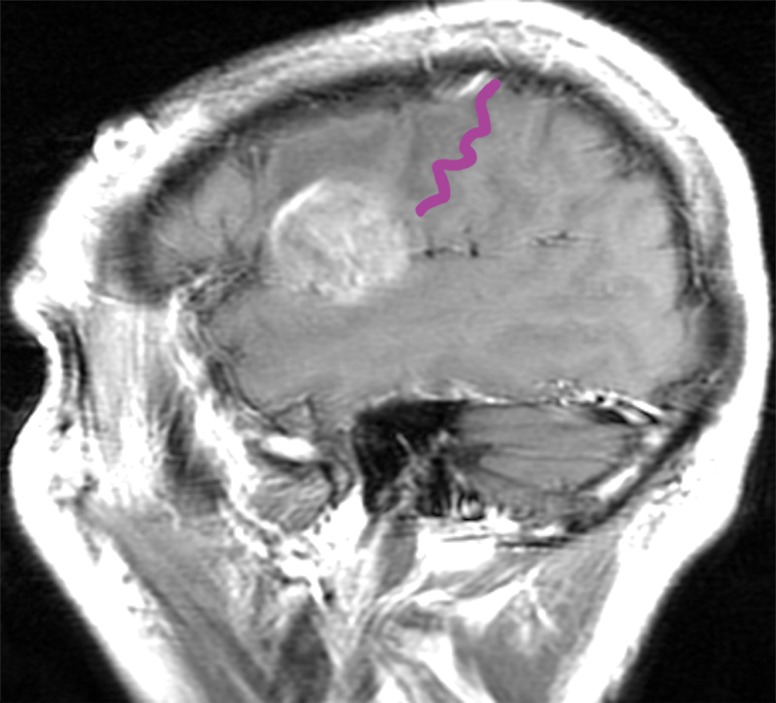
Figure 12a.

Mass effect causing difficulty in localization. A 52-year-old man presented with slurred speech due to a large left opercular glioblastoma in the lateral part of BA 6. (a) Sagittal unenhanced T1-weighted MR image shows a large glioblastoma, possibly in the primary motor area of the tongue, consistent with the patient’s dysarthria. (b) Confocal volume-rendered MR image with embedded deformable anatomic template of the corticospinal tract (see Fig 6 for color coding key) shows close relationship of the glioblastoma to the tract. No deformation for tumor mass effect was performed. (c) Postoperative sagittal gadolinium-enhanced T1-weighted MR image shows the small surgical cavity in the opercular part of BA 6.
