Abstract
Gaseous sulfide intrusion into seagrasses growing in sulfidic sediments causes little or no harm to the plant, indicating the presence of an unknown sulfide tolerance or detoxification mechanism. We assessed such mechanism in the seagrass Zostera marina in the laboratory and in the field with scanning electron microscopy coupled to energy dispersive X-ray spectroscopy, chromatographic and spectrophotometric methods, and stable isotope tracing coupled with a mass balance of sulfur compounds. We found that Z. marina detoxified gaseous sediment-derived sulfide through incorporation and that most of the detoxification occurred in underground tissues, where sulfide intrusion was greatest. Elemental sulfur was a major detoxification compound, precipitating on the inner wall of the aerenchyma of underground tissues. Sulfide was metabolized into thiols and entered the plant sulfur metabolism as well as being stored as sulfate throughout the plant. We conclude that avoidance of sulfide exposure by reoxidation of sulfide in the rhizosphere or aerenchyma and tolerance of sulfide intrusion by incorporation of sulfur in the plant are likely major survival strategies of seagrasses in sulfidic sediments.
Introduction
Seagrasses colonize coastal sediments characterized by low oxygen concentrations and high concentrations of toxic, reduced substances such as iron, manganese, and sulfide [1]. The effect of sulfide on growth and health of seagrasses is particularly puzzling. Sulfide is toxic to eukaryotic cells including seagrass cells even at concentrations as low as 1 to 10 μmol L-1 [2,3,4]. However, seagrasses can thrive in sediments with sulfide concentrations in the millimolar range [5]. The Mediterranean seagrass Posidonia oceanica (Linnaeus) Delile tolerates only low concentrations of pore water sulfide [6], whereas Zostera marina Linnaeus, the most common seagrass in temperate waters [7], can tolerate high concentrations of up to 4 mmol L-1 sulfide both in situ and in vitro [6,8,9].
Field and laboratory studies show that oxygen and sulfide dynamics in the water column, sediment, and plant tissues are key factors for seagrass growth, colonization, and survival [8,10,11]. Seagrasses avoid root anoxia and sulfide intrusion by leakage of oxygen from the roots (radial oxygen loss, ROL) [12,13]: photosynthetically derived oxygen diffuses via the aerenchyma to the respiring underground tissues [14], where it leaks out through the root tips to maintain an oxic rhizosphere [15]. Remarkably, the zone behind the root-tip region contains barriers to avoid ROL and sulfide intrusion [15,16]. Molecular oxygen derived from ROL probably also oxidizes toxic compounds such as sulfide to non-toxic compounds such as sulfate or elemental sulfur (S0), before reaching the root surfaces [15,17,18]. Oxidation of sulfide occurs both inside the plant in the aerenchyma [13] and externally in the rhizosphere [15,19,20]. In the dark, the photosynthetic oxygen pool in the aerenchyma is depleted in a few minutes [21] and the plant internal oxygen partial pressure is maintained by oxygen diffusion from the water column [10]. Thus, darkness combined with events of hypoxia or anoxia in the water column can lead to sulfide intrusion in the plants [13]. When oxygen returns into the plant, intruded sulfide can be oxidized: this has been observed in Z. marina, where gaseous sulfide in the aerenchyma disappears as oxygen is produced when photosynthesis commences in the morning [10,13].
In addition to microelectrode and optode studies, intrusion of sediment sulfide into seagrass tissues has been demonstrated by studies using stable sulfur isotopes [5]. High sulfur accumulation caused by sulfide intrusion has been found in most seagrasses studied to date and is correlated with environmental factors such as sediment sulfide concentration, water-column hypoxia, and shading [11,22,23]. In Z. marina, sulfur accumulates as S0 and other yet-unidentified sulfur compounds [11,24]. These unknown sulfur compounds accumulate to a high extent in rhizomes exposed to sulfide intrusion and, together with S0, are speculated to play a role in sulfide detoxification in the plant [6,11]. So far, S0 has been found and located by scanning electron microscopy with energy dispersive X-ray spectroscopy (SEM-EDX) in the vascular tissue of terrestrial plants [25]; in seagrasses, S0 has been found in underground tissues [22] but not yet localized. Despite these efforts, the mechanisms and metabolic pathways that cause S0 formation and accumulation remain unclear [26], as do their consequences for the seagrass.
Sediment sulfide intrudes in gaseous form via the roots of the plant [13] due to the membrane permeability of sulfide [27,28], and spreads by gas diffusion through the aerenchyma to the rhizome [21]. Once intruded, sulfide is available for enzymatic assimilation, possibly into thiol compounds (e.g., cysteine and glutathione) as occurs in wetland [29] and terrestrial [30,31] plants. Both terrestrial and wetland plants can incorporate and detoxify considerable amounts of sulfide, taken up by either the leaves or the underground tissues, resulting in elevated levels of sulfur compounds such as sulfate, thiols, and total sulfur in plant tissue without alteration of growth rates [31]. When sulfide uptake in terrestrial plants is foliar, the sulfur compounds are deposited in only the tissues exposed to sulfide and not translocated to other, more distal plant tissues [32]. However, soil or sediment sulfide uptake via roots typically results in higher accumulation of sulfur in the rhizome than the roots [11,31].
Terrestrial plants (e.g., Alium cepa (Linnaeus) and Brassica oleracea (Linnaeus)) can utilize gaseous sulfide as a source of sulfur, decreasing sulfur demand [32–34]. However, sulfate, which is considered to be the main source of sulfur nutrition, is mainly taken up by the roots, transported and stored in vacuoles throughout the plant [35,36]. In cases of sulfate deprivation in terrestrial plants, foliar uptake of sulfide can be an important sulfur source [32–34]. In contrast to terrestrial systems, the coastal marine environment offers three major sulfur sources for seagrasses: sulfate in the pore water, sulfate in the water column—both have roughly the same sulfate concentration and isotopic composition (γ34S around +20‰)—and sediment sulfide, which has a distinctly different isotopic composition (γ34S around −20‰) [37,38]. However, the contribution of each sulfur pool to sulfur nutrition in seagrasses has not been explored.
This study aims to characterize yet-unknown sulfur compounds and their origins in Z. marina subject to sulfide intrusion. We hypothesized that intruding sediment sulfide is metabolized and/or precipitated, and that sulfide is detoxified to non-toxic compounds inside Z. marina.
Material and Methods
Field study
We conducted a field study to assess the natural levels of sulfide intrusion in Z. marina and therefore collected Z. marina plants at a water depth of one meter from three intact seagrass meadows at Svenstrup Strand (+55°28'07", +9°45'18"), Kertinge Nor (+55°26'55", +10°33'27"), and Dalby Bay (55°31'07", +10°37'05"), Denmark, during July and August 2012. Plants were collected haphazardly by harvesting turfs (n = 5) with intact ramets. The salinity was 18. The water temperature was 18.6 ± 0.3°C in July and 18.1 ± 0.4°C in August, respectively.
The plants were gently and thoroughly rinsed with sea water and deionized water to remove sediment, root precipitates, and salts. Macroscopic epiphytes were removed by hand and plants were separated into the two youngest leaves, rhizome segments, and root bundles, then snap-frozen in liquid nitrogen. All samples were frozen (-80°C) until lyophilization for 48 h and homogenized in a ball mill before further processing. Svenstrup strand, Kertinge Nor and Dalby bay are public area, and in Denmark no specific permission is required for scientific research in this public areas. No endangered or protected species were involved in the study. This article does not report studies with human or animal subjects.
Mesocosm study
To determine the effect and fate of sulfide in Z. marina, we performed a mesocosm experiment in which we caused sulfide intrusion by artificially increasing pore water sulfide concentrations and measured its effects on plant sulfur compounds.
We collected medium sand sediment with low organic matter content and low iron pools [39] adjacent to a seagrass meadow at Svenstrup Strand in October 2012 at a water temperature of 15.2°C. The sediment was passed through a 1 mm sieve and placed in pots (11 cm in diameter and 12 cm height). To enhance sulfate reduction and hence sulfide concentration in the rhizosphere, we enriched half of the pots with caramelized glucose (720 g m-2; high sulfide, HS) at a sediment depth of 5 cm. The other pots acted as controls (C). After 10 days, apical shoots were collected from Svenstrup Strand as described previously. Senescent leaves, rhizome parts older than five internodes, and epiphytes were gently removed. Ten shoots were transplanted into each pot, yielding a shoot density of 1052 shoots m-2 (reflecting field densities) and placed in a mesocosm filled with seawater. Two shoots of each replicate were marked to estimate leaf growth, as described by Sand-Jensen [40]. The salinity (18) and temperature (15°C) were kept constant and half of the water was exchanged every week. Artificial illumination (Phillips SONT-T Agro 400W) was set to a diel cycle of 12:12 h at an intensity of 180–200 μmol photons m-2 s-1 at canopy level. The water column was aerated during the light period, but hypoxic during darkness to achieve sulfide intrusion following the method of Mascaro et al. [11], who ceased aeration during darkness resulting in oxygen levels at canopy height of around 40% of air saturation at the end of the dark cycle. After three weeks of exposure, we harvested the plants, separated them into leaves, rhizome, and root tissue, and preserved them as described above.
Plant sulfur extraction and analysis
Sulfate in plant tissue (leaf, rhizome, and root) was measured in the supernatant after a hot water extraction (20 mg plant tissue in 2 mL H2O, 95°C for 2 h, then centrifuged 5 min at 16000 × g, filtered through 0.45 μm pore size) on an ion chromatograph (DIONEX ICS-1500, DIONEX, Sunnyvale, CA, USA; IonPac AS22, 4.5 mM Na2CO3/1.4 mM NaHCO3). The remaining pellet was snap-frozen in liquid nitrogen and later lyophilized, and represents the water insoluble fraction (IF). S0 in plant tissues (10 mg extracted in 5 mL methanol for 24 h in darkness) was measured according to the method of Zopfi et al. [41] on an UV-RPHPLC (Agilent 1100 Series HPLC with UV detector (265 nm), Agilent Technologies Inc., Santa Clara, CA, USA). Concentrations of thiols (soluble, non-protein organo-sulfur compounds characterized by a chemical formula of R-SH) were estimated spectrophotometrically using a modified protocol of de Kok et al. [42] and lyophilized instead of fresh tissue. We tested the modification for glutathione recovery and found no significant differences between lyophilized and fresh tissues (data not shown). Samples of the whole plant organs (bulk S), the IF of the different tissues, and plant internal sulfate were analyzed for total sulfur (TS) and stable sulfur isotope ratio (δ34S) by elemental analyzer combustion continuous flow isotope ratio mass spectroscopy (EA-C-CF-IRMS; Thermo Scientific Delta V Advantage plus Flash EA 1112, Thermo Fisher Scientific Inc., Waltham, MA, USA). Plant internal δ 34S-sulfate and TS were measured as δ 34S and TS of BaSO4 after a hot water extraction (50 mg dry weight (DW) mL-1) for 1 h at 95\°C followed by precipitation with 1 M BaCl2 added to the filtered (0.45 μm) supernatant. The δ34S signal of the organic sulfur (TS-(sulfate+S0)) fraction was calculated according to the following isotope mass balance (Eq 1):
| (1) |
where δ34S represents the sulfur isotope ratio, m the mass; o the organic sulfur, b the bulk sulfur value, s the sulfate value, and i the IF. The mass of organic sulfur was assessed by the difference between bulk sulfur and IF + sulfate.
Localization of sulfur
The distribution of sulfur inside the aerenchyma and in parenchymatous tissue was obtained by SEM-EDX as described by Williams et al. [43]. Cross (1 mm thick) and longitudinal sections (1–4 mm long) through the two youngest root bundles of the plants from the mesocosms were obtained with a dichloromethane-washed scalpel. The sections were mounted on an adhesive carbon disc on an aluminum stub. Instant cryofixation was applied to avoid wash out of S0 [44–46] followed by lyophilization for 12 h [43]. SEM-EDX analysis was done without coating to avoid alteration of the elemental composition by the coating material. SEM-EDX spectra were measured on a PHILIPS XL 20 using ZAF quantification under 20 kV to assess the relative abundance of sulfur in the sample using the Gensis Software (EDAX, San Francisco, USA).
Sediment and water analysis
Pore water, extracted by rhizons (5 cm, rhizosphere.com, Wageningen, NL) utilizing vacuetts, and the water column were sampled weekly. Five milliliter pore water was immediately frozen for later analysis of nutrients and one milliliter immediately preserved with 100 μL 1 M zinc acetate for measuring sulfide concentrations [47]. Pore water sulfate was measured on an ion chromatograph (DIONEX, IonPac AS22). Ammonium [48] and phosphate [49] were analyzed spectrophotometrically. The δ34S of pore water and water column sulfate was measured as δ34S of BaSO4, as described above. To assess the isotopic signal of sediment, sediment cores (2.6 cm in diameter and 5 cm in depth) were taken at the start and the end of the mesocosm experiment. The sediment was preserved in 1 M zinc acetate (vol:vol) and frozen until later analysis. The sediment was distilled following the two-step method of Fossing and Jørgensen [50], where the first step extracts acid-volatile sulfide (AVS), consisting of FeS and H2S, and the second step chromium-reducible sulfur (CRS), consisting of Fe2S and S0. Sulfide was precipitated with Ag2S and measured for δ34S by EA-C-CF-IRMS.
Stable sulfur isotope tracing of sulfur compounds
We expected δ34S of sediment sulfide to increase following enrichment with glucose, caused by decreased discrimination of 34S at high rates of sulfate reduction; in contrast, δ34S of pore water and water column sulfate were not expected to be affected [6,11,51]. The difference in δ34S between HS and C is given as Δδ34S. Further, we used Δδ34S-sulfide between HS and C to trace which plant sulfur compounds originated from sediment sulfide. Another advantage of applying the Δδ34S is that all temporal variations in δ34S-sulfur sources during the experiment—e.g., diurnal variations that are the same for HS and C—are removed. This enabled us to exclusively trace the fate of intruding sediment sulfide in the sulfur compounds of seagrass tissues.
Statistical procedures
All figures were plotted in GraphPad Prism version 6 for Mac (GraphPad Software, La Jolla, CA, USA). Statistical analyses were conducted in JMP, Version 10 (SAS Institute Inc., Cary, NC, USA, 1989–2013). Analysis of variance (ANOVA) and the t-test were used if test assumptions were fulfilled. For results of ANOVA that showed significant effects, the Tukey’s honestly significant difference post-hoc procedure was used to determine significantly different means. If ANOVA assumptions were not achieved then non-parametric tests (Kruskal-Wallis and post-hoc Dunn’s test) were applied. Analysis of covariance (ANCOVA) was used to compare slopes of two or more regression lines if test assumptions were fulfilled. Data were adjusted for outliers using the Grubbs’ test. All mean values are presented with standard error of mean (mean ± SEM).
Results
Sediment sulfur analysis
Glucose enrichment of the sediments (HS) triggered a higher pore water sulfide concentration: 2.8 ± 0.4 mmol L-1 compared with 0.9 ± 0.3 mmol L-1 in C. In contrast, pore water sulfate, ammonium, and phosphate concentrations did not respond to glucose enrichment (S1 Table), indicating no influence of nutrients on seagrass performance in this study. As expected, the δ34S of sediment sulfide shifted in the glucose treatment (p < 0.0001; δ34S AVS: 6.9 ± 1.5‰ for HS and −16 ± 1.3% for C; δ34S CRS: −12.7 ± 1.5‰ for HS and −22.6 ± 0.5‰ for C), reflecting the expected increase in δ34S due to glucose enrichment. Of the three possible sulfur sources for seagrasses, the δ34S values of two—the pore water sulfate (20.9 ± 0.8‰) and the water column sulfate (20.4 ± 1.2‰)—did not respond to treatment (p > 0.05), whereas the third, the δ34S of sediment sulfide (AVS), shifted (p < 0.0001) by a Δδ34S of 22.9 ± 0.4‰. Consequently, every observed change between HS and C in the δ34S (Δδ34S) of plant sulfur compounds must have originated from intrusion of sediment sulfide.
Plant sulfur analysis
Plants in both treatment groups showed similar growth rates (HS: 16 ± 1 mm m-1 d-1 vs. C: 19 ± 2 mm m-1 d-1) (p > 0.05) and survival rates (83 ± 3.9% for HS and 95 ± 2.7% for C; p > 0.05), although there was a trend for lower values in the HS treatment group.
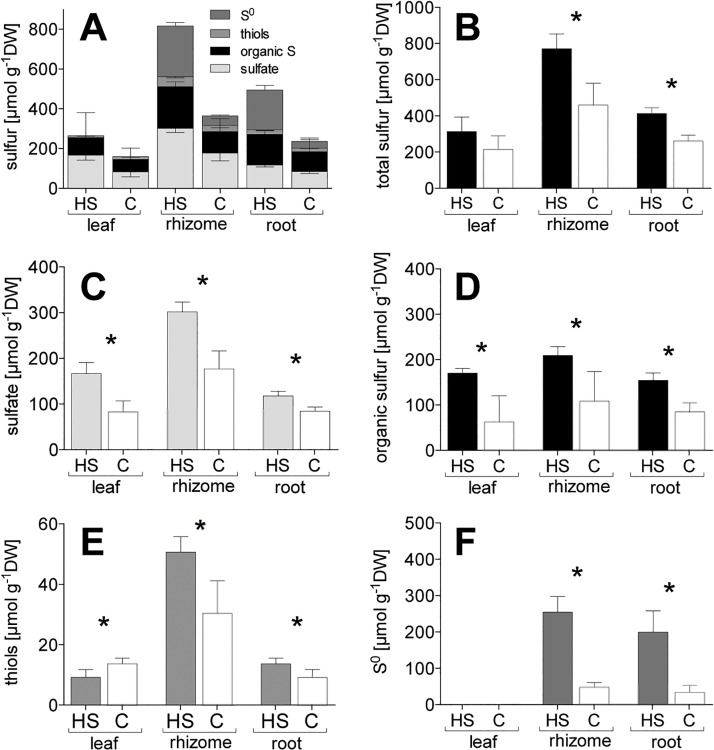
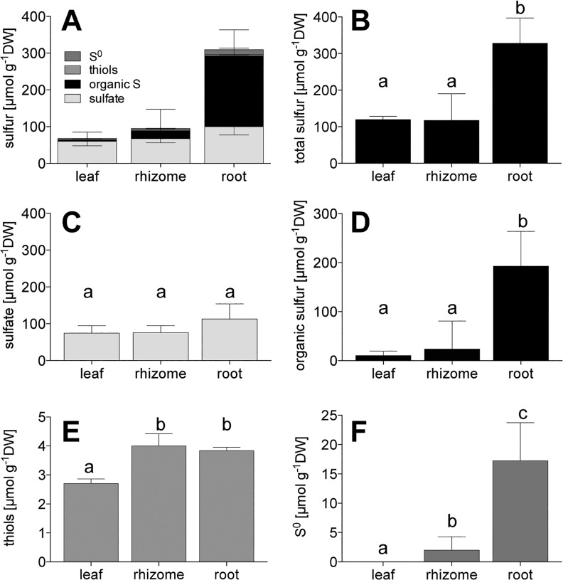
High sediment sulfide levels (HS) caused an accumulation of sulfur compounds in the roots, rhizomes, and leaves of Z. marina (Fig 1A): (i) The total sulfur content was highest in the rhizome followed by the roots, whereas there was no increased accumulation in the leaves (Fig 1B). (ii) The levels of sulfate and organic sulfur increased in all three tissues (Fig 1C and 1D). (iii) The levels of thiols increased in underground tissues and decreased in the leaves (Fig 1E). (iiii) There was high accumulation of S0 in the underground tissues and, remarkably, a total absence in leaves (Fig 1F). There are methodological problems for the detection of S0 in the leaves by UV-RPHPLC due to interference of chlorophyll with the S0 signal [22]. To overcome this issue, we assessed the amount of sulfur in the IF, which includes S0, and found no accumulation of sulfur in the leaves of C or HS (data not shown). However, the sulfur in the IF of underground tissues is exclusively S0. Therefore, we refer to δ34S-S0 when presenting the δ34S of the IF.
Fig 1. Sulfur budget of different fractions of Zostera marina in vitro (A) stacked values of all fractions, (B) total sulfur, (C) sulfate, (D) organic sulfur, (E) thiols, (F) S0.
Values represent mean ± SEM; n = 6. C, control; DW, dry weight; HS, high sulfide. Columns connected by the asterisk indicate significant intra-tissue differences (ANOVA and Tukey’s test, p < 0.05).
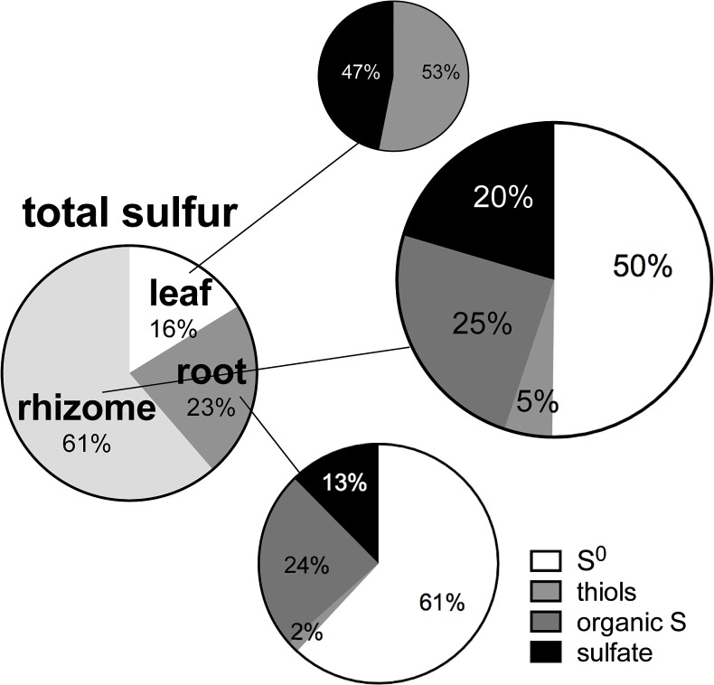
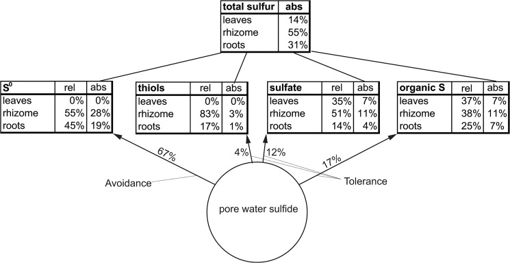
The net accumulation of total sulfur and sulfur compounds in HS plants (HS minus C) shows a major sulfur accumulation in underground tissues (84% in roots + rhizomes, 16% in leaves) (Fig 2) with a prominent share of S0 (45%). The proportion of S0 of TS in the rhizome was 61% and in the roots 50%.
Fig 2. Relative net accumulation of total sulfur in Zostera marina (HS minus C) including the assemblage total sulfur composition of sulfur compounds in the different tissues (n = 6).
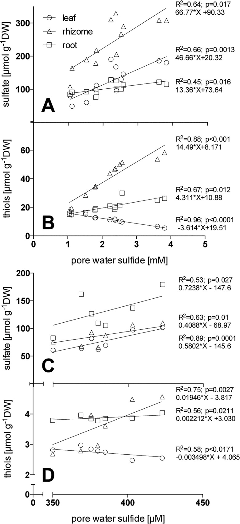
Plant sulfate levels were driven by pore water sulfide concentrations, showing a positive linear relationship: slopes were similar for roots and rhizomes but steeper for leaves (Fig 3A). Thiols in the plants were primarily influenced by pore water sulfide concentrations; related positively in underground tissues (with a steeper slope for the rhizome) and negatively in leaves (Fig 3B).
Fig 3. Linear regressions of pore water sulfide and Zostera marina tissue sulfate (A, C) and thiol concentration (B, D) from plants exposed to high pore water sulfide concentrations in experimental conditions (A, B) and from field plants (C, D); n = 12 for A, B and 9 for C, D.
DW, dry weight.
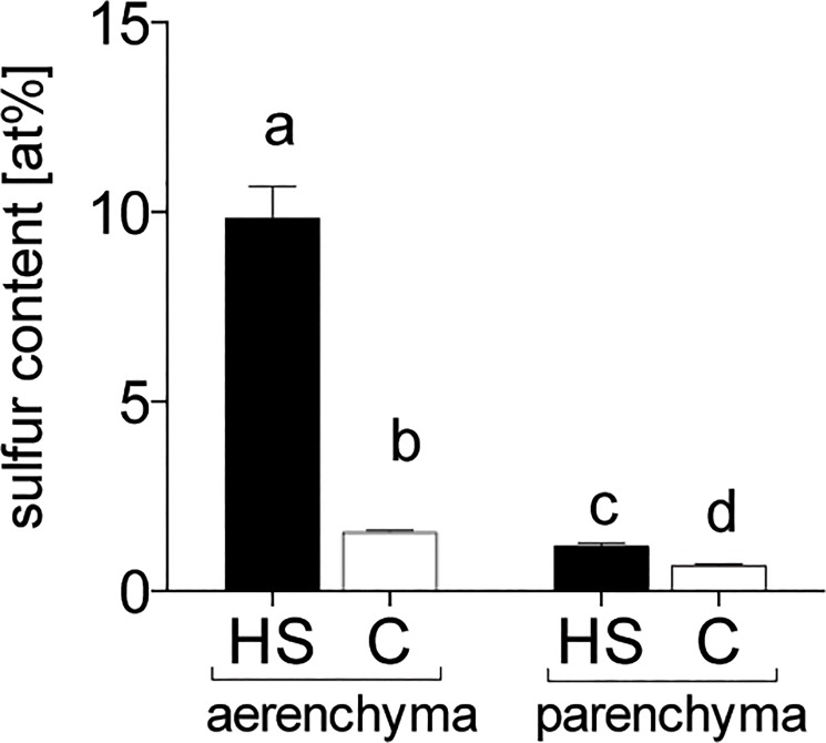
SEM-EDX analysis revealed sulfur accumulation to be higher in the root arenchyma than the parenchyma (Fig 4). HS treatment led to 6.4-fold higher sulfur content in aerenchymatous tissue and 1.7-fold higher in parenchymatous tissue compared with C.
Fig 4. Sulfur content in atomic-percent [at%] by SEM-EDX in Zostera marina roots exposed to sulfide intrusion.
Letters indicate significant differences (Dunn's test p < 0.05); n = 48. HS = high sulfide; C = control.
Stable isotope results
High pore water sulfide concentrations consistently increased the δ34S values of all sulfur compounds in the plants (Table 1), with higher values in the underground tissues than the leaves.
Table 1. Sulfur isotopic composition (δ34S, mean ± SEM) in per mille [‰] of bulk sulfur, insoluble sulfur compounds (S0), organic sulfur, and sulfate in different Zostera marina tissues cultivated under high sediment sulfide concentrations (HS) and control conditions (C); n = 6.
| Treatment | Tissue | δ34S [‰] | |||
|---|---|---|---|---|---|
| bulk S | S0 | organic sulfur | sulfate | ||
| HS | Leaf | 3.61 ± 0.62*A,b | 0.24 ± 0.49*B,c | 3.2 ± 0.67*A,b | 4.47 ± 0.28*A,a |
| Rhizome | 8.37 ± 0.99*A,a | 6.07 ± 0.61*A,a | 7.88 ± 1.06*A,a | 5.46 ± 0.3*A,a | |
| Root | 7.24 ± 0.99*A,a | 2.77 ± 0.67**B,b | 6.75 ± 1*A,a | 5.22 ± 0.27*AB,a | |
| C | Leaf | -2.12 ± 0.85*A,a | -4.03 ± 0.61*A,a | -1.9 ± 0.88*A,a | -2.76 ± 1.25*A,a |
| Rhizome | -7.29 ± 0.5*AB,b | -8.65 ± 0.51*B,b | -7.07 ± 0.51*AB,b | -5.11 ± 0.93*A,a | |
| Root | -5.6 ± 0.68*AB,b | -7.98 ± 0.61*B,b | -5.35 ± 0.7*A,b | -3.78 ± 0.49*A,a | |
Note that S0 was assessed from insoluble sulfur compounds. Asterisks indicate significant differences between HS and C. Capital letters indicate significant differences between compounds within a tissue; lower case letters indicate significant differences between the tissue types in a specific compound. ANOVA and post hoc Tukey’s test; p < 0.05.
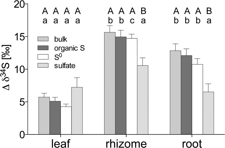
The Δδ34S of all plant sulfur compounds except sulfate were higher in the underground tissues than in the leaves (Fig 5). Δδ34S-sulfate did not vary between tissues. In the leaves, all sulfur compounds shifted to the same extent, whereas in the underground tissues, Δδ34S of bulk sulfur, organic sulfur, and S0 increased more than sulfate (Fig 5), indicating that sulfide is a precursor to these compounds. Δδ34S-sulfide explains to a high extent the increased Δδ34S levels of all sulfur compounds in underground tissues, and organic sulfur and sulfate in the leaves, respectively (S1 Fig, p < 0.01, R2 > 0.89); with similar slopes for the sulfur compounds within a tissue (p > 0.05; pooled slopes: leaf = 1.72, rhizome = 1.67, root = 1.62). In contrast, the Δδ34S of the other sulfur sources—pore water sulfate and water column sulfate—did not explain the increase in Δδ34S at all (linear regression; p > 0.05).
Fig 5. Sulfur isotopic response in sulfur compounds in Zostera marina upon sulfide intrusion.
Shift of average δ34S values (± SEM) in per mille [‰] of bulk sulfur and different sulfur compounds from Zostera marina between high sulfide concentrations (HS) and control conditions (C); n = 6. Capital letters indicate significant differences between compounds in a tissue and lower case letters indicate differences between tissues for a specific compound (ANOVA and Tukey’s test, p < 0.05).
Field study
The pore water sulfide concentrations ranged from 321 to 423 μmol L-1 at all sites, which are moderately high levels for sediments in coastal Denmark during the summer months. The δ34S value of sediment sulfide was not significantly different between sites and varied between −24.9‰ and −12.8‰ for AVS and −30.3‰ and −22.8‰ for CRS.
The highest accumulation of sulfur in field plants was found in the roots (Fig 6A and 6B), primarily caused by significantly higher accumulation of sulfate and organic sulfur (Fig 6D and 6F) (p < 0.0001). The levels of other sulfur compounds (thiols and S0) did not vary between tissues and sites (Fig 6E and 6F). The sulfate concentration in the plant tissues was positively linearly related to the pore water sulfide concentration, with similar slopes for root, rhizome, and leaves (p > 0.05; pooled slope = 0.57) (Fig 3C) and independent of site. Thiols in the plant were linearly related to pore water sulfide concentrations, positively in the underground tissues (with a steeper slope for the rhizome) and negatively in the leaves (Fig 3B). The δ34S of bulk sulfur, organic sulfur, and S0 in plants were most negative in the roots followed by rhizomes and leaves with the exception of δ34S-sulfate, which did not vary between tissues (Table 2) (p < 0.05).
Fig 6. Sulfur budget of different fractions of Zostera marina in situ (A) stacked values of all fractions, (B) total sulfur, (C) sulfate, (D) organic sulfur, (E) thiols, (F) S0.
Values represent mean ± SEM. Lower case letters indicate differences between tissues (ANOVA and Tukey’s test, p < 0.05); n = 12.
Table 2. Sulfur isotopic composition (δ34S, mean ± SEM) in per mille [‰] of bulk S, insoluble sulfur compounds (S0), organic sulfur, and sulfate in different Zostera marina tissues in situ; n = 6.
| Tissue | δ34S [‰] | |||
|---|---|---|---|---|
| bulk S | S0 | organic sulfur | sulfate | |
| Leaf | 1.71 ± 0.86A,a | 1 ± 0.98A,a | 3.02 ± 1.25A,a | 2.15 ± 0.55A,a |
| Rhizome | 0.61 ± 1AB,a | -2.17 ± 0.82B,a | 5.01 ± 1.38C,a | 2.14 ± 0.27A,a |
| Root | -10.24 ± 0.76A,b | -9.11 ± 0.96A,b | -11.27 ± 1.4A,b | 0.89 ± 0.73B,a |
Note that S0 was assessed from insoluble sulfur compounds. Capital letters indicate significant differences between compounds within a tissue; lower case letters indicate significant differences between the tissue types in a specific compound. ANOVA and post hoc Tukey’s test; p < 0.05.
Discussion
Identification of sulfur compounds
High levels of sulfide intrusion lead to increased sulfur pools in Z. marina, consisting of several sulfur compounds including S0, sulfate, organic sulfur, and thiols. S0 accumulated inside the aerenchyma, a pattern that was probably caused by chemical reoxidation of toxic sulfide and its precipitation as non-toxic S0 on the inner wall of the aerenchyma. This mechanism is indicated by high and similar accumulation of sulfur on the inner wall of the root aerenchyma (detected by SEM-EDX) and S0 in underground tissues exposed to sulfide intrusion. Both increased at similar rates—6.4-fold in the aerenchyma and 6.6-fold in the roots, respectively, but only 1.7-fold in the parenchyma. This is similar to the 1.4-fold increased sulfate accumulation in roots indicating that most S0 accumulates in the aerenchyma and most sulfate in the parenchyma. S0 seems unlikely to be present in the parenchyma. Roots and rhizomes exposed to high levels of sulfide can accumulate considerable amounts of S0, whereas S0 is not detectable in the leaves [6,11], suggesting that, even under high pore water sulfide concentrations coupled with hypoxic conditions and darkness, toxic gaseous sulfide does not reach the leaves. This also suggests that the sulfide-vulnerable leaf meristem, located at the basal part of the leaf, remains unexposed to sulfide. Similarly, Pedersen et al. [13] did not detect sulfide in the meristematic region of Z. marina at water column oxygen saturation levels of above 35%. The detoxification of sulfide to S0 in Z. marina supports previous studies proposing this mechanism [11,52,53] and we speculate that the precipitated S0 in the aerenchyma is permanently deposited and represents a sink of oxidized intruding sediment sulfide. Some terrestrial plants actively metabolize S0, which functions against fungal pathogens [25], but this is probably not the case for Z. marina, as the accumulation of S0 is due to passive chemical oxidation (by oxygen) rather than an active process. Further, the antifungal function seems unlikely because of the low impact of fungi in seagrass systems [54,55]. Thus, S0 presumably functions exclusively as a detoxification compound in underground tissues and can be used as an indicator of sulfide presence in the aerenchyma.
In contrast to S0, sulfate accumulated in both underground and above-ground tissues of Z. marina exposed to sulfide intrusion. Theoretically, three possible sulfate sources are present in seagrass beds: 1) sulfate derived from oxidation of sulfide in the leaves; 2) sulfate taken up from pore water and/or the water column; and 3) sulfate derived from oxidation of sulfide in underground tissue followed by translocation to the leaves. In this study, the first mechanism can be discounted due to the absence of sulfide in the leaves. Concentration- and demand-dependent uptake of sulfate from pore water and the water column can be discounted in this study due to the constant and high concentrations of the sulfur pools in all treatments; also, there is no expected increase in sulfur demand upon sulfide intrusion [56]. In addition, root sulfate uptake plays a minor role in plants exposed to gaseous sulfide [56] and we expect a similar lack of response for seagrasses. These findings favor the third hypothesis: that sulfate accumulation in Z. marina upon sulfide intrusion originates from direct oxidation of intruding sediment sulfide. This is further supported by the positive linear relation between pore water sulfide and tissue sulfate concentration, suggesting a positive coupling between sediment sulfide and plant sulfate accumulation. However, the excess sulfate derived from oxidized sulfide is probably surplus to demand and consequently transferred to vacuoles, where it can be stored [35]. Sulfate stored in the vacuoles might represent a possible end product of sulfide detoxification, similar to the function of S0.
Incorporation of sediment sulfide
The increased thiol content in underground tissues of Z. marina indicates that intruding sulfides penetrating the tissue are incorporated into thiol compounds. Sulfide is probably enzymatically incorporated directly into cysteine and subsequently metabolized into organic sulfur compounds, as observed in terrestrial plants [32,57]. The thiols cysteine and glutathione represent important precursors of all organic sulfur compounds [58], feeding the synthesis of organic sulfur in Z. marina and pinpointing the relevance of the small but reactive thiol pool in controlling the sulfur metabolism in plants [59]. The thiol content in underground tissues increased linearly with sulfide concentration, suggesting no limitation of the sulfide incorporation mechanism under the experimental conditions. Similarly Westerman et al. [60] found linearity between atmospheric sulfide levels and the increase in thiol and cysteine concentration in the terrestrial plant B. oleracea. This linearity indicates a constant sulfide incorporation mechanism in Z. marina that cannot be controlled by the plant and that facilitates sulfide detoxification in seagrass.
The organic sulfur content increased in leaves, roots, and rhizomes upon sulfide intrusion and accounts for a substantial amount (29%) of the increase in total sulfur content. The accumulation of organic sulfur could be a result of thiol metabolism, thereby bypassing the “normal” assimilatory sulfur uptake pathway of sulfate via the roots or leaves. This suggests a possible role of sediment sulfide in the sulfur nutrition in Z. marina, as has been found for terrestrial plants exposed to sulfide fumigation [33,34].
Stable isotope tracing
We traced the Δδ34S signal of sediment sulfide in the leaf, rhizome, and root tissues of Z. marina exposed to sediment sulfide, to assess the source of the accumulating sulfur compounds. Roots and rhizomes exposed to sulfide intrusion accumulated sulfur compounds characterized by a Δδ34S close to that of sediment sulfide, implying that sediment sulfide was the sole source of accumulated sulfur. Thus, a high share of the detoxification of sediment sulfide (86%) takes place in the underground tissues. In contrast, the leaves are characterized by a lower Δδ34S implying a lower share of sediment sulfide and thereby a lower contribution of the leaves to sulfide detoxification (14%).
The precipitation of sulfide as S0 is by far the dominant detoxification mechanism (67%), avoiding sulfide intruding from the aerenchyma into parenchymatous tissues. The remaining sulfide (33%) radially intruding from the aerenchyma into parenchymatous tissue is seized by a tolerance mechanism in which sulfides are incorporated into thiol compounds followed by metabolism to organic sulfur. Avoidance and tolerance mechanisms act in concert to avoid intruding sediment sulfide reaching the vulnerable leaf meristem.
Organic sulfur and S0 formed in the underground tissues upon sulfide intrusion are not translocated to the leaf, as indicated by the different Δδ34S of the tissues. In contrast, the similarity of roots, rhizome, and leaves in Δδ34S of plant sulfate accumulating upon sulfide intrusion implies that sulfate is the only compound that is translocated from underground to above ground tissues. This supports Rennenberg’s [35] suggestion that sulfate is the main sulfur transport form for plants in general. The mechanism lowers sulfur accumulation in underground tissues by translocating sulfate derived from toxic sulfide to the leaves, probably avoiding harmful hyperaccumulation of sulfate [35] in underground tissues.
We propose a conceptual model of relative and absolute sulfide detoxification capacities, based on the contribution of sulfur sources to the different plant sulfur compounds after sulfide intrusion (Fig 7). It is noteworthy to mention that the mechanisms described above occur only upon high rates of sediment sulfide intrusion.
Fig 7. Conceptual model of sulfide detoxification capacities of Zostera marina exposed to sulfide intrusion based on stable isotope tracing and mass balance modeling.
The numbers (percentage) next to the arrows indicate the contribution of a sulfur compound to the detoxification process. The numbers (percentage) under ‘rel’ indicate the relative distribution of a sulfur compound between the tissues; the numbers under ‘abs’ indicate the share of the net total sulfur accumulation upon sulfide intrusion of the compounds in a tissue. The total sulfur table indicates the share of the different tissues in detoxification of sulfide. Avoidance is indicated by preventing sulfide intrusion by precipitation of sulfide to S0 and tolerance by standing sulfide intrusion by incorporating sulfide into thiols, organic sulfur, and sulfate.
Sulfur compounds and intrusion under natural conditions
Under natural conditions and moderate levels of pore water sulfide, roots accumulate the most sulfur, with the most negative δ34S, followed by rhizomes and leaves (Table 2) [24,52]. This indicates that sulfide intrusion is constant in the roots, occasional in the rhizomes, and non-existent in leaves, which is further supported by the presence of S0 in roots and rhizomes, albeit at low concentrations [24,52]. However, even at low sulfide levels, sulfide is incorporated and metabolized via thiols into organic sulfur. This is indicated by the δ34S signal of organic sulfur ranging in between those of sediment sulfide and pore water sulfate, suggesting mixed sulfur uptake from both sources. This suggests that under natural conditions—without hypoxia events—ROL is sufficient to protect the leaf meristem from sulfide intrusion. Moderate sulfide intrusion constantly happens during the night but the sulfide is completely detoxified in the roots and only marginal amounts reach the rhizome, without harming the plants. Although field studies are not directly comparable with mesocosm studies, the approximately five-times-lower sulfide concentrations in the field provide a lower sulfide pressure that most likely results in lower levels of sulfide intrusion. This may explain the lower amounts of sulfur compounds in the field plants, similar to previous findings in field plants [6,11,52].
Detoxification pathway
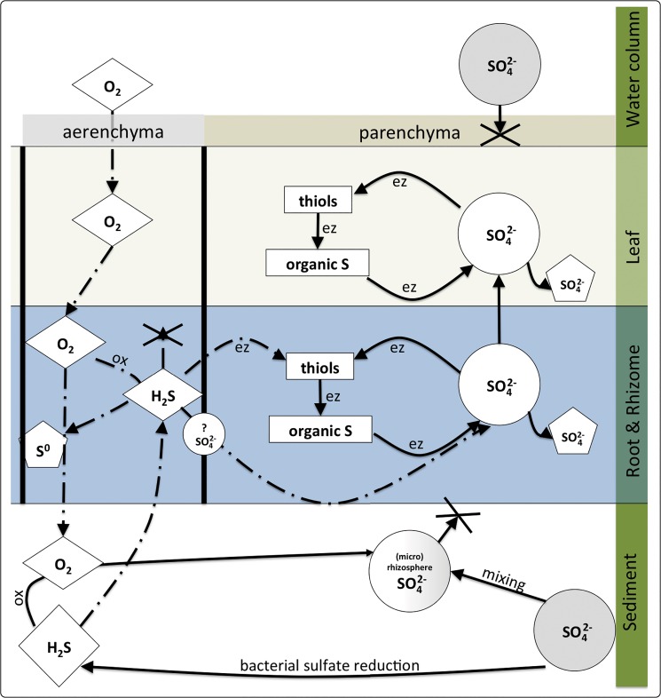
The results of our quantification of sulfur compounds and stable isotope tracing are summarized in a conceptual model in Fig 8. Sulfate in the pore water is reduced by sulfate-reducing bacteria to sulfide, which is depleted in 34S due to isotopic fractionation. The sulfide is subsequently oxidized to sulfate by ROL in the oxic halo around the roots; this process may be facilitated by sulfide oxidizing bacteria [20,61]. However the resulting sulfate has a δ34S similar to sediment sulfide, and is mixed with the bulk pore water pool of sulfate, resulting in a δ34S somewhere between those of sulfide and sulfate. During events of hypoxia and darkness ROL decreases and sediment-derived sulfide may intrude into the aerenchyma of roots and rhizomes. The intruding sulfide is chemically oxidized by oxygen to S0, precipitating on the inner wall of the aerenchyma, or oxidized to sulfate. The remaining sulfide radially penetrates the parenchymatous tissue and undergoes enzyme-facilitated incorporation into thiols. Thiols are further metabolized into organic sulfur or oxidized to sulfate. Thus, the oxidation of sulfide to sulfate occurs either in the aerenchyma or inside the tissue. Sulfate formed in the underground tissues is stored or translocated to the leaves, where it is stored and/or fuels sulfur metabolism. Under oxic water column conditions, the detoxification mechanisms prevent the intruding sulfide from penetrating the meristematic region in the leaf, whereas under anoxic or hypoxic conditions the sulfide may enter and lead to toxicity.
Fig 8. Conceptual model of sediment-sulfide intrusion, sulfate uptake, and possible sulfide accumulation/detoxification patterns in Zostera marina.
Dash-dotted lines = gas diffusion; dashed lines = liquid diffusion; circles = inorganic sulfur; rectangles = organic sulfur; diamonds = gaseous compounds; pentagons = detoxified and permanently stored sulfur; ox = oxidation; ez = enzymatic pathway. Symbols marked with a question mark indicate an unknown reaction. Symbols shaded in grey indicate sulfur pools enriched in 34S. White symbols indicate depleted 34S sulfur pools and symbols with a grey/white gradient indicate mixed δ34S pools.
Conclusions
Seagrasses can thrive in sulfidic sediments due to two major detoxification strategies: avoidance, by oxidation of sulfide; and tolerance, by incorporation of sulfide into plant tissues. Sulfide is oxidized and precipitated as S0 in the aerenchyma or is oxidized to sulfate inside or outside the plant. Further sulfide is incorporated as thiols and sulfate. S0 and thiols are stored in rhizomes and roots, whereas sulfate is transported from under- to above-ground tissues. Underground tissues possess the highest detoxification capacity (86%), and the rhizome (61%) acts as the primary buffer in detoxifying toxic sediment sulfide and protecting the vulnerable meristematic tissue in the leaves. Future climate scenarios predicting higher surface water temperatures [62], higher frequencies of hypoxic events [63] and increasing sediment sulfide levels [64] will lead to higher sulfide pressure on seagrass ecosystems. Such conditions might exceed the sulfide tolerance and detoxification capacities. Our findings open a discussion of how seagrasses cope with sulfidic sediments in future climate scenarios and how detoxification mechanisms vary between seagrass species and growth stages.
Supporting Information
R2, p, and the linear regression equation are presented next to the plot. asnf = assumptions for linear regression not fulfilled. n = 6.
(DOCX)
n = 12.
(DOCX)
Acknowledgments
We are in debt to Dr. Marieluise Weidinger and MSc. Daniela Gruber from the Core Facility Cell Imaging and Ultrastructure Research (CIUS), University of Vienna, Austria for assistance with the SEM-EDX analysis. Many thanks to Dr. Jens Borum and Dr. Max Castorani for critical reviews on earlier versions of this manuscript and fruitful discussion that substantially improved this paper. We are thankful to the staff of the University of Southern Demark for support during field work and lab analysis.
Data Availability
All relevant data are within the paper, its Supporting Information files or available from the public repository Figshare: http://dx.doi.org/10.6084/m9.figshare.1378709.
Funding Statement
Harald Hasler-Sheetal was funded by the Danish Council of Independent Research (grant no. 09-067485), Marianne Holmer was founded by the Danish National Research foundation (grant no. 12-127012). The funders had no role in study design, data collection and analysis, decision to publish, or preparation of the manuscript.
References
- 1. Terrados J, Duarte CM, Kamp-Nielsen L, Agawin NSR, Gacia E, Lacap D, et al. (1999) Are seagrass growth and survival constrained by the reducing conditions of the sediment? Aquat Bot 65: 175–197. [Google Scholar]
- 2. Fenchel T, Finlay BJ (1995) Ecology and evolution in anoxic worlds; May RM, Harvey PH, editors. New York: Oxford University Press Inc. [Google Scholar]
- 3. Lamers LPM, Govers LL, Janssen ICJM, Geurts JJM, Van der Welle MEW, Van Katwijk MM, et al. (2013) Sulfide as a soil phytotoxin—A review. Front Plant Sci 4: 1–14. 10.3389/fpls.2013.00001 [DOI] [PMC free article] [PubMed] [Google Scholar]
- 4. Garcias-Bonet N, Marba N, Holmer M, Duarte CM (2008) Effects of sediment sulfides on seagrass Posidonia oceanica meristematic activity. Mar Ecol Prog Ser 372: 1–6. [Google Scholar]
- 5. Holmer M, Hasler-Sheetal H (2014) Sulfide intrusion in seagrasses assessed by stable sulfur isotopes – A synthesis of current results. Front Mar Sci 1:64. [Google Scholar]
- 6. Frederiksen MS, Holmer M, Perez M, Invers O, Ruiz JM, Knudsen BB (2008) Effect of increased sediment sulfide concentrations on the composition of stable sulfur isotopes (delta S-34) and sulfur accumulation in the seagrasses Zostera marina and Posidonia oceanica . J Exp Mar Biol Ecol 358: 98–109. [Google Scholar]
- 7. Short FT, Carruthers T, Dennison W, Waycott M (2007) Global seagrass distribution and diversity: A bioregional model. J Exp Mar Biol Ecol 350: 3–20. [Google Scholar]
- 8. Dooley FD, Wyllie-Echeverria S, Roth MB, Ward PD (2013) Tolerance and response of Zostera marina seedlings to hydrogen sulfide. Aquat Bot 105: 7–10. [Google Scholar]
- 9. Koch MS, Schopmeyer SA, Nielsen OI, Kyhn-Hansen C, Madden CJ (2007) Conceptual model of seagrass die-off in Florida Bay: Links to biogeochemical processes. J Exp Mar Biol Ecol 350: 73–88. [Google Scholar]
- 10. Borum J, Pedersen O, Greve TM, Frankovich TA, Zieman JC, Fourqurean JW, et al. (2005) The potential role of plant oxygen and sulphide dynamics in die-off events of the tropical seagrass, Thalassia testudinum . J Ecol 93: 148–158. [Google Scholar]
- 11. Mascaro O, Valdemarsen T, Holmer M, Perez M, Romero J (2009) Experimental manipulation of sediment organic content and water column aeration reduces Zostera marina (eelgrass) growth and survival. J Exp Mar Biol Ecol 373: 26–34. [Google Scholar]
- 12. Jensen SI, Kühl M, Priemé A (2007) Different bacterial communities associated with the roots and bulk sediment of the seagrass Zostera marina . FEMS Microbiol Ecol 62: 108–117. [DOI] [PubMed] [Google Scholar]
- 13. Pedersen O, Binzer T, Borum J (2004) Sulphide intrusion in eelgrass (Zostera marina L.). Plant Cell Environ 27: 595–602. [Google Scholar]
- 14. Armstrong W, Brandle R, Jackson MB (1994) Mechanisms of flood tolerance in plants. Acta Bot Neerl 43: 307–358. [Google Scholar]
- 15. Frederiksen MS, Glud RN (2006) Oxygen dynamics in the rhizosphere of Zostera marina: A two-dimensional planar optode study. Limnol Oceanogr 51: 1072–1083. Limnol Oceanogr 51: 1072–1083. [Google Scholar]
- 16. Armstrong J, Armstrong W (2001) Rice and Phragmites: Effects of organic acids on growth, root permeability, and radial oxygen loss to the rhizosphere. Am J Bot 88: 1359–1370. [PubMed] [Google Scholar]
- 17. Luther GW, Findlay AJ, MacDonald DJ, Owings SM, Hanson TE, Beinart RA, et al. (2011) Thermodynamics and kinetics of sulfide oxidation by oxygen: A look at inorganically controlled reactions and biologically mediated processes in the environment. Front Microbiol 2. [DOI] [PMC free article] [PubMed] [Google Scholar]
- 18. Cline JD, Richards FA (1969) Oxygenation of hydrogen sulfide in seawater at constant salinity, temperature and pH. Environ Sci Technol 3: 838–843. [Google Scholar]
- 19. Jensen SI, Kühl M, Glud RN, Jørgensen LB, Priemé A (2005) Oxic microzones and radial oxygen loss from roots of Zostera marina . Mar Ecol Prog Ser 293: 49–58. [Google Scholar]
- 20. van der Heide T, Govers LL, de Fouw J, Olff H, van der Geest M, van Katwijk MM, et al. (2012) A three-stage symbiosis forms the foundation of seagrass ecosystems. Science 336: 1432–1434. 10.1126/science.1219973 [DOI] [PubMed] [Google Scholar]
- 21. Sand-Jensen K, Pedersen O, Binzer T, Borum J (2005) Contrasting oxygen dynamics in the freshwater isoetid Lobelia dortmanna and the marine seagrass Zostera marina . Ann Bot 96: 613–623. [DOI] [PMC free article] [PubMed] [Google Scholar]
- 22. Holmer M, Frederiksen MS, Mollegaard H (2005) Sulfur accumulation in eelgrass (Zostera marina) and effect of sulfur on eelgrass growth. Aquat Bot 81: 367–379. [Google Scholar]
- 23. Holmer M, Kendrick G (2013) High sulfide intrusion in five temperate seagrasses growing under contrasting sediment conditions. Estuaries and Coasts 36: 116–126. [Google Scholar]
- 24. Borum J, Raun AL, Hasler-Sheetal H, Pedersen MØ, Pedersen O, Holmer M (2013) Eelgrass fairy rings: sulfide as inhibiting agent. Mar Biol 161: 351–358. [Google Scholar]
- 25. Cooper RM, Williams JS (2004) Elemental sulphur as an induced antifungal substance in plant defence. J Exp Bot 55: 1947–1953. [DOI] [PubMed] [Google Scholar]
- 26. Rausch T, Wachter A (2005) Sulfur metabolism: a versatile platform for launching defence operations. Trends Plant Sci 10: 503–509. [DOI] [PubMed] [Google Scholar]
- 27. Mathai JC, Missner A, Kügler P, Saparov SM, Zeidel ML, Lee JK, et al. (2009) No facilitator required for membrane transport of hydrogen sulfide. Proc Natl Acad Sci U S A 106: 16633–16638. 10.1073/pnas.0902952106 [DOI] [PMC free article] [PubMed] [Google Scholar]
- 28. Raven JA, Scrimgeour CM (1997) The influence of anoxia on plants of saline habitats with special reference to the sulphur cycle. Ann Bot 79: 79–86. [Google Scholar]
- 29. Fürtig K, Ruegsegger A, Brunold C, Brandle R (1996) Sulphide utilization and injuries in hypoxic roots and rhizomes of common reed (Phragmites australis). Folia Geobot Phytotaxon 31: 143–151. [Google Scholar]
- 30. de Kok LJ, Stahl K, Rennenberg H (1989) Fluxes of atmospheric hydrogen sulphide to plant shoots. New Phytol 112: 533–542. [DOI] [PubMed] [Google Scholar]
- 31. Weber M, Brändle R (1996) Some aspects of the extreme anoxia tolerance of the sweet flag, Acorus calamus L. Folia Geobot 31: 37–46. [Google Scholar]
- 32. Durenkamp M, de Kok LJ (2004) Impact of pedospheric and atmospheric sulphur nutrition on sulphur metabolism of Allium cepa L., a species with a potential sink capacity for secondary sulphur compounds. J Exp Bot 55: 1821–1830. [DOI] [PubMed] [Google Scholar]
- 33. Buchner P, Stuiver CEE, Westerman S, Wirtz M, Hell R, Hawkesford MJ, et al. (2004) Regulation of sulfate uptake and expression of sulfate transporter genes in Brassica oleracea as affected by atmospheric H2S and pedospheric sulfate nutrition. Plant Physiol 136: 3396–3408. [DOI] [PMC free article] [PubMed] [Google Scholar]
- 34. de Kok LJ, Stuiver CEE, Rubinigg M, Westerman S, Grill D (1997) Impact of atmospheric sulfur deposition on sulfur metabolism in plants: H2S as sulfur source for sulfur deprived Brassica oleracea L. Bot Acta 110: 411–419. [Google Scholar]
- 35. Rennenberg H (1984) The fate of excess sulfur in higher plants. Annu Rev Plant Physiol 35: 121–153. [Google Scholar]
- 36. Buchner P, Takahashi H, Hawkesford MJ (2004) Plant sulphate transporters: co-ordination of uptake, intracellular and long-distance transport. J Exp Bot 55: 1765–1773. [DOI] [PubMed] [Google Scholar]
- 37. Rees CE, Jenkins WJ, Monster J (1978) The sulphur isotopic composition of ocean water sulphate. Geochim Cosmochim Acta 42: 377–381. [Google Scholar]
- 38. Fry B, Scalan RS, Winters JK, Parker PL (1982) Sulfur uptake by salt grasses, mangroves, and seagrasses in anaerobic sediments. Geochim Cosmochim Acta 46: 1121–1124. [Google Scholar]
- 39. Valdemarsen T, Kristensen E, Holmer M (2009) Metabolic threshold and sulfide-buffering in diffusion controlled marine sediments impacted by continuous organic enrichment. Biogeochemistry 95: 335–353. [Google Scholar]
- 40. Sand-Jensen K (1975) Biomass, net production and growth dynamics in an eelgrass (Zostera marina L) population in Vellerup Vig, Denmark. Ophelia 14: 185–201. [Google Scholar]
- 41. Zopfi J, Kjaer T, Nielsen LP, Jorgensen BB (2001) Ecology of Thioploca spp.: nitrate and sulfur storage in relation to chemical microgradients and influence of Thioploca spp. on the sedimentary nitrogen cycle. Appl Environ Microbiol 67: 5530–5537. [DOI] [PMC free article] [PubMed] [Google Scholar]
- 42. de Kok LJ, Buwalda F, Bosma W (1988) Determination of cysteine and its accumulation in spinach leaf tissue upon exposure to excess sulfur. J Plant Physiol 133: 502–505. [Google Scholar]
- 43. Williams JS, Hall SA, Hawkesford MJ, Beale MH, Cooper RM (2002) Elemental sulfur and thiol accumulation in tomato and defense against a fungal vascular pathogen. Plant Physiol 128: 150–159. [PMC free article] [PubMed] [Google Scholar]
- 44. Vetter RD (1985) Elemental sulfur in the gills of three species of clams containing chemoautotrophic symbiotic bacteria: a possible inorganic energy storage compound. Mar Biol 88: 33–42. [Google Scholar]
- 45. Lechaire J- P, Frébourg G, Gaill F, Gros O (2006) In situ localization of sulphur in the thioautotrophic symbiotic model Lucina pectinata (Gmelin, 1791) by cryo-EFTEM microanalysis. Biol Cell 98: 163–170. [DOI] [PubMed] [Google Scholar]
- 46. Himmel D, Maurin LC, Gros O, Mansot J-L (2009) Raman microspectrometry sulfur detection and characterization in the marine ectosymbiotic nematode Eubostrichus dianae (Desmodoridae, Stilbonematidae). Biol Cell 101: 43–54. 10.1042/BC20080051 [DOI] [PubMed] [Google Scholar]
- 47. Cline JD (1969) Spectrophotomitric determination of hydrogen sulfide in natural waters. Limnol Oceanogr 14: 454–458. [Google Scholar]
- 48. Solorzano L (1969) Determination of ammonia in natural waters by the phenolhypochlorite method. Limnol Oceanogr 14: 799–801. [Google Scholar]
- 49. Hansen HP, Koroleff F (2007) Determination of nutrients In: Grasshoff K, Kremling K, Ehrhardt M, editors. Methods of Seawater Analysis. 3rd ed. Weinheim, Germany: Wiley-VCH Verlag GmbH; pp. 159–228. [Google Scholar]
- 50. Fossing H, Jørgensen BB (1989) Measurement of bacterial sulfate reduction in sediments: Evaluation of a single-step chromium reduction method. Biogeochemistry 8: 205–222. [Google Scholar]
- 51. Canfield DE (2001) Isotope fractionation by natural populations of sulfate-reducing bacteria. Geochim Cosmochim Acta 65: 1117–1124. [Google Scholar]
- 52. Frederiksen MS, Holmer M, Borum J, Kennedy H (2006) Temporal and spatial variation of sulfide invasion in eelgrass (Zostera marina) as reflected by its sulfur isotopic composition. Limnol Oceanogr 51: 2308–2318. [Google Scholar]
- 53. Holmer M, Nielsen RM (2007) Effects of filamentous algal mats on sulfide invasion in eelgrass (Zostera marina). J Exp Mar Biol Ecol 353: 245–252. [Google Scholar]
- 54. Blum L, Mills A (1991) Microbial growth and activity during the initial stages of seagrass decomposition. Mar Ecol Prog Ser 70: 73–82. [Google Scholar]
- 55. Nielsen SL, Thingstrup I, Wigand C (1999) Apparent lack of vesicular–arbuscular mycorrhiza (VAM) in the seagrasses Zostera marina L. and Thalassia testudinum Banks ex König . Aquat Bot 63: 261–266. [Google Scholar]
- 56. Tausz M, Weidner W, Wonisch A, De Kok LJ, Grill D (2003) Uptake and distribution of 35S-sulfate in needles and roots of spruce seedlings as affected by exposure to SO2 and H2S. Environ Exp Bot 50: 211–220. [Google Scholar]
- 57. de Kok LJ, Bosma W, Maas FM, Kuiper PJC (1985) The effect of short-term H2S fumigation on water-soluble sulphydryl and glutathione levels in spinach. Plant Cell Environ 8: 189–194. [Google Scholar]
- 58. Heldt HW, Piechulla B (2011) Sulfate assimilation enables the synthesis of sulfur containing compounds Plant Biochemistry (Fourth Edition). San Diego: Academic Press; pp. 323–335. [Google Scholar]
- 59. Hawkesford MJ, de Kok LJ (2006) Managing sulphur metabolism in plants. Plant Cell Environ 29: 382–395. [DOI] [PubMed] [Google Scholar]
- 60. Westerman S, de Kok LJ, Stuiver CEE, Stulen I (2000) Interaction between metabolism of atmospheric H2S in the shoot and sulfate uptake by the roots of curly kale (Brassica oleracea). Physiol Plant 109: 443–449. [Google Scholar]
- 61. García-Martínez M, López-López A, Calleja ML, Marbà N, Duarte CM (2009) Bacterial community dynamics in a deagrass (Posidonia oceanica) meadow sediment. Estuaries and Coasts 32: 276–286. [Google Scholar]
- 62.IPCC (2007) Climate Change 2007: The physical science basis. Contribution of working group I to the fourth assessment report of the intergovernmental panel on climate change. Cambridge. 996 p.
- 63. Orth RJ, Carruthers TJB, Dennison WC, Duarte CM, Fourqurean JW, Heck KL, et al. (2006) A Global Crisis for Seagrass Ecosystems. Bioscience 56: 987–996. [Google Scholar]
- 64. García R, Holmer M, Duarte CM, Marbà N (2013) Global warming enhances sulphide stress in a key seagrass species (NW Mediterranean). Global Change Biol 19: 3629–3639. [DOI] [PubMed] [Google Scholar]
Associated Data
This section collects any data citations, data availability statements, or supplementary materials included in this article.
Supplementary Materials
R2, p, and the linear regression equation are presented next to the plot. asnf = assumptions for linear regression not fulfilled. n = 6.
(DOCX)
n = 12.
(DOCX)
Data Availability Statement
All relevant data are within the paper, its Supporting Information files or available from the public repository Figshare: http://dx.doi.org/10.6084/m9.figshare.1378709.


