Abstract
Gallstone ileus is an uncommon complication of cholelithiasis. Most patients affected by gallstone ileus are elderly and have multiple comorbidities. Symptoms are vague and insidious, which may delay the correct diagnosis for days. Here we are reporting an uncommon complication of gallstone ileus. We report on a 70-year-old man with small bowel obstruction at the jejunum due to an impacted stone, which led to necrosis and perforation of the proximal bowel wall. Laparoscope-assisted small bowel resection with enterolithotomy was used to successfully treat the patient's perforation and obstruction. His recovery was uneventful. Gallstone ileus commonly presents with bowel obstruction, but intestinal perforation occurs very rarely. A laparoscopic approach can provide both diagnostic and therapeutic roles in management.
Key words: Gallstone ileus, Jejunal perforation, Laparoscopic surgery, Intestinal obstruction
Gallstone ileus is characterized by intestinal obstruction due to intraluminal impaction of one or more gallstones. It is an uncommon but serious complication of cholelithiasis and accounts for 1% to 3% of cases of patients who undergo surgery for bowel obstruction.1,2 A cholecystoduodenal fistula is the most common tract.3 Most patients are elderly and female, and the average age range is 65 to 75 years. In spite of treatment, the mortality rate ranges from 10% to 20%.4,5
Gallstone ileus with proximal small bowel perforation is rare, and fewer than 10 cases have been reported in the medical literature.6 We describe a case of gallstone ileus with jejunum perforation that was successfully treated with laparoscopically assisted surgery.
Case Report
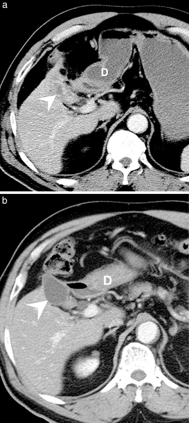
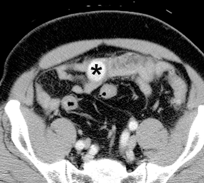
A 70-year-old man presented with a 1-day history of nausea, vomiting, and periumbilical abdominal pain. The patient had type 2 diabetes mellitus, cardiovascular disease, hypertension, and a history of gallstones. He also had a history of gallstone-related acute cholecystitis 2 years prior to this episode. Acute cholecystitis was successfully managed medically. This episode was quite different: this time the patient developed severe, persistent, and unrelenting abdominal pain, accompanied by biliary vomiting all in a single day. The patient had normal bowel movements and denied having any abdominal symptoms in recent months. His hemodynamics were stable, and the results of laboratory tests revealed leukocytosis. Abdominal palpation produced tenderness over the epigastric area, with equivocal peritoneal signs. Abdominal radiograms showed an area of mild ileus in the upper abdomen. A subsequent computed tomographic (CT) scan of the abdomen demonstrated a collapsed gallbladder with thick walls (Fig. 1a). In addition, the jejunum was obstructed, and there was proximal bowel dilatation by an impacted gallstone. No signs of bowel wall necrosis or free air were seen (Fig. 2). A tentative preoperative diagnosis of gallstone ileus was made.
Fig. 1.
(a) Abdominal CT scan upon admission. Note the inflammatory change around the entire gallbladder without gallstones. D, duodenum; arrowhead, gallbladder. (b) Abdominal CT scan 1 year after surgery. Note the lack of inflammation around the gallbladder. D, duodenum; arrowhead, gallbladder.
Fig. 2.
The missing stone was found in the lumen of the small intestine over the lower abdomen. Asterisk, gallstone.
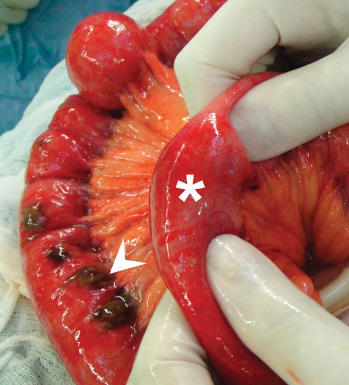
Laparoscopically assisted enterolithotomy was planned. However, after entering and exploring the abdomen, several patches of necrosis were found on the wall of the proximal jejunum. A small amount of dirty ascites was noted around the left upper abdomen. One segmental area of the small bowel containing a gallstone was identified; further exploration showed that another part of the small bowel distal to the obstructed segment was normal. The umbilical port wound was extended to about 8 cm in length, long enough to extract the bowel. The stone could be palpated easily and was located 150 cm from the ligament of Treitz. There were 4 areas of necrotic wall with purulent coating in the jejunum about 50 cm proximal to the stone (Fig. 3). Small bowel resection and anastomosis with removal of the stone were performed. The stone was 3.4 cm in length.
Fig. 3.
The stone (asterisk) impacted in the lumen could be easily palpated. Note the 4 areas of necrotic wall (arrowhead) on the left in this photo, and some purulent coating in the jejunum.
The patient had an uncomplicated recovery and was discharged 12 days after surgery. Final pathologic studies showed several small bowel perforations on the necrotic walls. The patient had no episodes of cholangitis or cholecystitis in the following year. An abdominal CT scan performed after an attack of sigmoid diverticulitis 1 year later showed that the inflammation around the gallbladder had subsided (Fig. 1b).
Discussion
Gallstone ileus is an uncommon disease and accounts for 1% of cases of mechanical intestinal obstruction. Bowel perforation after gallstone ileus is very rare. To our knowledge, only 10 other cases have been reported in the literature. The perforated site can be located in the jejunum, ileum, or sigmoid.6–15 The possible mechanism is that mucosal ulcer with bowel perforation can develop after direct high-pressure compression by an impacted stone. The impacted stone also can cause severe bowel obstruction, leading to high intraluminal pressure in the jejunum. Finally, such high pressure can result in bowel ischemic necrosis or perforation, especially in patients with jejunal diverticula.7–9 Our patient had rapidly progressed within 1 day to having acute abdominal pain, total bowel obstruction, and rapidly elevated intraluminal pressure compromising the perfusion of bowel to several patches of wall necrosis. The reason for such rapidly progressing disease was associated with his underlying cardiovascular disease and a huge stone in the small lumen. The former influenced the tissue perfusion and the latter caused severe obstruction.
Despite the improvement of diagnostic tools and perioperative care over the past decade, the surgical mortality rate is still high, ranging from 10% to 20%. There are two mainstream surgical procedures for gallstone ileus. The one-stage operation consists of enterolithotomy, cholecystectomy, and closure of the fistula, whereas the two-stage operation involves enterolithotomy first and fistula repair later, if indicated. The best surgical procedure for gallstone ileus is still being debated. The one-stage operation has led to higher mortality and morbidity rates in these mostly elderly patients.3–5 As a result, most doctors prefer using a simple procedure to minimize surgical risks. In the United States, 62% of patients receive enterolithotomy only, and there is a 5% mortality rate.2 Although enterolithotomy is a simple procedure, it has a 5% recurrence rate, and most of the recurrences are reported within 6 months.4 Management of gallstone ileus should be individualized. For example, our patient had only one large stone impacted in the jejunum, without a residual stone in the gallbladder. Removal of an impacted stone after resection of necrotic small bowel without closure fistula may be the best approach.
Laparoscopic management for gallstone ileus is increasingly popular and is used in 10% of cases in the United States.2 Both pure laparoscopic and laparoscopic-assisted enterolithotomy are described in the recent literature, with an acceptable conversion rate of 11%.16–18 The benefits of assisted laparoscopic enterolithotomy include the ease of performing extracorporeal stone removal and intestinal suturing. Operative time is almost the same as with the open method.18 Compared with the traditional open method, a laparoscopic approach can provide a smaller wound and less surgical trauma for a patient having gallstone ileus with bowel necrosis and perforation.
Conclusion
Gallstone ileus with jejunal perforation is a severe and very rare condition. Enterolithotomy alone is an easy and safe method for treating it. It can be performed with a laparoscopic approach, which has both diagnostic and therapeutic roles in management and also involves less trauma.
Acknowledgments
Informed consent was obtained from the patient's family by telephone for publication of this case report and any accompanying images.
References
- 1.Halabi WJ, Kang CY, Ketana N, Lafaro KJ, Nguyen VQ, Stamos MJ, et al. Surgery for gallstone ileus: a nationwide comparison of trends and outcomes. Ann Surg. 2014;259(2):329–335. doi: 10.1097/SLA.0b013e31827eefed. [DOI] [PubMed] [Google Scholar]
- 2.Chen XZ, Wei T, Jiang K, Yang K, Zhang B, Chen ZX, et al. Etiological factors and mortality of acute intestinal obstruction: a review of 705 cases. Zhong Xi Yi Jie He Xue Bao. 2008;6(10):1010–1016. doi: 10.3736/jcim20081005. [DOI] [PubMed] [Google Scholar]
- 3.VanLandingham SB, Broders CW. Gallstone ileus. Surg Clin North Am. 1982;62(2):241–247. doi: 10.1016/s0039-6109(16)42683-6. [DOI] [PubMed] [Google Scholar]
- 4.Reisner RM, Cohen JR. Gallstone ileus: a review of 1001 reported cases. Am Surg. 1994;60(6):441–446. [PubMed] [Google Scholar]
- 5.Ayantunde AA, Agrawal A. Gallstone ileus: diagnosis and management. World J Surg. 2007;31(6):1292–1297. doi: 10.1007/s00268-007-9011-9. [DOI] [PubMed] [Google Scholar]
- 6.Browning LE, Taylor JD, Clark SK, Karanjia ND. Jejunal perforation in gallstone ileus - a case series. J Med Case Rep. 2007;1:157. doi: 10.1186/1752-1947-1-157. [DOI] [PMC free article] [PubMed] [Google Scholar]
- 7.Martin-Perez J, Delgado-Plasencia L, Bravo-Gutierrez A, Burillo-Putze G, Martinez-Riera A, Alarco-Hernandez A, et al. Gallstone ileus as a cause of acute abdomen: importance of early diagnosis for surgical treatment. Cir Esp. 2013;91(8):485–489. doi: 10.1016/j.ciresp.2013.01.021. [DOI] [PubMed] [Google Scholar]
- 8.Martin-Perez J, Bravo-Gutierrez A, Delgado-Plasencia L, Hernandez-Leon CN, Medina-Arana V. Jejunal diverticular perforation due to gallstone ileus. Rev Esp Enferm Dig. 2012;104(9):503–505. doi: 10.4321/s1130-01082012000900015. [DOI] [PubMed] [Google Scholar]
- 9.Taira A, Yamada M, Takehira Y, Kageyama F, Yoshii S, Murohisa G, et al. A case of jejunal perforation in gallstone ileus. Nihon Shokakibyo Gakkai Zasshi. 2008;105(4):578–582. [PubMed] [Google Scholar]
- 10.Gupta M, Goyal S, Singal R, Goyal R, Goyal SL, Mittal A. Gallstone ileus and jejunal perforation along with gangrenous bowel in a young patient: a case report. N Am J Med Sci. 2010;2(9):442–443. doi: 10.4297/najms.2010.2442. [DOI] [PMC free article] [PubMed] [Google Scholar]
- 11.Hayes N, Saha S. Recurrent gallstone ileus. Clin Med Res. 2012;10(4):236–239. doi: 10.3121/cmr.2012.1079. [DOI] [PMC free article] [PubMed] [Google Scholar]
- 12.Limjoco UR. Gallstone jejunal perforation: surgical implications. Mil Med. 1990;155(1):42–44. [PubMed] [Google Scholar]
- 13.Mukaramov AM, Mukaramov OM. Perforation of the jejunum by a gallstone. Klin Khir. 1990;(2):56–57. [PubMed] [Google Scholar]
- 14.Schoofs C, Vanheste R, Bladt L, Claus F. Cholecystocolonic fistula complicated by gallstone impaction and perforation of the sigmoid. JBR-BTR. 2010;93(1):32. doi: 10.5334/jbr-btr.107. [DOI] [PubMed] [Google Scholar]
- 15.Van Kerschaver O, Van Maele V, Vereecken L, Kint M. Gallstone impacted in the rectosigmoid junction causing a biliary ileus and a sigmoid perforation. Int Surg. 2009;94(1):63–66. [PubMed] [Google Scholar]
- 16.Gupta RA, Shah CR, Balsara KP. Laparoscopic-assisted enterolithotomy for gallstone ileus. Indian J Surg. 2013;75(suppl 1):497–499. doi: 10.1007/s12262-013-0895-3. [DOI] [PMC free article] [PubMed] [Google Scholar]
- 17.Zygomalas A, Karamanakos S, Kehagias I. Totally laparoscopic management of gallstone ileus–technical report and review of the literature. J Laparoendosc Adv Surg Tech A. 2012;22(3):265–268. doi: 10.1089/lap.2011.0375. [DOI] [PubMed] [Google Scholar]
- 18.Moberg AC, Montgomery A. Laparoscopically assisted or open enterolithotomy for gallstone ileus. Br J Surg. 2007;94(1):53–57. doi: 10.1002/bjs.5537. [DOI] [PubMed] [Google Scholar]


