CLINICAL IMAGE IN HEMATOLOGY
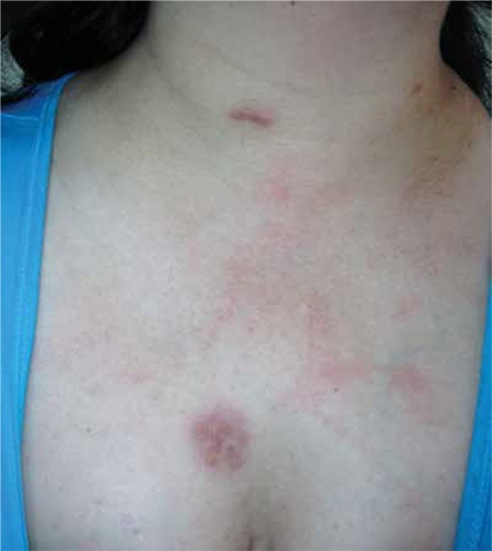
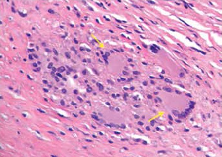
Analysis of a skin biopsy obtained from a 42-year-old female presenting with multiple yellowish to reddish-brown nodules and plaques on the arm, chest (Figure 1), and abdomen showed that she had generalized necrobiotic xanthogranuloma (Figure 3). Informed consent was obtained.
Figure 1. Dermatological lesions.

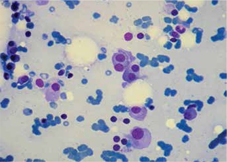
Figure 2. Morphological analysis of the bone marrow aspirate demonstrated abnormal plasma cells.

A week later, she was diagnosed with IgG-kappa multiple myeloma (Figure 2).
Figure 3. Skin biopsy studies revealed 2 granulomas containing Touton giant cells and foamy histiocytes.

The laboratory workup showed the following: Hb: 109 g/L, Hct: 0.34 L/L, WBC: 9.5x10⁹/L, Plt: 194x109/L, with normal differential white cell count. Other studied parameters included immunoglobulin IgG:1790 mg/dL (normal range: 750-1750), IgA: 279 mg/dL (normal range: 90-450), IgM: 40 mg/dL (normal range: 70-280); and kappa light chains: 1830 mg/dL (normal range: 629-1320 mg/dL).
The coexistence of paraproteinemias and necrobiotic xanthogranuloma is well described (above all, monoclonal gammopathy of unknown significance), and it would seem reasonable to recommend performing at least serum electrophoresis for patients affected by these rare conditions [1].
Conflict of Interest Statement
The authors of this paper have no conflicts of interest, including specific financial interests, relationships, and/or affiliations relevant to the subject matter or materials included.
References
- 1.Rose A, Robinson M, Kamino H, Latkowski JA. Necrobiotic xanthogranuloma. Dermatol Online J. 2012;18:30–30. [PubMed] [Google Scholar]


