

Abstract
Popliteal cysts are characterized by enlargement of the gastrocnemius-semimembranosus bursa. The pathogenesis includes a valvular opening between the knee joint and the bursa, and associated intra-articular pathology may give rise to knee effusion. The mainstay of treatment is conservative. If popliteal cysts are symptomatic, analgesia, aspiration, and steroid injection therapy may be considered, but most recur rapidly. In the past, open excision was an option if they remained symptomatic, but the associated recurrence rate was high. One important reason was that the intra-articular pathology causing the knee effusion was not treated. We present an alternative minimally invasive arthroscopic treatment using dye (methylene blue) directly injected into the cyst, which will leak from the cyst into the joint, to identify the valvular opening. The thickened valve is opened using a basket forceps and then enlarged using a motorized shaver to disrupt the 1-way mechanism between the joint and bursa, as well as to establish an unobstructed freeway connection between them. We also present a safe technique to create a direct posterior portal. Intracystic debridement of the fibrous membrane, nodules, and septa through this portal will decrease the recurrence rate of the popliteal cyst.
Popliteal cysts (also termed “Baker cysts”) were first described over a century ago by Adams 1 and later by Baker.2 They are most frequently characterized by enlargement of the gastrocnemius-semimembranosus bursa, which is one of several bursae around the knee. Studies of the pathogenesis of popliteal cysts have shown that they are connected to the knee joint by means of a valvular opening.3,4 The presence of a valve, along with the existence of an effusion, creates unidirectional flow of the synovial fluid from the articular cavity to the cyst and is one of the fundamental factors responsible for the appearance and persistence of the cyst.
Treatments for popliteal cysts have included conservative approaches or open resection.5-7 Because the cysts are often asymptomatic and resolve spontaneously, many are treated by observation alone. If they are symptomatic, analgesia, aspiration, and steroid injection therapy may be considered, but most recur rapidly. In the past, open excision was an option if they remained symptomatic; however, an associated recurrence rate as high as 42% to 63% has been reported.6,7 Several studies have reported frequently associated intra-articular pathologies with the cysts and warned of a high recurrence rate if the intra-articular pathologic condition is not addressed.8-12
Arthroscopic intervention is favored over open excision because of the successful outcomes of arthroscopic treatment for conditions associated with popliteal cysts. Arthroscopy is minimally invasive, is associated with a lower amount of risk, directly addresses both intra-articular pathology and the cyst, and allows early aggressive rehabilitation. Recent advances in arthroscopic techniques have been made to effectively address the intra-articular pathology, particularly the orifice of the cyst, which has a valvular mechanism.
It has been agreed that the fibrous membrane, nodules, and septa within the cysts may be factors associated with a postoperative recurrence after arthroscopic cystectomy. Ahn et al.13 established the posteromedial cystic portal on the medial side of the skin overlying the cyst, using the outside-in technique. One of the critical issues of concern regarding the posteromedial aspect is injury to the saphenous nerve and vessels.14
In this study we propose an inside-out technique to create a direct posterior portal in the knee. The benefit of this technique is the avoidance of injury to the saphenous nerve, as well as the popliteal vessel, using transillumination.
Surgical Technique
A standard leg holder is used with the knee easily flexed at 90° in most patients. A tourniquet is placed high on the operative thigh, and the patient's gluteal fold is brought to the end of the bed. The nonoperative leg is placed into a well-padded leg holder in the lithotomy position. This position allows the surgeon and assistants adequate space to perform surgery. The operative leg is placed into an arthroscopic leg holder at the level of the thigh tourniquet and adjusted so that the femur is parallel to the floor. The entire extremity is prepared from the holder to the toes. A stockinet is placed over the foot and distal leg and secured with an elastic bandage. A sterile U-drape is placed around the thigh, and a standard arthroscopic lower-extremity drape is used.
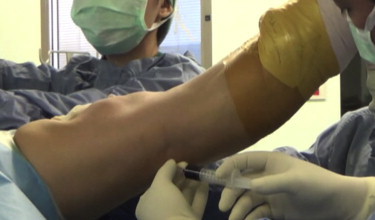
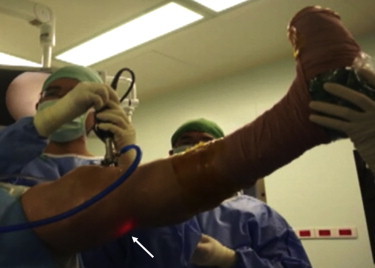
After general or spinal anesthesia is administered, the popliteal cyst fluid is aspirated as much as possible (Fig 1). The dye (methylene blue) is injected through the skin into the cyst. In this way the site of the opening, which is sometimes obliterated by a thin and pliant fibrous septum, is well evidenced (Fig 2, Table 1). Routine arthroscopic examination of the knee joint is performed through the standard anterolateral portal with a 30° arthroscope. Treatment of associated intra-articular pathology (e.g., removal of loose bodies, partial meniscectomy, or chondroplasty) is then performed.
Fig 1.

Aspiration of popliteal cyst fluid from left knee with 18-gauge needle.
Fig 2.
The dye (methylene blue) is injected through the skin into the cyst.
Table 1.
Key Points
| The dye (methylene blue) aids in finding the site of the opening of the popliteal cyst, which is sometimes obliterated by a thin and pliant fibrous septum. |
| The posteromedial fold is resected and enlarged to establish an unobstructed freeway connection between the popliteal cyst and the joint. |
| The direct posterior portal, for intracystic debridement of the fibrous membrane, nodules, and septa, can be created safely by the inside-out technique. |
The knee is kept in 90° of flexion to move the sartorial branch of the saphenous nerve posteriorly. The arthroscope is placed through the anterolateral portal into the posteromedial compartment through the intercondylar notch between the medial femoral condyle and the posterior cruciate ligament (Fig 3). Passage is facilitated by gentle but firm posterior pressure on the arthroscope as far medially as possible. The long saphenous nerve is identified by transillumination. The soft spot between the medial collateral ligament, the medial head of the gastrocnemius, and the tendon of the semimembranosus is palpated. The posteromedial portal is localized with a spinal needle at this soft spot just above the joint line. A superficial longitudinal skin incision is made, and a hemostat is used to bluntly dissect through subcutaneous tissue and penetrate the joint capsule.
Fig 3.

Arthroscopic findings from the anterolateral portal in the left knee show the entry point for the arthroscope to the posteromedial compartment between the medial femoral condyle and posterior cruciate ligament.
By use of a probe through the posteromedial portal, the opening of the cyst is identified by inferiorly displacing the overlying capsular fold located on the posteromedial side of the medial head of the gastrocnemius (Fig 4). Once the opening connection of the cyst has been identified, the capsular fold is resected partially by inserting a set of basket forceps (Acufex; Smith & Nephew Endoscopy, Andover, MA) through the posteromedial portal (Fig 5). The valvular opening of the posterior capsule is enlarged with a motorized shaver. After excision of the capsular fold, a 30° arthroscope is advanced into the cystic wall through the opening, which is posteromedial to the medial head of the gastrocnemius. If a fibrous membrane, nodules, and septa (Fig 6) are present within the cysts, the direct posterior portal will facilitate intracystic debridement. The leg is straightened. By use of a transillumination technique to mark the position of the direct posterior portal, injury to the popliteal vessel is avoided (Fig 7). The arthroscope is withdrawn from the sheath, and a Wissinger rod (4.3 mm; Smith & Nephew Endoscopy) is then inserted. The Wissinger rod can be palpated from the skin, and under direct vision, the direct posterior portal is created by an inside-out technique (Fig 8). The shaver is then introduced through the direct posterior portal to debride the fibrous membrane, nodules, and septa (Fig 9). A complete arthroscopic cystectomy is performed by shaving the inner wall of the popliteal cyst. A suction drain is inserted, and a compressive dressing is used. Full weight bearing and active-passive motion are permitted according to the protocol for associated intra-articular pathology (Fig 10, Video 1).
Fig 4.

Arthroscopic findings from the anterolateral portal in the left knee show the capsular fold located on the posteromedial side of the medial head of the gastrocnemius tendon. (The dotted lines represent the border of the capsular fold.)
Fig 5.

Arthroscopic findings from the anterolateral portal in the left knee show partial resection of the posteromedial fold with a basket forceps through the posteromedial portal.
Fig 6.

Arthroscopic findings from the anterolateral portal in the left knee show the fibrous membrane, nodules, and septa within the popliteal cyst, which may be factors associated with a postoperative recurrence after arthroscopic cystectomy.
Fig 7.
Transillumination technique to avoid injury to popliteal vessels. The arrow indicates that the arthroscopic camera is within the cyst and close to the skin.
Fig 8.

Inside-out technique to create direct posterior portal. One should note that the position of the direct posterior portal (white arrow) is separated from the posteromedial portal (black arrow).
Fig 9.

Introduction of motorized shaver through direct posterior portal (arrow). The inset shows the motorized shaver through this portal, which facilitates intracystic debridement.
Fig 10.

Flowchart for arthroscopic treatment of popliteal cyst.
Discussion
Although the operative techniques for popliteal cysts differ from author to author, arthroscopic resection is being actively attempted. The popliteal cyst is almost never an isolated pathology in an adult knee. Open surgical excision cannot be considered a definitive solution in most patients. Cho15 believe that addressing the opening or rather the connection between the joint cavity and cyst is a key procedure for completely excising the cyst. The arthroscopic approach to a popliteal cyst would provide an effective solution for managing not only the cyst, by arthroscopic decompression, but also the associated intra-articular pathology, with treatment of combined intra-articular lesions causing chronic synovitis.
Anatomic studies have shown a communication or opening between popliteal cysts and the joint cavity.4,15 This connection is usually formed between the joint and semimembranosus bursa. The septum dividing the 2 structures becomes thinner and more fragile, ultimately forming a communication. Therefore elimination of the unidirectional flow of effusion from the knee joint to the cyst is a target. Some specialists have tried to achieve this by closing the valvular opening with a suture,8,17,18 but the buildup of intra-articular fluid pressure during normal knee flexion and extension is difficult to resist.16 Moreover, closure of the valvular opening between the cyst and the knee joint is not necessary because such communications actually exist in 50% of normal adults without any clinically evident popliteal cysts.19 More recently, Kim et al.20 described the relation between the type of arthroscopic anatomy in the posteromedial capsule and the popliteal cyst. They believe that the posteromedial capsular fold is associated with the pathogenesis of popliteal cysts.
Thus it is important to re-establish the normal bidirectional communication between the cyst and the knee joint. Takahashi and Nagano12 reported in a technical note that arthroscopic correction of the valvular mechanism through the posteromedial portal is the most pathologically reasonable procedure to treat popliteal cysts surgically. Similarly, Ahn et al.13 advocated arthroscopically assisted popliteal cyst decompression, performed by removal of the capsular fold through the posteromedial portal. All previous arthroscopic studies reported removing the valvular opening of the cyst. However, only 2 studies reported an intracystic fibrous or septal membrane. Kanekasu et al.21 presented good results using a shaver and performing cystectomy and intra-articular synovectomy in popliteal cysts associated with rheumatoid arthritis. It was agreed that the fibrous membrane, nodules, and septa within the cysts may be factors associated with a postoperative recurrence after arthroscopic cystectomy. Ahn et al.13 established the posteromedial cystic portal on the medial side of the skin overlying the cyst, using the outside-in technique. One of the critical issues of concern regarding the posteromedial aspect is injury to the saphenous nerve and vessels. We propose an inside-out technique to create a direct posterior portal in the knee. The benefit of this technique is the avoidance of injury to the saphenous nerve and vessels.
Concerning safety, the primary neurovascular structure at risk during the procedure is the popliteal neurovascular bundle. Keser et al.22 showed that arteries are pushed laterally by the popliteal cyst. Therefore, when the 30° arthroscope is advanced into the cystic wall through the opening, the direct posterior portal can be created safely by the inside-out technique (Fig 11). The Wissinger rod can also be palpated from the skin to make sure that no structure stands between the knife and the Wissinger rod. The neurovascular bundle is also avoided by keeping the instruments in constant view and avoiding crossing the midline of the knee joint. As long as the instruments are kept medial to the midline, the neurovascular structures are safe. Regarding the limitations of our technique, we need knee magnetic resonance imaging scans of the patients before surgery. Because anatomic variations may occur, careful reading of magnetic resonance imaging scans in terms of the location of the cyst is very important. Other limitations relate to patient selection. For example, a patient with tricompartmental osteoarthritis in addition to a popliteal cyst should not be selected to undergo this technique because the target of treatment will be pain from osteoarthritis, not from the popliteal cyst. Moreover, this technique cannot be used in patients in whom the arthroscope cannot be advanced to the posteromedial compartment through the anterolateral portal.
Fig 11.

An axial-view magnetic resonance imaging scan of the left knee shows that the neurovascular structures are being pushed laterally by the popliteal cyst. The gray tube and blue arrow show the direction of the arthroscopic sheath and the Wissinger rod, which are distant from the neurovascular structures (red area).
Footnotes
The authors report that they have no conflicts of interest in the authorship and publication of this article.
Dr. Kongmalai is currently affiliated with the Department of Orthopaedics, Faculty of Medicine, Srinakharinwirot University, 62, Moo 7, Nakhon Nayok, 26120, Thailand.
Supplementary Data
A 46-year-old man with a large popliteal cyst in the left knee. Sagittal and axial T2-weighted magnetic resonance imaging is shown. Surgery was performed with the patient in the supine position with a standard leg holder.
References
- 1.Adams R. Chronic rheumatic arthritis of the knee joint. Dublin J Med Sci. 1840;17:520–522. [Google Scholar]
- 2.Baker W.M. On the formation of the synovial cysts in the leg in connection with disease of the knee joint. St Barth Hosp Rep. 1877;13:245–261. [Google Scholar]
- 3.Jayson M.I.V., Dixon A.S.J. Valvular mechanism in juxtaarticular cysts. Ann Rheum Dis. 1970;29:415–420. doi: 10.1136/ard.29.4.415. [DOI] [PMC free article] [PubMed] [Google Scholar]
- 4.Rauschning W. Anatomy and function of the communication between the knee joint and popliteal bursae. Ann Rheum Dis. 1980;39:354–358. doi: 10.1136/ard.39.4.354. [DOI] [PMC free article] [PubMed] [Google Scholar]
- 5.Chatzopoulos D., Moralidis E., Markou P., Makris V., Arsos G. Baker’s cysts in knees with chronic osteoarthritic pain: A clinical, ultrasonographic, radiographic and scintigraphic evaluation. Rheumatol Int. 2008;29:141–146. doi: 10.1007/s00296-008-0639-z. [DOI] [PubMed] [Google Scholar]
- 6.Chen J.C., Lu C.C., Lu Y.M. A modified surgical method for treating Baker’s cyst in children. Knee. 2008;15:9–14. doi: 10.1016/j.knee.2007.10.004. [DOI] [PubMed] [Google Scholar]
- 7.Fritschy D., Fasel J., Imbert J.C., Bianchi S., Verdonk R., Wirth C.J. The popliteal cyst. Knee Surg Sports Traumatol Arthrosc. 2006;14:623–628. doi: 10.1007/s00167-005-0028-z. [DOI] [PubMed] [Google Scholar]
- 8.Childress H.M. Popliteal cysts associated with undiagnosed posterior lesions of the medial meniscus. The significance of age in diagnosis and treatment. J Bone Joint Surg Am. 1970;52:1487–1492. [PubMed] [Google Scholar]
- 9.Rupp S., Seil R., Jochum P. Long-term results after excision of a popliteal cyst. Unfallchirurg. 2001;104:847–851. doi: 10.1007/s001130170056. [in German] [DOI] [PubMed] [Google Scholar]
- 10.Sansone V., De Ponti A. Arthroscopic treatment of popliteal cyst and associated intra-articular knee disorders in adults. Arthroscopy. 1999;15:368–372. doi: 10.1016/s0749-8063(99)70053-8. [DOI] [PubMed] [Google Scholar]
- 11.Stone K.R., Stoller D., De Carli A., Day R., Richnak J. The frequency of Baker’s cysts associated with meniscal tears. Am J Sports Med. 1996;24:670–671. doi: 10.1177/036354659602400518. [DOI] [PubMed] [Google Scholar]
- 12.Takahashi M., Nagano A. Arthroscopic treatment of popliteal cyst and visualization of its cavity through the posterior portal of the knee. Arthroscopy. 2005;21:638.e1–638.e4. doi: 10.1016/j.arthro.2005.02.007. [DOI] [PubMed] [Google Scholar]
- 13.Ahn J.H., Lee S.H., Yoo J.C. Arthroscopic treatment of popliteal cysts: Clinical and magnetic resonance imaging results. Arthroscopy. 2010;26:1340–1347. doi: 10.1016/j.arthro.2010.02.012. [DOI] [PubMed] [Google Scholar]
- 14.Cosgarea A.J., Kramer D.E., Bahk M.S., Totty W.G., Matava M.J. Proximity of the popliteal artery to the PCL during simulated knee arthroscopy: Implications for establishing the posterior trans-septal portal. J Knee Surg. 2006;19:181–185. doi: 10.1055/s-0030-1248103. [DOI] [PubMed] [Google Scholar]
- 15.Cho J.H. Clinical results of direct arthroscopic excision of popliteal cyst using a posteromedial portal. Knee Surg Relat Res. 2012;24:235–240. doi: 10.5792/ksrr.2012.24.4.235. [DOI] [PMC free article] [PubMed] [Google Scholar]
- 16.Lindgren P.G. Gastrocnemio-semimembranosus bursa and its relation to the knee joint. III. Pressure measurements in joint and bursa. Acta Radiol Diagn (Stockh) 1978;19:377–388. doi: 10.1177/028418517801900213. [DOI] [PubMed] [Google Scholar]
- 17.Rauschning W. Popliteal cysts (Baker’s cysts) in adults. II. Capsuloplasty with and without a pedicle graft. Acta Orthop Scand. 1980;51:547–555. doi: 10.3109/17453678008990839. [DOI] [PubMed] [Google Scholar]
- 18.Hughston J.C., Baker C.L., Mello W. Popliteal cyst: A surgical approach. Orthopedics. 1991;14:147–150. doi: 10.3928/0147-7447-19910201-09. [DOI] [PubMed] [Google Scholar]
- 19.Lindgren P.G., Willén R. Gastrocnemio-semimembranosus bursa and its relations to the knee joint. 1. Anatomy and histology. Acta Radiol Diagn (Stockh) 1977;18:497–512. doi: 10.1177/028418517701800501. [DOI] [PubMed] [Google Scholar]
- 20.Kim K.I., Lee S.H., Ahn J.H., Kim J.S. Arthroscopic anatomic study of posteromedial joint capsule in knee joint associated with popliteal cyst. Arch Orthop Trauma Surg. 2014;134:979–984. doi: 10.1007/s00402-014-2001-0. [DOI] [PubMed] [Google Scholar]
- 21.Kanekasu K., Nagashima K., Yamauchi D., Yamakado K. A clinical study of arthroscopic cystectomy on popliteal cysts associated with rheumatoid arthritis. Ryumachi. 1997;37:761–769. [in Japanese] [PubMed] [Google Scholar]
- 22.Keser S., Savranlar A., Bayar A. Anatomic localization of the popliteal artery at the level of the knee joint: A magnetic resonance imaging study. Arthroscopy. 2006;22:656–659. doi: 10.1016/j.arthro.2006.04.076. [DOI] [PubMed] [Google Scholar]
Associated Data
This section collects any data citations, data availability statements, or supplementary materials included in this article.
Supplementary Materials
A 46-year-old man with a large popliteal cyst in the left knee. Sagittal and axial T2-weighted magnetic resonance imaging is shown. Surgery was performed with the patient in the supine position with a standard leg holder.