Abstract
Mandible is the most frequently affected bone during head and neck irradiation. Late changes in the mandible may manifest in the form of reduced bone density, dental caries, loss of spongiosa trabeculations, delayed healing following dental extraction, pathologic fractures, osteoradionecrosis, trismus, growth defects in children or second malignancies. Pathologic fractures of mandibular bone are rare and may be spontaneous or traumatic (following dental extraction). We report the case of a 55-year lady, who had undergone surgery and adjuvant radiotherapy for carcinoma oral tongue T2N0M0 on a cobalt-60 unit and was disease-free. After a follow-up of 8 years post-irradiation, she presented with sudden onset oral pain and inability to open mouth. Pantomogram showed fracture at the junction of body and ramus of the mandible bilaterally.
Keywords: Mandible, osteoradionecrosis, radiotherapy, tongue carcinoma
INTRODUCTION
Head and neck irradiation almost always entails irradiation of at least a part of the mandible; hence it is the commonest bone manifesting various radiation effects. Asymptomatic bone changes could include decreased bone density in the irradiated region, delayed wound healing and cortical bone destruction, thereby increasing the possibility of serious side effects such as osteoradionecrosis (ORN) and fractures. We describe a late spontaneous pathologic fracture in a case of carcinoma tongue and the implications of such developments for radiation oncologists.
CASE REPORT
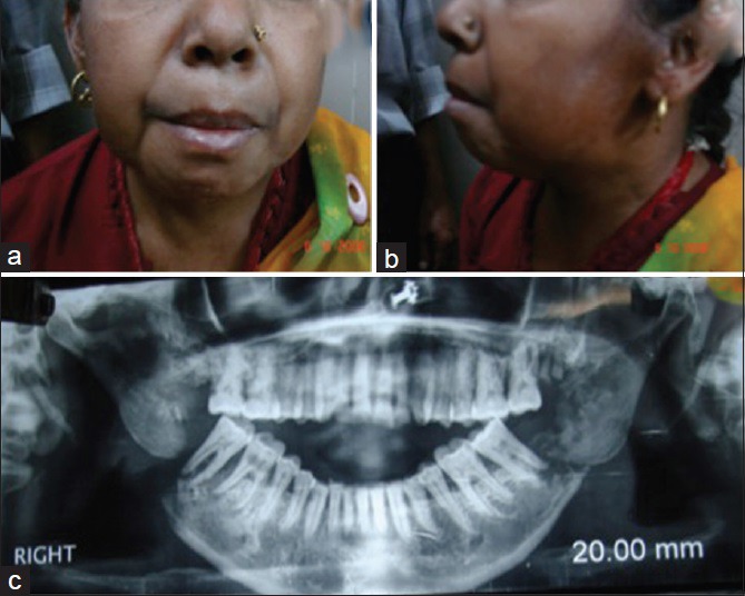
A 55-year-old lady, with no comorbid conditions and no history of tobacco or alcohol intake, was evaluated at our institute nearly 9 years ago for a 2-month ulcerating lesion over left lateral border of tongue, not healing with conservative management. Following examination and biopsy, a squamous cell carcinoma of oral tongue T2N0M0 was diagnosed. She underwent partial glossectomy and ipsilateral supraomohyoid neck dissection, revealing a 2.2 cm × 1.5 cm infiltrative growth with clear margins, depth of 6 mm, no perineural or lymphovascular invasion, and negative nodes (0/8). She was advised adjuvant irradiation and received 60 gray in 30 fractions, 5 fractions/week, by cobalt-60 unit using lateral opposed portals. She tolerated the treatment well and was on regular follow-up as per institutional protocol, with no specific events. Eight years after completion of her treatment, she reported to our clinic with the complaint of sudden onset oral pain and inability to open her mouth or eat. There was no history of any dental or oral procedures, oral infections or trauma preceding her complaints. On examination, there was fullness over both angles of lower jaw. Oral examination was painful but did not show any mucosal lesions or dental caries. Pantomogram was done, which showed a comminuted fracture at both angles of the jaw, just posterior to the last molars [Figure 1]. She was subsequently referred to the head and neck surgery department, where she underwent plate reconstruction for correction after initial conservative management.
Figure 1.

(a) and (b) Anterior and lateral views of the patient showing the facial appearance. (c) Pantomogram showing bilateral mandibular fracture lateral to the molars
DISCUSSION
The mandible is the longest bone in head and neck region, and commonest bone affected is head and neck irradiation due to its unique location bearing the lower set of teeth, high density, and poor vascular supply compared with other bones in this region. Vascular supply is through inferior alveolar and facial arteries. Complication risk is highest in the region of premolar, molar and retromolar trigone due to high density and low vascularity. It has an α/β ratio of 0.85, suggesting it is a late reacting tissue and high sensitivity to large doses per fraction. Most cases of osteonecrosis post-radiotherapy (RT) occur within first 2–3 years after treatment, but patients remain at indefinite risk due to ongoing changes in the bone due to age, altered oral microflora and dental infections.[1]
In early 1980s, Marx redefined the pathophysiology of ORN by proposing that RT induces endarteritis that results in tissue hypoxia, hypocellularity, which in turn causes tissue breakdown and chronic non-healing wounds.[2] Several factors that are associated with risk of bone injury or ORN include patient factors (deep parodontitis, bad oral hygiene, alcohol and tobacco abuse, bone inflammation, immune deficiencies, malnutrition, dental injury or extraction after RT), tumor factors (tumor size, stage, proximity of tumor or nodes to bone, tumor location) and treatment factors (pre-irradiation bone surgery, surgical handling of bone or its vascular supply, RT dose, biologically effective dose, photon energy, brachytherapy dose rate, combination of external beam irradiation and interstitial brachytherapy, field size, fraction size, volume of the mandible irradiated with a high dose).[3,4,5,6]
A diagnosis of ORN of bone mandates radiological evidence of bone necrosis within the radiation field, without any evidence of tumor recurrence. Incidence varies from 2% to 22% in various series, and is higher in dentate patients compared to edentulous patients. Clinical manifestations may include pain, orofacial fistulas, exposed necrotic bone, pathologic fractures or suppuration. Pathologic fractures may be spontaneous (35%), occurring usually within the first 2 years of irradiation, while traumatic fractures related to dental procedures are more common but occur late.
The highest incidence of ORN has been reported with brachytherapy of oral lesions if the RT source lies in close proximity to the bone. External beam RT, especially when using kilovoltage or low energy (1–4 MV) X-rays or electron beam, leads to high radiation absorption within the bone. This risk was greatest with the older techniques when use of large open parallel-opposed fields led to the inclusion of large areas of bone within the radiation portal, exposing it to high doses. Modern techniques have been able to circumvent this problem to a large extent by conforming the high dose areas to the tumor bed only while minimizing the dose delivered to normal structures including mandible.[7,8]
Given the severe quality of life implications of bone complications, preventive measures in the form of meticulous oral hygiene and dental evaluation before and after irradiation, improvement in RT techniques such as intensity modulation and development of reliable diagnostic and therapeutic procedures have gained importance in recent years.[9] Important considerations include completion of any necessary dental procedures such as extraction and management of periodontal disease prior to starting RT, education on lifelong use of high fluoride content dental gels and toothpastes, and minimal soft tissue trauma during extraction, and an interval of at least 2 weeks between extraction and RT commencement to allow healing.[10] RT technique should allow adequate sparing of salivary glands to prevent oral dryness, prevention and avoid high dose regions within mandible and teeth. Management of oral mucositis during treatment with soda-saline mouthwashes, antibiotics and antifungals, and use of spacers between mandible and brachytherapy source would reduce chances of oral infections and bone necrosis. Post-treatment regular dental examination, fluoride prophylaxis and delaying use or dentures till 9–12 months following RT would help prevent most instances or risks of ORN.
Bilateral mandibular pathologic post-radiation fracture is a very uncommon but serious late complication, with a huge impact on subsequent quality of life, and thus, preventive measures are necessary to minimize the risk.
Footnotes
Source of Support: Nil.
Conflict of Interest: None declared.
REFERENCES
- 1.Thorn JJ, Hansen HS, Specht L, Bastholt L. Osteoradionecrosis of the jaws: Clinical characteristics and relation to the field of irradiation. J Oral Maxillofac Surg. 2000;58:1088–93. doi: 10.1053/joms.2000.9562. [DOI] [PubMed] [Google Scholar]
- 2.Marx RE. Osteoradionecrosis: A new concept of its pathophysiology. J Oral Maxillofac Surg. 1983;41:283–8. doi: 10.1016/0278-2391(83)90294-x. [DOI] [PubMed] [Google Scholar]
- 3.Nabil S, Samman N. Risk factors for osteoradionecrosis after head and neck radiation: A systematic review. Oral Surg Oral Med Oral Pathol Oral Radiol. 2012;113:54–69. doi: 10.1016/j.tripleo.2011.07.042. [DOI] [PubMed] [Google Scholar]
- 4.Jereczek-Fossa BA, Orecchia R. Radiotherapy-induced mandibular bone complications. Cancer Treat Rev. 2002;28:65–74. doi: 10.1053/ctrv.2002.0254. [DOI] [PubMed] [Google Scholar]
- 5.Curi MM, Dib LL. Osteoradionecrosis of the jaws: A retrospective study of the background factors and treatment in 104 cases. J Oral Maxillofac Surg. 1997;55:540–4. doi: 10.1016/s0278-2391(97)90478-x. [DOI] [PubMed] [Google Scholar]
- 6.Vikram B, Deore S, Beitler JJ, Sood B, Mullokandov E, Kapulsky A, et al. The relationship between dose heterogeneity (“hot” spots) and complications following high-dose rate brachytherapy. Int J Radiat Oncol Biol Phys. 1999;43:983–7. [PubMed] [Google Scholar]
- 7.Ahmed M, Hansen VN, Harrington KJ, Nutting CM. Reducing the risk of xerostomia and mandibular osteoradionecrosis: The potential benefits of intensity modulated radiotherapy in advanced oral cavity carcinoma. Med Dosim. 2009;34:217–24. doi: 10.1016/j.meddos.2008.08.008. [DOI] [PubMed] [Google Scholar]
- 8.Nguyen NP, Vock J, Chi A, Ewell L, Vos P, Mills M, et al. Effectiveness of intensity-modulated and image-guided radiotherapy to spare the mandible from excessive radiation. Oral Oncol. 2012;48:653–7. doi: 10.1016/j.oraloncology.2012.01.016. [DOI] [PubMed] [Google Scholar]
- 9.Ben-David MA, Diamante M, Radawski JD, Vineberg KA, Stroup C, Murdoch-Kinch CA, et al. Lack of osteoradionecrosis of the mandible after intensity-modulated radiotherapy for head and neck cancer: Likely contributions of both dental care and improved dose distributions. Int J Radiat Oncol Biol Phys. 2007;68:396–402. doi: 10.1016/j.ijrobp.2006.11.059. [DOI] [PMC free article] [PubMed] [Google Scholar]
- 10.National Institute of Health Consensus Development Panel. National Institutes of Health Consensus Development Conference statement. Diagnosis and management of dental caries throughout life, March 26-28, 2001. J Am Dent Assoc. 2001;132:1153–61. doi: 10.14219/jada.archive.2001.0343. [DOI] [PubMed] [Google Scholar]


