

Abstract
Pyogenic granuloma (PG) is an inflammatory reactive hyperplasia of connective tissue. It usually arises in response to various stimuli such as low-grade local irritation, traumatic injury, hormonal factors or certain kinds of drugs. It predominantly occurs in the second decade of life in young females and rarely may cause significantly alveolar bone loss. It managed by conservative surgical excision and removal of causative irritants. This paper presents the case of a PG in a 55-year-old male with severe alveolar bone loss in the affected site, managed by surgical intervention.
Keywords: Gingival hyperplasia, hyperactive lesion, pyogenic granuloma
Introduction
The term “pyogenic granuloma (PG)” or “granuloma pyogenicum” was introduced by Hartzell in 1904.[1] PG is non neoplastic common tumor-like growth of oral cavity or skin. It is a reactive inflammatory lesion, commonly seen in the oral cavity with most common affected site gingiva followed by buccal mucosa, tongue and lips.[2] It is mostly presents as a painless, pendculated, or sessile mass of gingiva. Lesions more common on the maxillary gingiva than mandibular gingiva and anterior region of arches are more commonly affected than posterior regions.[3]
Case Report
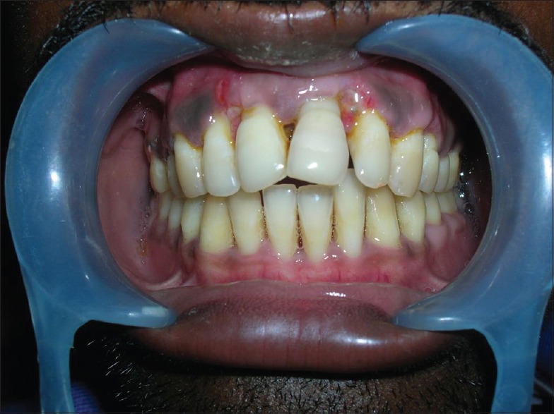
A 55-year-old male patient reported with a chief complaint of pain and swelling on gums at upper front region of the jaw since last 2 years, and swelling was gradually increasing in size, which caused masticatory problems on chewing foods and esthetic deformities because due to large size lips was not closed properly [Figure 1].
Figure 1.

Preoperative view
On clinical examination, a localized gingival swelling of 3 cm × 3 cm present in relation to the facial aspect of upper anterior region with moderate supra and subgingival calculus. Lesion was solitary red, exophytic and pedunculated with a broad base, which was hemorrhagic with bleeding on probing the area [Figure 2].
Figure 2.

Excised lesion
Medical history was no contributory and orthopantomograph showed severe alveolar bone loss in affected areas [Figure 3]. Based on above features, a provisional diagnosis of PG was made.
Figure 3.

Orthopantomograph showed severe alveolar bone loss in affected anterior areas
In the treatment, oral prophylaxis was done. Thereafter, it was decided to further treat the lesion with a surgical approach. After local anesthesia, the enlarged localized lesion was excised up to the base of the lesion, and It was ensured the that lesion was completely excised by trimming up the remnants of the soft tissue adjacent to the tooth to prevent recurrence of the lesion. The excised tissue was sent for histopathological examination, which showed connective tissue was loose fibrillar and comprised of numerous proliferative capillaries with dense mixed inflammatory infiltrate [Figure 4]. The histopathological examination confirmed diagnosis of PG.
Figure 4.

Histopathological examination showed loose fibrillar connective tissue and comprised of numerous proliferative capillaries with dense mixed inflammatory infiltrate
Discussion
Pyogenic granuloma may occur at all age groups but mostly seen in the second decade of life in young adult female, due to the vascular effects of female hormones.[4] But according to Epivatianos et al. the average patient age was 52 years with a peak incidence of occurrence in the sixth decade of life.[5] In the present case, patient was male with age of 55 years.
Pyogenic granuloma may occur as a result of hyperactive localized connective tissue reaction to a minor trauma or any underlying irritation, and these irritating factors may be poor oral hygiene and nonspecific infections, etc., In the present case, oral hygiene was poor due to which abundant plaque and calculus accumulated which produced chronic irritation and contributed to the development of PG.
Clinically PG seen as an exophytic lesion with a pedunculated or sessile base and their size varies from few millimeters to several centimeters in size but rarely seen >2.5 cm[6] and rarely, may cause significant bone loss.[7] In present cases, size of lesions attained a larger size >2.5 cm and caused significant bone loss in the affected area.
Treatment of PG involves a complete surgical excision,[8] but when not completely removes recurrence occurs. In present case postoperative 6 months recurrence was not found [Figure 5].
Figure 5.
Six months postoperative follow-up
Footnotes
Source of Support: Nil.
Conflict of Interest: None declared.
References
- 1.Hartzell MB. Granuloma pyogenicum. J Cutan Dis Syph. 1904;22:520–5. [Google Scholar]
- 2.Eversole LR. 3rd ed. Hamilton: BC Decker; 2002. Clinical Outline of Oral Pathology: Diagnosis and Treatment; pp. 113–4. [Google Scholar]
- 3.Neville BW, Damm DD, Allen CM, Bouquot JE. 2nd ed. Philadelphia: Saunders; 2002. Oral and Maxillofacial Surgery; pp. 447–9. [Google Scholar]
- 4.Sangamesh NC, Poornima B, Vidya KC, Sakri SB. Extragingival pyogenic granuloma: A rare case report. J Sci Soc. 2013;40:49–51. [Google Scholar]
- 5.Epivatianos A, Antoniades D, Zaraboukas T, Zairi E, Poulopoulos A, Kiziridou A, et al. Pyogenic granuloma of the oral cavity: Comparative study of its clinicopathological and immunohistochemical features. Pathol Int. 2005;55:391–7. doi: 10.1111/j.1440-1827.2005.01843.x. [DOI] [PubMed] [Google Scholar]
- 6.Verma PK, Srivastava R, Baranwal HC, Chaturvedi TP, Gautam A, Singh A. “Pyogenic granuloma - Hyperplastic lesion of the gingiva: Case reports”. Open Dent J. 2012;6:153–6. doi: 10.2174/1874210601206010153. [DOI] [PMC free article] [PubMed] [Google Scholar]
- 7.Goodman-Topper ED, Bimstein E. Pyogenic granuloma as a cause of bone loss in a twelve-year-old child: Report of case. ASDC J Dent Child. 1994;61:65–7. [PubMed] [Google Scholar]
- 8.Jafarzadeh H, Sanatkhani M, Mohtasham N. Oral pyogenic granuloma: A review. J Oral Sci. 2006;48:167–75. doi: 10.2334/josnusd.48.167. [DOI] [PubMed] [Google Scholar]