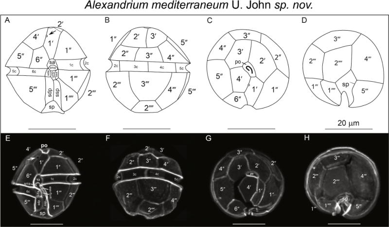
Figure 7.

Alexandrium mediterraneum U. John sp. nov. Line drawings (A–D), light micrographs of calcofluor-stained cells of type strain SZN01. (A) Ventral, (B) dorsal, (C) apical, (D) antapical (E) ventral, (F) dorsal, (G) apical and (H) antapical views. Arrows in panels A and E mark the ventral pore in the 1′ plate. Scale bars = 20 μm.
