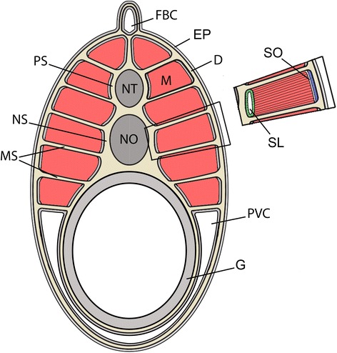
Figure 1.

Morphology of muscle segments in adult amphioxus. Schematic transverse section shows the muscle (M) segments (myomeres) in red and the axial and dermal extracellular connective tissue in tan. Because adult segments are chevron shaped, multiple myosepta (MS), which are present at their anterior and posterior borders of each segment, are observed in a transverse section. Mesothelial cells (grey) surround the muscle segments and enclose the fin box and perivisceral coeloms. Inset details the structure of the mesothelia surrounding muscle segments; the medial mesothelium (green) is double layered and encloses the sclerocoel (SL). The lateral mesothelium (blue) is single layered and separated from the muscle by the somitocoel (SO). Abbreviations: D, dermis; FBC, fin box coelom; M, muscle; MS, myoseptum; NO, notochord; NS, notochordal sheath; NT, neural tube; PS, perineural sheath; PVC, perivisceral coelom; SL, sclerocoel; SO, somitocoel; EP, epidermis.
