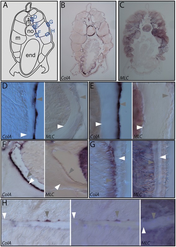
Figure 15.

Collagen A is expressed in the non-myotome somite derivatives in adults. In adults, all derivatives of the non-myotome somite cells are directly apposed to extracellular collagen layers and express the fibrillar collagen gene ColA, suggesting a connective tissue identity. (A) Schematic of transverse sections shown in (B, C); boxes indicate positions detailed in panels (D-H). (B, C) Adjacent transverse sections show expression of collagen A (ColA) and Myosin Light Chain (MLC), respectively. MLC marks the myotome and notochord cells, and ColA expression is observed the mesothelia that line the collagen layers including the perineural sheath, notochordal sheath, dermis, fin box coelom, and myosepta. Details in (D-H) show adjacent sections stained with ColA or MLC from regions boxed in (A). White arrowheads indicate extracellular collagen layers, and grey arrowheads indicate ColA-expressing mesothelial cells derived from somites. (D) Perineural sheath and the sclerotome-derived mesothelium (E) notochordal sheath and the sclerotome-derived mesothelium (F) notochord/gut boundary, position where the notochordal sheath collagen is thickened, and the sclerotome-derived mesothelium (G) dermis and the external cell layer-derived mesothelium. (H) Myoseptum with myoseptal cells lining it. Left panel shows brightfield; middle panel overlays a DAPI image to show nuclei of the fibroblast-like myoseptal cells. Myoseptal cells strongly express collagen (left and middle panels) but not MLC (right panel, which includes DAPI overlay), while the surrounding muscle stains for MLC (right panel). Note that muscle cell nuclei located laterally and are not found along myosepta, thus are not visible in these panels. In all panels, dorsal is up, medial is to the left. Micrographs were taken at 200× (B, C) or 1,200× (G, H) magnification. Abbreviations: end, endoderm; m, muscle; no, notochord; nt, neural tube.
