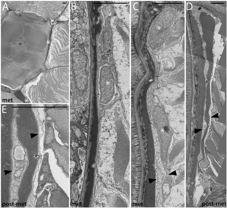
Figure 5.

Development of the non-myotome somite (3): metamorphosis through adult stages. (A) Lower power view, showing sclerotome-derived mesothelium beside the notochord and a portion of the neural tube. Sclerotome nuclei are marked by white asterisks in all panels. (B, C) Higher power views of the sclerotome-derived mesothelium beside the neural tube (B) and notochord (C). (D) The double-layered sclerotome beside the notochord is more easily visualized metamorphosis when it is often filled with fluid. Black arrowheads indicate the double layer at the notochord level in (C) and (D). (E) Detail of (D) showing an adherens junction between adjacent sclerotome cells (black asterisk). In all panels, dorsal is up, medial to the left; scale bars are 2 μm. Stage abbreviations: met, early metamorphosis, post-met, post-metamorphic juvenile.
