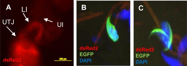
Figure 7.
Representative images of sperm inside the female reproductive tract after natural mating with transgenic males carrying Acr-EGFP and Ds-Red2. The photographs show spermatozoa migrating through the female reproductive tract detected by Ds-Red2 using an epi-fluorescence microscope (A). B-C: Representative confocal images of a cross section of the upper isthmus isolated 4 h after mating containing EQ (B) and AC (C) spermatozoon. UTJ: utero-tubal junction; LI: lower isthmus; UI: upper isthmus.

