Abstract
Background and Purpose
Iliotibial Band Syndrome (ITBS) has commonly been thought of as an overuse injury in runners. The exact etiology of ITBS is not well understood and there is no consensus on how to properly manage it. The purpose of this case series is to present a comprehensive model that utilizes a review of the current literature and the concept of regional interdependence as a foundation for the treatment of ITBS in runners.
Case Descriptions
The first subject was a 36‐year old female, referred from an orthopedic physician with the diagnosis of left iliotibial band friction syndrome. She reported a 9/10 stabbing pain on a visual analog scale (VAS) in the left lateral knee at mile two while running. The second subject was a 41‐year old female with a referral to physical therapy from an orthopedic surgeon for left iliotibial band tendinitis. She reported the symptoms beginning one month prior to her presentation to therapy, and that she would get an 8/10 stabbing pain on a VAS in the left lateral knee at mile three while running. Both subjects complained of the onset of lateral knee pain at a consistent mileage that forced them to stop running. Neither of them initially reported pain in adjoining regions, but did recall some low back stiffness from time to time when questioned further. The concept of regional interdependence, as well as neuromuscular re‐education, and strengthening interventions in conjunction with addressing the contributing factors of training errors, shoe wear, running surface, and program design were utilized.
Outcomes
At a six‐month follow‐up, subject one had successfully completed a half marathon without knee pain. At a nine‐month follow‐up, subject two was able to run five miles, twice weekly and 10 miles once weekly without pain or symptoms.
Discussion
These case reports demonstrate the successful management of ITBS in two subjects using a multifaceted approach based on the current literature and the concept of regional interdependence.
Keywords: Iliotibial band syndrome, manual therapy, physical therapy, regional interdependence, running injuries
Background and Purpose
Iliotibial Band Syndrome (ITBS)/Iliotibial Band Friction Syndrome (ITBFS) is considered to be one of the most common overuse injuries in the lower extremity, affecting anywhere from 7‐14% of the running population.1,2 It not only affects runners, but it can decrease performance in cyclists, soccer players, field hockey players, basketball players, and rowers.3 ITBS often leads to an inability to participate in a sport secondary to severe hip, lateral thigh, and knee pain.
Currently, the exact etiology of ITBS is not well understood. Orchard et al4 described an area of friction occurring between the iliotibial band and the lateral femoral condyle when the knee is flexed to around thirty degrees. The friction is said to lead to inflammation and pain. However, findings of cadaver studies and biopsies of the area are leading researchers to challenge this theoretical model.5,6 Fairclough et al5,6 suggest that there is an illusion of movement of the ITB on the condyle due to changing tension, and however, the tendon does not actually have the capability to slide across the bone. It instead exerts a compressive force on the joint when the fascia tightens. In addition to fascial restrictions, Ferber et al7 studied three hundred recreational athletes and identified decreased iliotibial band and iliopsoas extensibility in recreational athletes. Surgical examination of the area often reveals a lack of inflammatory response. In a few cases, there may be an underlying cyst or extension of the joint capsule laterally, but this has not been found consistently in all subjects. Even more infrequently, degeneration of the lateral femoral condyle has been discovered.3 On cadaver dissection, no bursa was seen distally at the knee in the region of where the iliotibial band inserts. There is a lateral recess of the knee that contains a highly vascularized fatty tissue visible on magnetic resonance image (MRI). It is most likely that the iliotibial band is not transferring loads properly and friction is not likely responsible for the painful presentation.5,6
Abnormal kinematics of the lower extremity have also been suggested as a contributing factor to ITBS. Multiple authors have studied the hip, knee, and ankle kinematics in runners with and without ITBS and found conflicting results.7,8 Noehren et al studied competitive female runners with a history of ITBS and found that they had increased hip internal rotation and adduction range of motion upon contact.8 Grau et al found that hip adduction at initial contact and maximal hip adduction in the control group were significantly less than in the group with ITBS.9 Orchard et al found that runners with ITBS landed with less knee flexion upon initial contact on the involved limb than uninjured runners.4 Noehren et al studied over 400 runners and found that the runners with ITBS had greater peak hip adduction, peak knee internal rotation and femoral external rotation moments and remained more adducted during stance.8 No difference was found in knee flexion and rearfoot eversion.8 Muscle weakness, specifically in the hip abductors, has also been suggested as a potential contributing factor in ITBS. Grau et al studied ten individuals with ITBS compared to ten controls and found no difference in hip abductor strength.9 Fredericson et al, however, studied a group of 24 runners with ITBS against 30 matched controls and found that hip abductor torque was significantly lower on the involved side in those with ITBS.10
Due to these varied results, the optimal treatment for athletes with ITBS remains undescribed. In the initial stages, anti‐inflammatory medications are frequently prescribed. However, this practice comes under question if the evidence from biopsies supports the lack of inflammation in the area.3 Physical therapy is often recommended. Interventions such static stretching, strengthening, manual therapy and neuromuscular re‐education have been researched. Pinshaw et al stressed the importance of addressing shoe wear and training schedules.11 Deep friction massage is often used, but outcomes research does not support it.12 In some chronic cases lasting greater than one year, resection of the lateral synovial recess have been performed. The purpose of this case series is to present a comprehensive model that utilizes a review of the current literature and the concept of regional interdependence as a foundation for the treatment of ITBS in runners.
Case Description: Subject History and Systems Review #1
Subject History
The subject was a 36‐year old female, referred from an orthopedic physician with the diagnosis of left iliotibial band friction syndrome. She reported a 9/10 stabbing pain on a verbal analog scale (VAS) in the left lateral knee at mile 2 two while running, that forced her to stop. X‐rays were negative and she decided not to take the anti‐inflammatories that had been prescribed. Self‐injury management included static iliotibial band stretching while standing and supine with a strap as well as rolling on a foam roller before and after running for 20‐minutes each. She ran on the road 60% of the run and on the sidewalk for the remaining 40% of the time. The Lower Extremity Functional Scale (LEFS) placed her at 89% function. She had previously been an avid runner, competing in two marathons per year prior to having children. At the time of this case report, her children were 10‐months and three years old, both delivered naturally. The subject's goal was to return to running a half marathon.
Systems Review
Details of the initial physical examination presented in Table 1.
Table 1.
Hip, Knee, and Ankle Examination findings.
| Measurement | Right | Left |
|---|---|---|
| Hip Flexion MMT | 5/5 | 4‐/5 |
| Hip Abduction MMT | 5/5 | 4/5 |
| Hip External Rotation MMT | 5/5 | 4/5 |
| Knee Flexion AROM | 135 ° degrees | 135 ° degrees |
| Knee Extension AROM | +5 ° degrees | +5 ° degrees |
| Ankle Dorsiflexion ROM | 15 ° | 7 ° |
MMT = Manual muscle test; AROM = active range of motion
Table 1: Hip, Knee and Ankle Examination findings, Subject 1
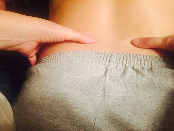
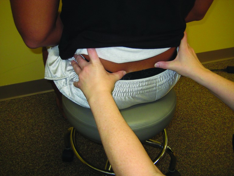
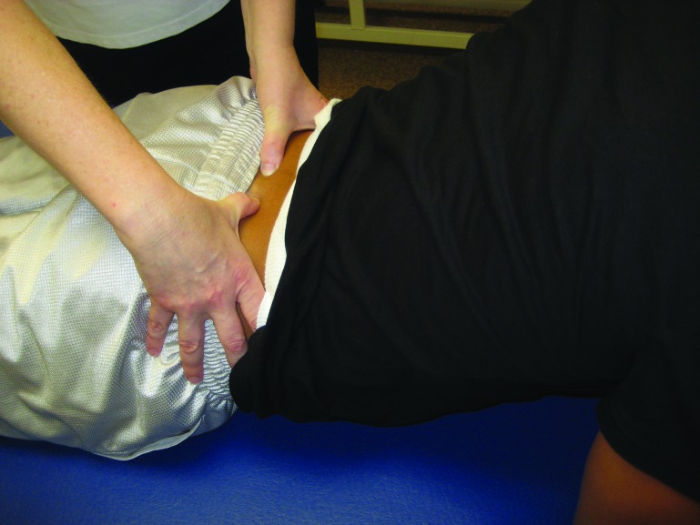
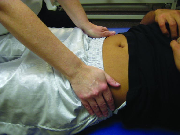
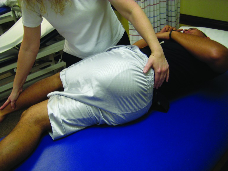
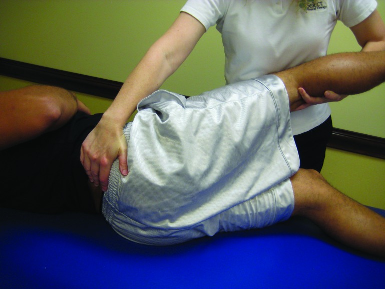
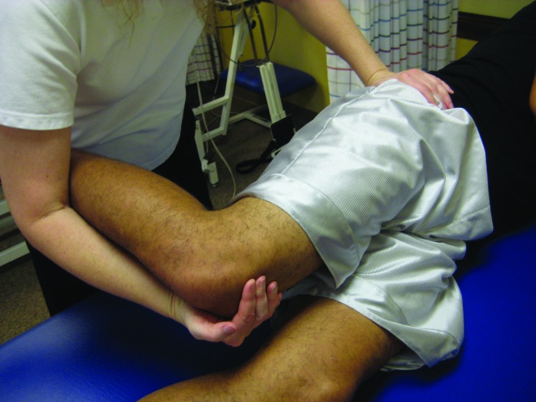
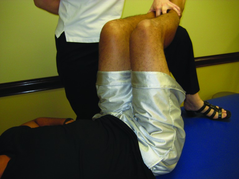
Genu recurvatum was noted and the subject was observed standing with her knees in a hyperextended position. The iliotibial band was moderately tender to palpation at the lateral knee and myofascial mobility in the area was decreased. Positional faults were found in the 5th lumbar vertebrae (flexed, rotated and side bent left; L5 FRLSL) (Figure 1), sacrum (Left rotated sacrum on a right oblique axis; L on R) (Figures 2 and 3), innominates (left innominate posteriorly rotated and outflared) (Figure 4), and a posterior fibular head at the superior tibiofibular joint. Increased muscle tone was present in the left psoas, piriformis and the lateral gastrocnemius muscles. Lumbar spine active range of motion in flexion was 75% with no segmental mobility occurring in L4‐5 and L5‐S1. Active left rotation was 90% and active side bending right was 90%. Active rotation right and active side bending left were both 75% of normal range of motion. Lumbar lordosis was increased and extension of the lumbar spine revealed a fulcrum at the level of L3. Straight leg raise testing was positive for neural tension at 75 ° degrees of hip flexion. Joint mobility testing through the foot and ankle were normal. Sensation and reflexes were intact and equal bilaterally. Mild pronation bilaterally was noted during gait analysis. She used a mild motion control shoe with a dual density sole present in only the rearfoot. The clinical impression was that the iliotibial band pain was a result of biomechanical and neuromuscular dysfunctions related to regional interdependence and that this subject would benefit from a multifaceted approach over a local approach that focused on the pain at the ITB only.
Figure 1.
Lumbar spine assessment for positional faults
Figure 2.
Sacral assessment, seated
Figure 3.
Sacral assessment, prone
Figure 4.
Innominate assessment, supine
Intervention
Table 2: Treatment Summary Subject 1
Table 2.
| Treatment Number | Day | Interventions | Exercises Added | Response on that day or presentation on the next visit |
|---|---|---|---|---|
| 1 | 1 | Muscle energy to positional faults Grade III joint mobilization superior tibiofibular joint Myofascial release of the ITB Detonification of the piriformis and lateral gastrocnemius muscle Counterstrain to the psoas |
Side lying: Upper trunk rotation and lower trunk rotation Isometric hip abduction and adduction exercises Prone opposite arm and leg raises Standing postural re‐education of knee position Dynamic stretching Running gait verbal retraining |
L Hip abduction strength 5‐/5 L Dorsiflexion 15 ° degrees Straight leg raise 95 ° degrees 100% Lumbar spine active range of motion No ITB tenderness Subjective pain level (SPL) on a (VAS) 0/10 Ran 3 miles without pain the next day |
| 2 | 3 | Counterstrain psoas | Side lying: hip abduction and external rotation exercises Lateral step downs without contacting floor Run 3x/week, increase by ½ mile increments |
No positional faults Normal muscle tone Worked up to running 5 miles painfree LEFS 100% |
| 3 | 13 | Add a long run that would not exceed 25% of the total weekly mileage | ||
| 6 months | Completed a ½ marathon |
Session One
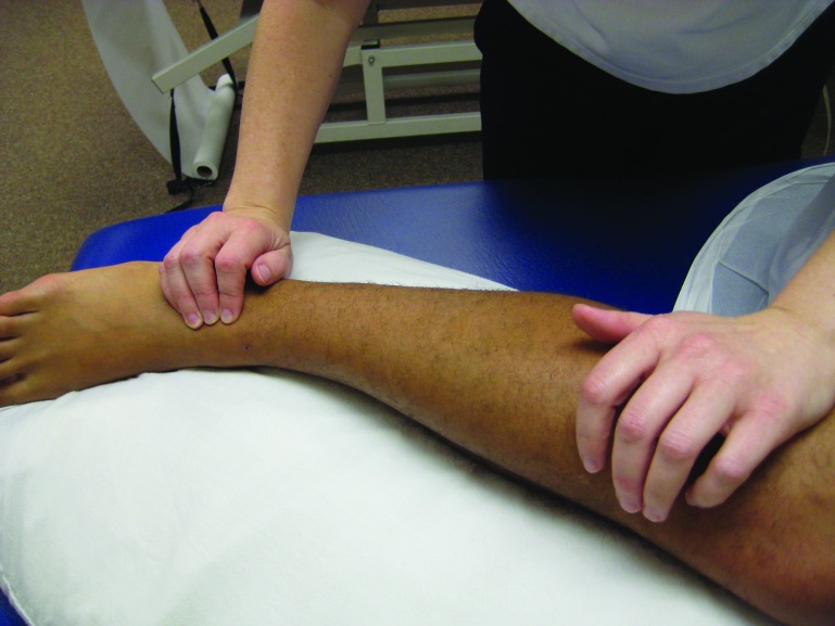
Following the evaluation, treatment was provided. First, the sacral torsion was treated with a muscle energy technique in right sidelying (Figure 5). The lumbar spine was then treated with a muscle energy technique in left sidelying position utilizing hip adduction of the top leg (Figure 6). The left posterior innominate was addressed in right sidelying with hip flexion (Figure 7). Hip abduction strength was then retested and improved from 4/5 to 5‐/5. The superior tibiofibular joint demonstrated a posterior fibular head and was mobilized with a Grade 3 joint glide anteriorly in a sidelying position followed by ligament articular strain fibular balance technique (Figure 8). The distal iliotibial band and lateral fascia were treated with grasping the fascia and gliding in a perpendicular anterior and posterior direction. A detonification technique consisting of grasping the muscle belly of the gastrocnemius and lengthening it laterally over a period of seven seconds was used. This combination of interventions increased her ankle dorsiflexion to 15 ° degrees to match the opposite side. The left psoas tone was reduced using counterstrain (Figure 9). The piriformis tone was addressed by applying a moderate pressure on the muscle in a shortened position and as the therapist passively lengthens the muscle, the pressure eases up. Once joint and soft tissue mobility were improved, the subject was instructed to perform upper trunk and lower trunk rotation in sidelying to promote segmental mobility as well as circulation and nutrition in the facet joints. Isometric hip abduction and adduction exercises were performed in supine hooklying. Prone opposite arm and leg raises were introduced to increase recruitment of the hip and back musculature and to reinforce the new length of the psoas. Her straight leg raise was performed and showed a 20 ° degree increase in range of motion from 75 ° to 95 ° degrees with negative neural tension. She was instructed how to stand with her knees in neutral and to maintain a neutral lumbar spine posture. The subject was advised to stop her static stretching and was taken through a walking warm up and dynamic stretching consisting of high knees, hip flexion with knee extension and hip internal and external rotation in midrange. While she was running on the treadmill, visual and verbal cues were used to normalize knee position, avoiding hyperextension and adduction. She was permitted to run 1.5 miles on even ground on the next day, but not on the cement.
Figure 5.
Muscle energy intervention for sacral torsion
Figure 6.
Muscle energy technique for the lumbar spine, L5 positional fault
Figure 7.
Muscle energy technique for posterior innominate
Figure 8.
Fibular balance technique
Figure 9.
Conterstrain technique for psoas
Session Two: Two days later
The subject reported that she ran past the 1.5 miles that were recommended but stopped at three miles just to be safe. Subjective pain level on a visual analog scale (VAS) was 0/10. Her lumbar spine AROM was full and there was no muscle guarding in the piriformis or gastrocnemius. Mild hypertonicity in the psoas with treated with counterstrain. The tenderness at the distal iliotibial band had resolved. The subject reported that she no longer felt the need to use her foam roller. Sidelying hip abduction exercises and clam shells for hip external rotation strength were added. Lateral step (dip) downs off the edge of a step without the leg touching the ground were utilized for neuromuscular re‐education of knee position. The subject was advised to run three times per week, every other day. She was to run three miles one more time and then increase by ½ mile increments up to five miles if there was no pain during or after the run.
Session Three: Ten days later
The subject reported that she had been compliant with her home program. She was gradually increasing her running mileage and had run five miles without lateral knee pain. Lower extremity functional scale (LEFS) score was 100%. She had full joint mobility in the spine and lower extremities. There was no abnormal tone. It was agreed that she would progress to one longer run on the weekend that was no more than 25% of her total weekly mileage. The long run could then progress every other week until her goal was met. The subject was discharged.
Six‐month follow up
The subject had successfully completed a half marathon without knee pain. She was signed up to run a marathon within the subsequent four months.
Case Description: Subject History and Systems Review #2
Subject History
The subject was a 41‐year old female referred for physical therapy by an orthopedic surgeon for left iliotibial band tendinitis. She reported the symptoms began one month prior and that she would experience stabbing pain, rated at 8/10 on a visual analog scale (VAS) in the left lateral knee at mile three while running. X‐rays of the left knee were negative. Self‐injury management had included static stretching of the iliotibial band, hamstrings and gastroc‐soleus complex. Once the pain had developed, she started using a foam roller along her iliotibial band, both before and after running. She had stopped running for two weeks, during which the pain subsided, but it returned the first time she tried to run after this break. She was running on the sidewalk. The subject reported a LEFS score of 86% function. After further questioning, the subject recalled a fall down her flight of stairs about three months prior to the onset of left knee pain. She had forgotten to mention it because the pain in her back pain had resolved. She reported that she was currently unable to exercise. Her goals included being able to run a 5k and to run for training multiple times a week without pain.
Systems Review
Physical examination summary findings are presented in Table 3.
Table 3.
Hip and Knee Examination findings, Subject 2
| Right | Left | |
|---|---|---|
| Hip Extension MMT | 5/5 | 3+/5 |
| Hip External Rotation MMT | 5/5 | 4/5 |
| Hip Abduction MMT | 5/5 | 4/5 |
| Knee Flexion AROM | 140 ° degrees | 140 ° degrees |
| Knee Extension AROM | 0 ° degrees | 0 ° degrees |
Table 3 Hip and Knee examination findings, Subject 2
The left distal iliotibial band was slightly tender to palpation and had decreased mobility anterior/posterior. Lumbar spine active range of motion was 80% of flexion, 60% of rotation and side bending bilaterally and 40% of extension with pain. Straight leg raise testing was positive for neural tension at 65 ° degrees on the left. Palpation and special tests led to the conclusion that she had a left on right sacral torsion, a left anterior innominate, and L5‐S1 flexed, rotated left, and sidebent left (L5 FRLSL). The proximal fibular head was posterior at the superior tibiofibular joint. There was increased tone in the bilateral psoas muscles, the left piriformis, the left erector spinae and the left lateral gastrocnemius. The subject lacked 30% of hip internal rotation PROM on the left compared to the right. Sensation and reflexes were intact. Gait analysis revealed that she was a late midstance pronator. A single leg squat test on the left revealed poor eccentric control with excessive hip internal rotation and adduction, also present during her running visual analysis on the treadmill. The clinical impression was that the iliotibial band pain was a result of biomechanical and neuromuscular dysfunction related to regional interdependence and that this subject would benefit from a multifaceted approach over an approach that focuses on the pain at the ITB only.
Intervention
Table 4 Treatment Summary, Subject 2
Table 4.
Treatment Summary, Subject 2
| Treatment | Day | Intervention | Exercises Added | Response that day or presentation on the next visit |
|---|---|---|---|---|
| 1 | 1 | Muscle energy to the positional faults Grade III mobilizations to the lumbar spine and superior tibiofibular joint Detonification to the erector spinae, piriformis and gastrocnemius Myofascial release to the ITB Counterstrain psoas |
Side lying: Upper trunk rotation, lower trunk rotation and hip external rotation Supine: Isometric hip abduction and adduction Squatting to ¼ depth Postural education for sitting No running at this time |
SLR 85 ° degrees Lumbar spine active range of motion 100% all planes Symmetrical hip internal rotation Pelvis was level |
| 2 | 3 | Muscle energy L5 Detonification of the erector spinae Counterstrain of the psoas |
Bridging Running gait training on the treadmill Dynamic stretching Run 2 miles every other day |
SPL on a verbal analog scale 0/10 Run 2 miles without pain the next day No positional faults in the lumbar spine |
| 3 | 10 | Muscle energy to the innominate Counterstrain psoas and iliacus Detonification of the erector spinae |
Prone opposite arm and leg raises Resisted walk backs Leg press Run 3 miles every other day |
Run 4 miles without pain No positional faults Normal tone |
| 4 | 24 | Side lying: hip abduction Lateral step downs without ground contact Run 3 times a week increasing in ½ mile increments once a week |
Run 5 miles without pain 100% lumbar spine active range of motion Hip and knee strength 4+/5 LEFS 100% |
|
| 5 | 38 | Increasing her mileage steadily | ||
| 9 months | Run 5 miles 2x/week and 10 miles on Saturday |
Session One
After the evaluation, the positional faults in the sacral and lumbar spine were addressed with muscle energy techniques in sidelying and were followed by ten grade 3 oscillations to L5‐S1 in prone until the surrounding soft tissue musculature softened. This resulted in increased passive hip internal rotation and active lumbar spine range of motion as well as an increase in the straight leg raise from 65 ° to 85 ° degrees with minimal neural tension. At superior tibiofibular joint the fibular head was mobilized anteriorly with ligament articular strain balancing technique. The lateral gastroc and the left erector spinae were detonified with lateral bending/displacement, and the distal iliotibial band was treated with myofascial techniques. Detonification was performed on the psoas muscles using a counterstrain technique and the piriformis with a passive pump technique with the subject in prone. The subject was instructed in sidelying upper trunk and lower trunk rotations to promote segmental mobility as well as joint circulation and joint nutrition. The subject was instructed in isometric hip abduction and adduction exercises supine. Hip external rotation strengthening was introduced with sidelying clam shells. Squatting with a focus on eccentric control and proper hip and knee position was used to re‐educate the muscles in closed chain function. Sitting posture and transfers were reviewed for good form with an emphasis on keeping the lower extremities symmetrical and not crossing the legs. The subject was asked not to run until after the next visit.
Session Two: Two days later
The subject returned with a positional fault at L5 (FRLSL), but the pelvis was level. The L5 positional fault was addressed with a muscle energy technique in left sidelying. The erector spinae and psoas muscles were slightly hypertonic so they were again detonified using the previously described techniques. Bridging was added to the home program in order to activate the gluteal muscles. Retraining neuromuscular control of knee position was conducted on the treadmill to ensure carry over. The subject was permitted to run two miles every other day on asphalt until the next visit. Dynamic stretches of the lower extremities to be used prior to running included high knee marching, butt kicks, marching with knee extension, and hip internal and external rotation walking.
Session Three: One week later
The subject reported that she had run pain free for two miles. The subject had slight increased psoas and iliacus tone on the left that was addressed using strain‐counterstrain techniques. Lumbosacral mobility was normal, however there was slight increase in tone of the left erector spinae. The increased tone was addressed with active pump techniques, combining soft tissue mobilization with active movement. The left anterior innominate was treated with a muscle energy technique. Isometric hip abduction and adduction strengthening were continued. Prone opposite arm and leg raises were added to the program to increase hip extensor and back musculature recruitment and to encourage a lengthened position of the psoas. The subject walked backwards while holding tubing at the chest level, and verbal and tactile cues were used to improve trunk position and proprioception. Leg press exercises were added to increase strength of the quadriceps and gluteals, as well as to work on hip and knee position and to provide equal weight bearing with symmetrical force through the pelvis. The subject was permitted to run three miles every other day until the next visit.
Session Four: Two weeks later
The subject reported that she was able to run three miles without pain and even went up to four miles without symptoms. She no longer felt the need to use her foam roller. Joint and soft tissue mobility had normalized. Hip abduction isolation exercises were advanced to the sidelying position, being careful to avoid sidebending of the lumbar spine. Squatting was advanced to lateral step (dip) downs off of the step without contacting the ground to reproduce the mechanics of support phase during running. A return to running program was discussed that included running only three days per week and increasing mileage only once weekly, by ½ mile increments.
Session Five: Two weeks later
The subject requested one additional visit to make sure that everything was progressing as expected. She was up to running five miles without pain. Her lumbar active range of motion was 100%, her hip and knee strength on the left had increased to 4+/5. LEFS score upon discharge was 100%.
Nine month follow–up
The subject was running five miles, twice weekly and 10 miles on Saturdays without pain or symptoms. She had participated in one 5k and was satisfied with her time.
Discussion
Both subjects had similar presentations. They each complained of the onset of lateral knee pain at a consistent mileage that forced them to stop running. Neither of them initially reported pain in adjoining regions, but did recall some low back stiffness from time to time when questioned further. They had limited lumbar spine active range of motion, decreased superior tibiofibular joint mobility (posterior fibular head) and increased tone in the psoas, piriformis or erector spinae, and gastrocnemius. There was decreased myofascial mobility in the distal one third of the iliotibial band. Strength of the involved lower extremity was decreased or inhibited in the hip flexors, external rotators, and abductors.
Muscle energy techniques and joint mobilization were the first interventions performed. Muscle energy was utilized to correct positional faults of the lumbosacral spine and Grade 3 mobilizations were used on the superior tibiofibular joint. This led to increased joint mobility, increased neural mobility and it contributed to reduced tone in the area. This is due to not only the biomechanical effect of manual therapy, but also the effects on the central nervous system.13,14 Additional soft tissue techniques were used by applying a moderate pressure on the muscles in a shortened position and as the muscle lengthens passively the pressure eased up to achieve normal tone throughout the lumbar spine and lower extremities.15 In both cases, utilizing the soft tissue techniques to normalize the tone led to a quick shift in pain levels.
Treatment that includes the lumbar spine and lower extremity below the knee is consistent with the model of regional interdependence for the treatment of iliotibial band syndrome. Joint dysfunction in the lumbar spine or extremities can precipitate or perpetuate injury regardless of how remote it is from the site of pain.16 Several authors17-20 reported success in subjects with lower extremity complaints when techniques to improve lumbosacral positional faults have been utilized. Vaughn reported using lumbosacral mobilizations on a subject with isolated knee pain that eliminated all pain in one visit.17 High velocity, low amplitude (HVLA) lumbopelvic manipulation has also been reported to not only decrease pain dramatically but to increase hip abductor and extension strength while increasing hip internal rotation range of motion on the affected side.18 Grassi et al reported they were able to normalize weight distribution through the lower extremities with a HVLA mobilization of the sacroiliac joint.19 Additionally, it was not until treatment commenced on the lumbar spine that a 70‐year old woman's knee pain resolved.20 Although diagnostic tests have been unable to support the presence of a positional fault, it is apparent that techniques targeting these areas of joint restriction may trigger the endogenous descending pain inhibition system and create a hypoalgesic affect. Manual therapy techniques may not only work at a biomechanical level but also at the neurophysiological level providing influence to the supraspinal level.13,14 It is also plausible that the cutaneous nerve distribution to the lateral knee is affected by the lumbosacral spine interventions.21 In our case series, this quick shift in symptoms also supports the notion that inflammation may not be the driving force in iliotibial band syndrome. Selkow et al22 promote the concept of utilizing muscle energy techniques to provide an immediate decrease in non‐specific low back pain. However, this effect is transient when used in isolation. Selkow et al22 felt that this subsequent decrease in pain levels lead to the ability to reeducate and strengthen inhibited muscles. Wilson et al23 had superior outcomes in their subjects with acute low back pain when muscle energy techniques were combined with neuromuscular reeducation and resistance training which is similar to the current suggested model of treatment.
In both cases, neuromuscular reeducation and strengthening exercises were introduced during the first visit. This approach is supported by Thein‐Nissenbaum et al24 who strengthened the core muscles and utilized treadmill retraining to alleviate hip and low back pain in a post partum runner. An eight‐week program of hip abduction and external rotator strengthening was reported by Khayambashi et al to decrease knee pain and increase function in females with patellofemoral pain.25 A one‐year follow up was provided that showed the positive results remained. An individual with piriformis syndrome was also successfully treated with an emphasis on hip muscle strengthening and movement reeducation instead of static stretching of the piriformis.26 A program of manual therapy, trunk and hip stabilization, and taping and orthoses led to positive outcomes in subjects with patellofemoral stress syndrome.27
Furthermore, on the first or second visit a running analysis was conducted with each of the two subjects, with cuing provided as needed. Providing same day feedback on running biomechanics was an important component to the successful long term return to running.28-30 Teran‐Yengle et al utilized treadmill training to significantly reduce sagittal plane knee extension.31 Crowell and Davis found that treadmill training could also reduce vertical ground reaction forces by over 30% and improve tibial and overall lower extremity position during running.32 These studies illustrate that having proper flexibility and strength in the absence of neuromotor control will not automatically create normal running mechanics.28-32
Also important to the long‐term success of these two runners was the information and education provided regarding training surface, shoes, and training schedules. All subjects were advised to run a surface that provided some shock absorption such as the road, a track, or grass (if level). They were to avoid cement at all times. Shoes were inspected for each subject in order to ensure that the footwear matched the foot type, they were not excessively worn and that foot position was stable and not excessively pronated during running. Kong et al determined that as little as 200 miles on a running shoe can lead to altered biomechanics.33 Butler et al were able to demonstrate that motion control shoes were able to control rearfoot motion better than cushion shoes.34 Schedule was important to review secondary to training errors accounting for at least 60% of all running related injuries.35 These subjects were provided with a program that outlined a four week progression, running three times weekly. Three times a week was chosen because of the correlation between the risk of injury and the number of days per week an individual runs.1,35
Case report research results are not generalizable. In addition, several factors were not controlled for or were not assessed using objective measures (e.g positional faults, MMT vs. hand‐held dynamometry). The ability to reproduce the results seen in these case reports may relate to the skill of the therapist in order to identify positional faults and the ability to normalize tone.
Conclusion
Treating athletes can be a challenging task due to their dedication to a sport. Most athletes, however, find that they cannot participate fully when they have ITBS. Utilizing a multifaceted treatment approach based on a review of the literature has been demonstrated to be quite effective. Incorporating lumbosacral spine and lower quarter joint mobilization techniques as indicated improved muscle tone and myofascial mobility. Additional soft tissue mobilization, neuromuscular reeducation, strengthening, and running form retraining with proper advice on surface, shoes and schedule, led to a successful return to running in these two subjects. These case reports demonstrate the successful management of ITBS using a multifaceted approach based on the current literature and regional interdependence. These two case reports present the rationale for the use of a multifaceted approach for not just this condition, but with the entire lower extremity. Physical therapists should incorporate a multifaceted approach with all of their subjects. Multiple individual techniques were performed which turned out to be effective. It is the authors’ opinion that the techniques are more effective combined then individually based on the reduced number of visits and the quick shift in pain levels that led to the return to pain free running. This quick shift in symptoms also supports the notion that inflammation may not be the driving force in this condition. Too often, in the clinical management of ITBS, the focus is ultrasound, static stretching, and myofascial techniques directly to the ITB. Physical therapists need to address underlying dysfunctions and compensatory patterns.
Future research needs to look at how Johansson described how the simulation of fascial mechanoreceptors may primarily lead to changes in gamma motor tone regulation.36,37 The fascia may have some microscopic sympathetic or contractile properties that benefited from the techniques used in this case series. In addition, future research should continue to investigate the descending pain inhibitory system and the most effective way to activate it to decrease pain levels. This may lead to implementing neuromuscular reeducation and strengthening interventions into the treatment plan, early on, for excellent outcomes.
References
- 1.McKean KA Manson NA Stanish WD. Musculoskeletal injury in the master's runners. Clin J Sports Med. 2006 Mar;16(2):149‐54. [DOI] [PubMed] [Google Scholar]
- 2.Taunton JE Ryan MB Clement DB McKenzie DC Llyod‐Smith DR Zumbo BD. A retrospective case‐control analysis of 2002 running injuries. Br J Sports Med. 2002 Apr;36(2):95‐101. [DOI] [PMC free article] [PubMed] [Google Scholar]
- 3.Lavine R. Iliotibial band friction syndrome. Curr Rev Musculoskeletal Med. 2010 Jul 20;3(1‐4):18‐22. doi: 10.1007/s12178‐010‐9061‐8. [DOI] [PMC free article] [PubMed] [Google Scholar]
- 4.Orchard JW Fricker PA Abud AT Mason BR. Biomechanics of iliotibial band friction syndrome in runners. Am J Sports Med. 1996 May‐June;24(3):375‐9. [DOI] [PubMed] [Google Scholar]
- 5.Fairclough J Hayashi K Toumi H Lyons K Bydder G Phillips N Best TM Benjamin M. The functional anatomy of the iliotibial band during flexion and extension of the knee: implications for understanding iliotibial band syndrome. J Anat. Mar 2006;208(3):309‐16. [DOI] [PMC free article] [PubMed] [Google Scholar]
- 6.Fairclough J Hayashi K Toumi H Lyons K Bydder G Phillips N Best TM Benjamin M. Is iliotibial band syndrome really a friction syndrome? J Sci Med Sport. 2007 Apr;10(2):74‐6; discussion 77‐8. Epub 2006 Sep 22. [DOI] [PubMed] [Google Scholar]
- 7.Ferber R Kendall KD McElroy L. Normative and critical criteria for iliotibial band and iliopsoas muscle flexibility. J Athl Train. 2010 Jul‐Aug;45(4):344‐8. doi:10.4085/1062‐6050‐45.4.344. [DOI] [PMC free article] [PubMed] [Google Scholar]
- 8.Noehren B Davis I Hamill J. ASB Clinical biomechanics award winner 2006 prospective study of the biomechanical factors associated with iliotibial band syndrome. Clin Biomech (Bristol, Avon) 2007 Nov;22(9):951‐6. [DOI] [PubMed] [Google Scholar]
- 9.Grau S Krauss I Maiwald C, et al. Hip abductor weakness is not the cause for iliotibial band syndrome. Int J Sports Med. 2008;79(7):579‐83. [DOI] [PubMed] [Google Scholar]
- 10.Fredericson M Cookingham CL Chaudhari AM Dowdell BC Oestreicher N Sahrmann SA. Hip abductor weakness in distance runners with iliotibial band syndrome. Clin J Sport Med. 2000 Jul;10(3):169‐75. [DOI] [PubMed] [Google Scholar]
- 11.Pinshaw R Atlas V Noakes TD. The nature and response to therapy of 196 consecutive injuries seen at a runners’ clinic. S Afr Med J. 1984 Feb 25;65(8):291‐8. [PubMed] [Google Scholar]
- 12.Schwellnus MP Mackintosh L Mee J. Deep transverse frictions in the treatment of iliotibial band friction syndrome in athletes: a clinical trial. Physiotherapy. 1992 Aug 10;78(8):564‐8. doi: 10.1016/s0031‐9406(10)61197‐2. [Google Scholar]
- 13.Vicenzino B Hing W Rivett DA Hall T. Mobilisation with Movement: The Art and the Science. 1st ed. Chatswood, NSM: Churchill Livingstone Australia; 2011. [Google Scholar]
- 14.Vicenzino B Paungmali A Teys P. Mulligan's mobilization‐with‐movement, positional faults and pain relief: current concepts from a critical review of the literature. Man Ther. 2007 May;12(2):98‐108. [DOI] [PubMed] [Google Scholar]
- 15.DiGiovanna EL Schiowitz S Dowling DJ. An Osteopathic Approach to Diagnosis and Treatment. 3rd ed. Philadelphia, PA: Lippincottt Williams & Wilkins; 2005, ISBN: 0781742935. [Google Scholar]
- 16.Wainner RS Whitman JM Cleland JA Flynn TW. Regional interdependence: a musculoskeletal examination model whose time has come. J Orthop Sports Phys Ther. 2007 Nov;37(11):658‐60. [DOI] [PubMed] [Google Scholar]
- 17.Vaughn DW. Isolated knee pain: a case report highlighting regional interdependence. J Orthop Sports Ther. 2008;38(10):616‐23. doi: 10.2519/jospt.2008.2759. [DOI] [PubMed] [Google Scholar]
- 18.Crowell MS Wofford NH. Lumbopelvic manipulation in subjects with patellofemoral pain syndrome. J Man Manip Ther. 2012 Aug;20(3):113‐20. doi: 10.1179/2042618612Y.0000000002. [DOI] [PMC free article] [PubMed] [Google Scholar]
- 19.Grassi Dde O de Souza MZ Ferrareto SB Montebelo MI Guirro EC. Immediate and lasting improvements in weight distribution seen in baropodometry following a high‐velocity, low‐amplitude thrust manipulation of the sacroiliac joint. Man Ther. 2011 Oct 16;16(5):495‐500. doi: 10.1016/j.math.2011.04.003. Epub 2011 May 14. [DOI] [PubMed] [Google Scholar]
- 20.Goncalves da Ropch RC #Nee R Hall T Chopard R. Treatment of persistent knee pain associated with lumbar dysfunction: a case report. NZ J Physio. 2006;34(1):30‐4. [Google Scholar]
- 21.Maigne R. Low back pain of thoracolumbar origin. Arch Phys Med Rehabil. 1980 Sept; 61(9):389‐95. [PubMed] [Google Scholar]
- 22.Selkow NM Grindstaff TL Cross KM Pugh K Hertel J Saliba S. Short‐term effect of muscle energy technique on pain in individuals with non‐specific low back pain: a pilot study. J Man Manip Ther. 2009;17(1):E14‐8. [DOI] [PMC free article] [PubMed] [Google Scholar]
- 23.Wilson E Payton O Donegan‐Shoaf L Dec K. Muscle energy technique in subjects with acute low back pain: a pilot clinical trial. J Orthop Sports Phys Ther. 2003 Sep; 33(9):502‐12. [DOI] [PubMed] [Google Scholar]
- 24.Thein‐Nissenbaum JM Thompson EF Chumanow ES Heiderscheit B. Low back and hip pain in a postpartum runner: applying ultrasound imaging and running analysis. J Orthop Phys Ther. 2012;42(7):615‐24. doi: 10.2519/jospt.2012.3941. Epub 2012 Mar 23. [DOI] [PubMed] [Google Scholar]
- 25.Khayambashi K Mohammadkhani Z Ghaznavi K Lyle MA Powers CM. The effects of isolated hip abductor and external rotator muscle strengthening on pain, health status, and hip strength in females with patellofemoral pain: a randomized controlled trial. J Orthop Sports Phys Ther. 2012 Jan;42(1):22‐9. doi: 10.2519/jospt.2012.3704. Epub 2011 Oct 25. [DOI] [PubMed] [Google Scholar]
- 26.Tonley JC Yun SM Kochevar RJ Dye JA Farrokhi S Powers CM. Treatment of an individual with piriformis syndrome focusing on hip muscle strengthening and movement reeducation: a case report. J Orthop Sports Phys Ther. 2010 Feb; 40(2):103‐11. doi: 10.2519/jospt.2010.3108. [DOI] [PubMed] [Google Scholar]
- 27.Lowry CD Cleland JA Dyke K. Management of subjects with patellofemoral pain syndrome using a multimodal approach: a case series. J Orthop Sports Phys Ther. 2008 Nov;38(11):691‐702. doi: 10.2519/jospt.2008.2690. [DOI] [PubMed] [Google Scholar]
- 28.Willy RW Scholz JP Davis IS. Mirror gait retraining for the treatment of patellofemoral pain in female runners. Clin Biomech (Bristol, Avon). 2012 Dec;27(10):1045‐51. doi: 10.1016/j.clinbiomech.2012.07.011. Epub 2012 Aug 20. [DOI] [PMC free article] [PubMed] [Google Scholar]
- 29.Willy RW Davis IS. The effect of a hip‐strengthening program on mechanics during running and during a single leg squat. J Orthop Sports Phys Ther. 2011 Sep;42(9):625‐32. doi: 10.2519/jospt.2011.3470. Epub 2011 Jul 12. [DOI] [PubMed] [Google Scholar]
- 30.Davis IS. Gait Retraining in Runners. Orthopaedic Practice. 17;2:05 Accessed April 24, 2015http://www.udel.edu/PT/PT%20Clinical%20Services/journalclub/caserounds/07_08/Dec07/GaitRetrain_OP.pdf [Google Scholar]
- 31.Teran‐Yengle P Emrick A Hare K Yack J Cole K. CSM 2012 Orthopaedic section platform presentations: Effects of Internal and External Focus of Attention in Correcting Knee Hyperextension in Young Women. J Orthop Sports Phys Ther. Jan 2012;42(1)A14‐40, OPL 4. doi:10.2519/jospt.2012.42.1.A14 [Google Scholar]
- 32.Crowell HP Davis IS. Gait retraining to reduce lower extremity loading in runners. Clin Biomech (Bristol, Avon). 2011 Jan;26(1):78‐83. doi: 10.1016/j.clinbiomech.2010.09.003. [DOI] [PMC free article] [PubMed] [Google Scholar]
- 33.Kong PW Candelaria NG Smith DR. Running in new and worn shoes: a comparison of three types of cushioning footwear. Br J Sports Med. 2009 Oct; 43(10):745‐9. doi: 10.1136/bjsm.2008.047761. Epub 2008 Sep 18. [DOI] [PubMed] [Google Scholar]
- 34.Butler RJ Davis IS Hamill J. Interaction of arch type and footwear on running mechanics. Am J Sports Med. 2006 Dec;34(12):1998‐2005. [DOI] [PubMed] [Google Scholar]
- 35.Buist I Bredeweg SW Lemmink KA Van Mechelen W Diercks RL. Predictors of running‐related injuries in novice runners enrolled in a systematic training program: a prospective cohort study. Am J Sports Med. 2010 Feb;38(2):273‐80. doi: 10.1177/0363546509347985. Epub 2009 Dec 4. [DOI] [PubMed] [Google Scholar]
- 36.Johansson H Sjolander P Sojka P. Receptors in the knee joint ligaments and their role in the biomechanics of the joint. Crit Rev Biomed Eng. 1991;18(5):341‐68. [PubMed] [Google Scholar]
- 37.Schleip R. Fascial plasticity – a new neurobiological explanation. Journal of Bodywork and Movement Therapies. 2003;7(1):11‐9. doi:10.1016/S1360‐8592(02)00067‐0. [Google Scholar]


