

Abstract
Objective
The aims of this study were to investigate the prevalence and distribution of dental anomalies in a group of Saudi subjects with cleft lip and palate (CLP), to examine potential sex-based associations of these anomalies, and to compare dental anomalies in Saudi subjects with CLP with published data from other population groups.
Design
This retrospective study involved the examination of pre-treatment records obtained from three CLP centers in Riyadh, Saudi Arabia, in February and March 2010. The pre-treatment records of 184 subjects with cleft lip and palate were identified and included in this study. Pre-treatment maxillary occlusal radiographs of the cleft region, panoramic radiographs, and orthodontic study models of subjects with CLP were analyzed for dental anomalies.
Results
Orthopantomographs and occlusal radiographs may not be reliable for the accurate evaluation of root malformation anomalies. A total of 265 dental anomalies were observed in the 184 study subjects. Hypodontia was observed most commonly (66.8%), followed by microdontia (45.6%), intra-oral ectopic eruption (12.5%), supernumerary teeth (12.5%), intra-nasal ectopic eruption (3.2), and macrodontia (3.2%). No gender difference in the prevalence of these anomalies was observed.
Conclusions
Dental anomalies were common in Saudi subjects with CLP type. This will complicate the health care required for the CL/P subjects. This study was conducted to epidemiologically explore the prevalence of dental anomalies among Saudi Arabian subjects with CLP.
Keywords: CLP, Dental anomalies, Cleft lip, Cleft palate
1. Introduction
Clefts of the lip and palate (CL/P) are currently the most common craniofacial birth defects. Most studies have suggested that 70% of CL/P cases are non-syndromic, and that the remaining 30% are associated with structural abnormalities outside the cleft region (Schutte and Murray, 1999; Cobourne, 2004; Lidral et al., 2008). Non-syndromic clefts affect one in every 700 live births, with ethnic and geographic variation (Dixon et al., 2011). Individuals with non-syndromic cleft lip and palate CL/P commonly exhibit various dental anomalies involving tooth shape, size, and position (Ranta, 1983; Kim and Baek, 2006; Da Silva et al., 2008). The extent of these dental anomalies varies according to sex, ethnicity, and cleft type (Aizenbud et al., 2005; Al Jamal et al., 2010; Pegelow et al., 2012; Matern et al., 2012; Paranaiba et al., 2013; Mikulewicz et al., 2014). For example, hypodontia was the most prevalent dental anomaly in a Brazilian CL/P population, followed by impacted teeth, supernumerary teeth, and microdontia (Paranaiba et al., 2013). Likewise, hypodontia was the predominant dental anomaly among individuals with CL/P in Sweden and Jordan; other dental anomalies present in these populations included impacted teeth, supernumerary teeth, microdontia, macrodontia, taurodontism, and dilaceration (Pegelow et al., 2012; Al Jamal et al., 2010).
These anomalies have deleterious effects on the dentition leading to esthetic problems, impairment of mastication, and improper phonation (Hardin-Jones and Jones, 2005). Knowledge of the presence of such anomalies in individuals with CL/P will aid orthodontists’ anticipation of malocclusion and other challenges when dealing with such cases in the clinic. Orofacial clefts and associated malocclusion contribute substantially to long-term disability in children, as well as tremendous emotional and financial stresses for affected individuals and families.
The current study was conducted in view of these considerations, and to address the paucity of reported epidemiological studies of craniofacial anomalies in Saudi Arabia (Kumar et al., 1991; Tahir, 1998; Al-Balkhi, 2008). The prevalence and distribution of dental anomalies (abnormalities in tooth number, size, shape, and location) were investigated in a group of Saudi subjects with CLP type, and the possible existence of gender-based associations with these anomalies was examined. Furthermore, the study aimed to compare dental anomalies in Saudi subjects with CLP with published data from other population groups.
2. Materials and methods
The records of 184 subjects with CLP were collected in this retrospective study; 138 subjects were from an orthodontic clinic at a university hospital and 62 subjects were from orthodontic clinics at two other hospitals, in Riyadh, Saudi Arabia. The study was conducted in February and March 2010, and included medical records from the period of November 1993 to October 2009 were collected and examined. Pre-treatment maxillary occlusal radiographs of the cleft region, panoramic radiographs, and orthodontic study models of subjects with CLP were analyzed. This is to investigate the presence of dental anomalies, and to evaluate the differences between gender, and between unilateral and bilateral CLP. The ethics committee of the College of Dentistry Research Center, King Saud University, approved this study in January 2010 (NF 2156).
The inclusion criteria were: (1) non-syndromic CLP; age 6–30 years to ensure complete calcification of all permanent tooth crowns, which occurs at around 6–7 years of age. (2) Availability of good-quality pre-treatment records.
Data were obtained by visual evaluation of occlusal and panoramic radiographs. The presence of dental anomalies was confirmed by evaluating subjects’ orthodontic casts. Patients’ treatment records were also studied to eliminate the possibility of premature tooth loss or extraction. The number, size, and shape of permanent dentition affected by hypodontia, microdontia, macrodontia, and ectopic eruption, as well as supernumerary teeth, were determined using panoramic and occlusal radiographs and recorded using the World Dental Federation index of tooth numbering and regional location (Nilsson and Ash, 2010). The investigated dental anomalies were defined as follows.
2.1. Abnormalities of tooth number
Diagnoses of hypodontia and supernumerary teeth in the cleft area were established according to the criteria reported by Damante (1972): hypodontia was defined as the absence of the lateral incisor and a supernumerary tooth was defined as any additional tooth mesial or distal to the cleft area in the presence of the lateral incisor. Outside of the cleft area, these anomalies were diagnosed according to the criteria of Gravey et al. (1999): hypodontia or tooth agenesis was diagnosed when the tooth or tooth bud was absent on radiographs, resulting in a deficient dental developmental series, and a supernumerary tooth was diagnosed based on the identification of an additional tooth germ or calcification (beyond the normal dental developmental series) on radiographs in any region of the dental arch.
2.2. Abnormalities of crown morphology
Macrodontia and microdontia refer to teeth that are substantially larger and smaller, respectively, than the average normal size, or larger and smaller, respectively, than the contralateral homolog or a tooth in the sample group from the opposing arch (D’Souza et al., 2006). Microdontia also refers to a tooth that does not fill its space in the dental arch, or appears small because of the absence of expected shape (D’Souza et al., 2006).
2.3. Root malformation
Dilaceration was defined as a bend in a root at any level along its length, as observed on panoramic and occlusal radiographs. A short root was identified by comparison with the root of the contralateral tooth on radiographs, based on the normal crown to root ratio (D’Souza et al., 2006).
2.4. Ectopic eruption
Intra-oral ectopic eruption was diagnosed when a tooth had not erupted in its normal position/site in the oral cavity. Intra-nasal ectopic eruption was diagnosed when a tooth had erupted through the floor of the nasal cavity.
2.5. Statistical analysis
Data were analyzed and are presented in two main parts: evaluation of methodological error and calculation of descriptive and analytical statistics. Analyses were conducted using the Statistical Package for the Social Sciences (version 16.0; SPSS Inc., Chicago, IL, USA).
Data on the presence and absence of dental anomalies, as determined by evaluation of radiographs and study models, were analyzed. A single examiner conducted evaluations to avoid variation in examination criteria due to differences in personal interpretation. To ensure acceptable intra-examiner reliability, the same examiner analyzed a random sample of 10 sets of radiographs and study models twice, with evaluations separated by a 2-week interval. Errors of identification were evaluated by testing reliability using Kappa statistics, which reflects the reproducibility of results of the measurement procedure (Viera and Garrett, 2005). Methodological error was assessed using kappa statistics (Viera and Garrett, 2005).
Standard Pearson chi-squared tests were used for all comparisons of dental anomalies, in addition to specific dental anomalies in relation to gender except when the number of observations was five or fewer, in which case Fisher’s exact test was used. A P value <0.05 was considered to be significant.
3. Results
Inter-examiner reliability scores were high for the evaluation of hypodontia, supernumerary teeth, and crown malformations, moderate (acceptable) for ectopic eruption, and low (poor) for root malformations. Root malformation variables were thus excluded from further statistical analysis (Table 1). The results revealed very high intra-examiner reliability, ranging from 1 (perfect agreement) to 0.61 (substantial agreement). The results were considered to be valid according to Viera and Garret’s (2005) interpretation. The exception was the root malformation variable, which showed low reliability (κ = 0.35).
Table 1.
Error of the method.
| Variable | Kappa statistic |
|---|---|
| Hypodontia | 1 |
| Supernumerary teeth | 1 |
| Crown malformations | 1 |
| Ectopic eruption | .61 |
| Root malformations | .35 |
The study sample consisted of 184 subjects (122 [66.3%] males, 62 [33.7%] females) with CLP; 115 (62.5%) subjects had unilateral cleft lip and palate (UCLP; 62 [54%] left, 53 [46%] right) and 60 (37.5%) subjects had bilateral cleft lip and palate (BCLP). A total of 265 dental anomalies were observed in 168 (91.3%) subjects with CLP; many subjects had more than such anomaly (Table 2).
Table 2.
The prevalence of dental anomalies and distribution in relation to gender.⁎
| Male CLP No. (%) |
Female CLP No. (%) |
Total CLP No. (%) |
S-value | P-value | |
|---|---|---|---|---|---|
| Hypodontia | 82 (66.8%) | 41 (66%) | 123 (66.8%) | 0.82 | NS |
| Microdontia | 58 (47.5%) | 26 (42%) | 84 (45.6%) | 0.44 | NS |
| Intraoral ectopic eruption | 15 (12.3%) | 8 (13%) | 23 (12.5%) | 0.39 | NS |
| Intranasal ectopic eruption | 6 (5%) | 0 (0%) | 6 (3.2%) | NA | NA |
| Supernumerary | 14 (11.5%) | 9 (14.5%) | 23 (12.5%) | 0.68 | NS |
| Macrodontia | 3 (2.5%) | 0 (0%) | 6 (3.2%) | NA | NA |
CLP = cleft lip and palate.
Pearson chi-square test was performed; NS = non-significant; NA = not applicable to statistical testing due to small sample.
The most common dental anomaly was hypodontia, which occurred in 123 (66.8%) subjects. The prevalence of missing teeth was highest in subjects with UCLP (n = 71) in comparison to BCLP (n = 52); the left side (38 [28.3%] subjects) was more affected than the right side (33 [24.7%] subjects; Table 3).
Table 3.
The prevalence of dental anomalies and distribution in relation to cleft side.∗
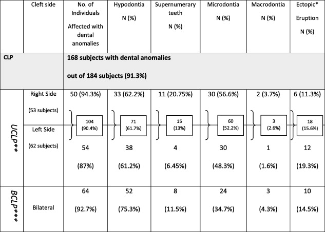 |
∗Ectopic eruption represents intraoral ectopic eruption and intranasal ectopic eruption.
∗∗The total number of UCLP = 115 subjects.
∗∗∗The total number of BCLP = 69 subjects.
Microdontia was the second most commonly observed dental anomaly, occurring in 84 (45.6%) subjects. This anomaly was more prevalent in subjects with UCLP (n = 60 [66%]) than in those with BCLP (n = 24 [26%]; Table 3).
Ectopic eruption was the third most commonly observed dental anomaly, occurring in 29 (15.7%) subjects. Intra-oral ectopic eruption was seen in 23 (12.5%) subjects; intra-nasal ectopic eruption was very rare, affecting only 6 (3.2%) subjects (Table 2).
Supernumerary teeth were found in 23 (12.5%) subjects (Table 2). This anomaly occurred more frequently in subjects with UCLP (n = 15 [57.4%]) than in those with BCLP (n = 8 [31%]); it was observed in 11 (42%) individuals with right UCLP and 4 (15.4%) individuals with left UCLP (Table 3). Macrodontia was observed in six (3.2%) subjects (Table 2). No significant difference between sexes in the prevalence of any dental anomaly was observed (Table 2).
4. Discussion
In the error of identification, the root malformation variable, which showed low reliability (κ = 0.35). This finding suggests that the use of orthopantomographs and/or occlusal radiographs to evaluate root malformation does not produce accurate or reproducible results; standardized full-mouth periapical radiographs should be required for this purpose. Such radiographs were not obtained for all patients with CLP in the present sample; root malformation variables were thus eliminated from analyses.
Gender differences in the prevalence of oral clefts have been reported previously; in comparison with females, males are affected more often and show more severe clefting (Fogh-Andersen, 1967; Natsume et al., 1988; Conway and Wagner, 1966; Christensen, 1999; Cooper et al., 2000; Al-Balkhi, 2008).
The absence of a gender-based difference in the prevalence of dental anomalies in the present study is in agreement with the findings of others (Ranta, 1983, 1986; Shapira et al., 1999; Ribeiro et al., 2003). According to Demirjian et al. (1973), mechanisms controlling dental development are independent of somatic and sexual maturity, but may be influenced by the same factors that cause clefting.
Hypodontia, the most frequently observed dental anomaly, was more prevalent in the present study (66.8%) than in some previous reports (45.5% [Bohn, 1950], 23.8% [Hellquist et al., 1979], and 33.5% [Lopes et al., 1991]), but less prevalent than reported in other studies (77% [Shapira et al., 1999] and 70.2% [Tereza et al., 2010]). These differences in prevalence could be attributed to differences in sample size, inclusion criteria, and methodology. All of these values for hypodontia of the lateral incisors, however, are significantly higher than in the general population (1–11%; Silva Meza, 2003; Stamatiou and Symons, 1991; Fekonja, 2005; Pinho et al., 2005; Altug-Atac and Erdem, 2007; Celikoglu et al., 2012). In cases of severe clefting, the embryonic structures that give rise to the tissue in this region may become severely impaired as early as the dental development phase; this impairment may explain the higher prevalence of hypodontia in subjects with CLP, in agreement with results reported by Fishman (1970), Hellquist et al. (1979), and Vichi and Franchi (1995).
Microdontia, the second most common dental anomaly, was more prevalent in the present study (47.5%) than reported by Dewinter et al. (2003; 32%). The results clearly show that morphological irregularities of dental crowns, especially microdontia, occur throughout the entire dentition in subjects with CLP; they are not limited to maxillary units in the immediate area of the cleft.
Intra-oral ectopic eruption was found in 12.3% of the current sample. Lai et al. (2009) observed ectopic eruption of the maxillary incisors and canines in subjects with CLP. Numerous studies have examined ectopic eruption of the permanent maxillary first molars, but little information on the ectopic eruption of permanent maxillary incisors has been reported. Larson et al. (1998) reported that ectopic eruption of the permanent maxillary first molars was seen in 45% of subjects with large clefts and in (31%) subjects with small clefts. Menezes and Viera (2008) found no ectopically erupted tooth in their sample of subjects with CL/P, but they defined ectopic eruption exclusively as transposition of teeth, which differs from the definition used in this study.
The presence of foreign objects in the oral and nasal structures of children has been widely reported; however, teeth in the nasal cavity are rare (Ranalli et al., 1990). In the present study, intra-nasal teeth were found in 3% of the sample; they were thus more prevalent than reported by Medeiros et al. (2000; 0.48%). The etiology of intra-nasal teeth is obscure. Incomplete union of embryonic processes (developmental disturbances) has been proposed as the probable etiology for the ectopic displacement of the tooth germ (King and Lee, 1987).
The prevalence of supernumerary teeth in our sample (12.5%) was slightly higher than reported by Dewinter et al. (2003; 10.6%) and Tereza et al. (2010; 11.7%). Other researchers have shown a higher prevalence of supernumerary teeth, including Dahloff et al. (1989; 18%), Lopez et al. (1991; 16%), and Tahir (1998; 15.8%). In the present study, supernumerary teeth were observed more frequently in males than in females (14 vs. 9 subjects), which was in agreement with the findings of Tahir (1998) and Tereza et al. (2010).
The prevalence of macrodontia in the current study was higher than that reported by Tahir (1998; 2.6%). Other investigators have reported similar findings (Aizenbud et al., 2005; Al Jamal et al., 2010; Pegelow et al., 2012; Matern et al., 2012; Paranaiba et al., 2013; Mikulewicz et al., 2014). The low prevalence of macrodontia relative to other dental anomalies could be attributed to the relation of CLP to tissue deficiency, rather than excessive tissue development.
The prevalence of dental anomalies varies among ethnic/population groups (Aizenbud et al., 2005; Al Jamal et al., 2010; Pegelow et al., 2012; Matern et al., 2012; Paranaiba et al., 2013; Mikulewicz et al., 2014). The prevalence of hypodontia in a Jordanian population with CLP (66.7%) was comparable to that observed in our sample of Saudi subjects (Al Jamal et al., 2010). Similarly, Aizenbud et al. (2005) found that hypodontia occurred in 67.6% of the Israeli population with CL/P, and Matern et al. (2012) reported a prevalence of 63% in subjects with CLP in Strasbourg, France. Conversely, only 20% of a Swedish sample of subjects with CLP presented with hypodontia (Pegelow et al., 2012). Paranaiba et al. (2013) found that hypodontia occurred in 20% of a sample of Brazilian subjects with CLP (Table 4). These contradictory findings may be attributed to selection criteria and methodological variations as well as ethnic/regional differences.
Table 4.
Dental anomalies among different population/ethnic groups.
| Current study | Al Jamal et al. (2010) | Aizenbud et al. (2005) | Matern et al. (2012) | Paranaiba et al. (2013) | Pegelow et al. (2012) | Mikulewicz et al. (2014) | |
|---|---|---|---|---|---|---|---|
| Population | Saudi | Jordanian | Israeli | French | Brazilian | Swedish | Polish |
| Total participants no. | 184 | 78 | 179 | 124 | 296 | 129 | 202 |
| Cleft types | UCLP, BCLP | UCLP, BCLP | CL, CLA, CLP, CP | CL, CLA, CLP, CP | CL, CLP, CP, CL ± P | CL, CLA, CLP | CLP, CLA, CP |
| Control group | Not included | Not included | Not included | Not included | Not included | Not included | Not included |
| Male participants | 122 | Included but not specified | 101 | 81 | 165 | 72 | 121 |
| Female participants | 62 | Included but not specified | 78 | 43 | 131 | 27 | 81 |
| Total dental anomalies (%) | 91.3 | Not reported | 67.6 | 63 | 39.9 | Not reported | 19.3 |
| Hypodontia (%) | 66.8 | 66.7 | 67.6 | 63 | 22.3 | 30.6⁎ | 19.3 |
| Microdontia (%) | 45.6 | 37 | Not reported | Not reported | 8.1 | 12.4⁎ | Not reported |
| Intraoral ectopic eruption (%) | 12.5 | Not reported | Not reported | Not reported | Not reported | Not reported | Not reported |
| Intranasal ectopic eruption (%) | 3.2 | Not reported | Not reported | Not reported | Not reported | Not reported | Not reported |
| Supernumerary (%) | 12.3 | 16.7 | Not reported | Not reported | 3 | 17⁎ | Not reported |
| Macrodontia (%) | 3.2 | 70.5 | Not reported | Not reported | 3.7 | Not reported | Not reported |
| Transposition (%) | Not reported | 30.8 | Not reported | Not reported | Not reported | Not reported | Not reported |
| Dilacerated roots (%) | Not reported | 19.2 | Not reported | Not reported | Not reported | Not reported | Not reported |
| Hypoplastic teeth (%) | Not reported | 30.8 | Not reported | Not reported | 3 | 26.3⁎⁎ | Not reported |
Percentages of findings related to the permanent lateral incisors.
Percentages of findings related to the permanent central incisor.
Microdontia was more prevalent in the current Saudi Arabian sample of subjects with CLP (45%) than among those in Jordan (37%), Sweden (12.4%), and Brazil (8.1%) (Al Jamal et al., 2010; Pegelow et al., 2012; Paranaiba et al., 2013). Al Jamal et al. (2010) reported a high frequency of macrodontia among Jordanian subjects with CLP. Conversely, macrodontia was reported in 3.7% of a Brazilian sample (Paranaiba et al., 2013), which is similar to our findings. Sample size, cleft type, and ethnic/regional characteristics differed among these studies, which may explain the contradictory prevalence rates.
Generally, subjects with CL/P require extensive dental care; the large amount of required health care interventions is complicated by the presence of various dental anomalies. This study was conducted to epidemiologically explore the prevalence of dental anomalies among Saudi Arabian subjects with CLP. Unfortunately, the sample was small and only included subjects with CLP. A larger multi-center investigation including different cleft types and various regions of Saudi Arabia is needed.
Conflict of interest
The authors have no conflict of interest to declare.
Footnotes
Peer review under responsibility of King Saud University.
Contributor Information
Ghada H. Al-Kharboush, Email: drghada.h.k@gmail.com.
Khalid M. Al-Balkhi, Email: kalbalkhi@ksu.edu.sa.
Khalid Al-Moammar, Email: kalmoammar@ksu.edu.sa.
References
- Aizenbud D., Camasuvi S., Peled M., Brin I. Congenitally missing teeth in the Israeli cleft population. Cleft Palate Craniofac. J. 2005;42(3):314–317. doi: 10.1597/03-126.1. [DOI] [PubMed] [Google Scholar]
- Al-Balkhi K. The distribution and classification of clefts in patients attending a cleft lip and palate clinic in Riyadh, Saudi Arabia. Saudi Med. J. 2008;29:739–742. [PubMed] [Google Scholar]
- Al Jamal G., Hazza’a A., Rawashdeh M. Prevalence of dental anomalies in a population of cleft lip and palate patients. Cleft Palate Craniofac. J. 2010;47(4):413–420. doi: 10.1597/08-275.1. [DOI] [PubMed] [Google Scholar]
- Altug-Atac A.T., Erdem D. Prevalence and distribution of dental anomalies in orthodontic patients. Am. J. Orthod. Dentofacial Orthop. 2007;131:510–514. doi: 10.1016/j.ajodo.2005.06.027. [DOI] [PubMed] [Google Scholar]
- Bohn A. Anomalies of the lateral incisor in cases of harelip and cleft palate. Acta Odontol. Scand. 1950;9(1):41–59. doi: 10.3109/00016355009087225. [DOI] [PubMed] [Google Scholar]
- Celikoglu M., Kamak H., Yildirim H., Ceylan I. Investigation of the maxillary lateral incisor agenesis and associated dental anomalies in an orthodontic patient population. Med. Oral Patol. Oral Cir. Bucal. 2012;17(6):e1068–e1073. doi: 10.4317/medoral.17767. [DOI] [PMC free article] [PubMed] [Google Scholar]
- Christensen K. The 20th Century Danish facial cleft population-epidemiological and genetic-epidemiological studies. Cleft Palate Craniofac. J. 1999;36(2):96–104. doi: 10.1597/1545-1569_1999_036_0096_tcdfcp_2.3.co_2. [DOI] [PubMed] [Google Scholar]
- Conway H., Wagner K. Incidence of clefts in New York City. Cleft Palate J. 1966;3:284–290. [PubMed] [Google Scholar]
- Cooper M., Stone R., Liu Y., Hu D., Melnick M., Marazita M. Descriptive epidemiology of non-syndromic cleft lip with or without cleft palate in Shanghai, China from 1980 to 1989. Cleft Palate Cranio J. 2000;37(3):274–280. doi: 10.1597/1545-1569_2000_037_0274_deoncl_2.3.co_2. [DOI] [PubMed] [Google Scholar]
- Cobourne M. The complex genetics of cleft lip and palate. Eur. J. Orthod. 2004;26:7–16. doi: 10.1093/ejo/26.1.7. [DOI] [PubMed] [Google Scholar]
- Dahllof G., Ussisoo-Joandi R., Ideberg M., Modeer T. Caries, gingivitis and dental abnormalities in preschool children with cleft lip and/or palate. Cleft Palate J. 1989;26(3):233–237. discussion 237–238. [PubMed] [Google Scholar]
- Damante, J.H., 1972. Anomalies de Numero na Area da Fenda em Portadores de Malformações Congĕnitas Lăbio-Palatais Bauru Faculdade de Odontologia de Bauru (dissertation), Universidade de Săo Paulo.
- da Silva A.P., Costa B., de Carvalho Carrara C. Dental anomalies of number in the permanent dentition of patients with bilateral cleft lip: radiographic study. Cleft Palate Craniofac. J. 2008;45(5):473–476. doi: 10.1597/06-099.1. [DOI] [PubMed] [Google Scholar]
- Demirjian A., Goldstein H., Tanner J.M. A new system of dental age assessment. Hum. Biol. 1973;45:211–227. [PubMed] [Google Scholar]
- Dewinter G., Quirynen M., Heidbuchel K., Verdonck A., Willems G., Carels C. Dental abnormalities, bone graft quality, and periodontal conditions in patients with unilateral cleft lip and palate at different phases of orthodontic treatment. Cleft Palate Craniofac. J. 2003;40(4):343–350. doi: 10.1597/1545-1569_2003_040_0343_dabgqa_2.0.co_2. [DOI] [PubMed] [Google Scholar]
- Dixon M.J., Marazita M.L., Beaty H.T., Murray J.C. Cleft lip and palate: understanding genetic and environmental influences. Nature. 2011;12:167–178. doi: 10.1038/nrg2933. [DOI] [PMC free article] [PubMed] [Google Scholar]
- D’Souza R., Kapadia H., Vieira A. Teeth. In: Stevenson R.E., Hall J.G., editors. Human malformations and related anomalies. Oxford University Press; New York: 2006. [Google Scholar]
- Fekonja A. Hypodontia in orthodontically treated children. Eur. J. Orthod. 2005;27:457–460. doi: 10.1093/ejo/cji027. [DOI] [PubMed] [Google Scholar]
- Fishman L.S. Factors related to tooth number, eruption time, and tooth position in cleft palate individuals. ASDC J. Dent. Child. 1970;37(4):303–306. [PubMed] [Google Scholar]
- Fogh-Andersen P. Genetic and non-genetic factors in the etiology of facial clefts. Scand. J. Plast. Reconstr. Surg. 1967;1:25–35. [Google Scholar]
- Gravey M.T., Barry H.J., Blake M. Supernumerary teeth-an overview of classification, diagnosis and management. J. Can. Dent. Assoc. 1999;65(11):612–616. [PubMed] [Google Scholar]
- Hardin-Jones M.A., Jones D.L. Speech production of preschoolers with cleft palate. Cleft Palate Craniofac. J. 2005;42(1):7–13. doi: 10.1597/03-134.1. [DOI] [PubMed] [Google Scholar]
- Hellquist R., Linder-Aronson S., Norling M., Ponten B., Stenberg T. Dental abnormalities in patients with alveolar clefts, operated upon with or without primary periosteoplasty. Eur. J. Orthod. 1979;1:169–180. doi: 10.1093/ejo/1.3.169. [DOI] [PubMed] [Google Scholar]
- Kim N.Y., Baek S.H. Cleft sidedness and congenitally missing or malformed permanent maxillary lateral incisors in Korean patients with unilateral cleft lip and alveolus or unilateral cleft lip and palate. Am. J. Orthod. Dentofacial Orthop. 2006;130(6):752–758. doi: 10.1016/j.ajodo.2005.02.029. [DOI] [PubMed] [Google Scholar]
- King N.U., Lee A.M. An internasal tooth in a patient with a cleft lip and palate: report of a case. JADA. 1987;114:475–478. doi: 10.14219/jada.archive.1987.0124. [DOI] [PubMed] [Google Scholar]
- Kumar P., Hussain M.T., Cardoso E., Hawary M.B., Hassanain J. Facial clefts in Saudi Arabia: an epidemiologic analysis in 179 patients. Plast. Reconstr. Surg. 1991;88(6):955–958. doi: 10.1097/00006534-199112000-00002. [DOI] [PubMed] [Google Scholar]
- Lai M.C., King N.M., Wong H.M. Abnormalities of maxillary anterior teeth in Chinese children with cleft lip and palate. Cleft Palate Craniofac. J. 2009;46(1):58–64. doi: 10.1597/07-077.1. [DOI] [PubMed] [Google Scholar]
- Larson M., Hellquist R., Jacobsson O.P. Dental abnormalities and ectopic eruption in patients with isolated cleft palate. Scand. J. Plast. Reconstr. Surg. Hand Surg. 1998;32:203–212. doi: 10.1080/02844319850158831. [DOI] [PubMed] [Google Scholar]
- Lidral A.C., Moreno L.M., Bullard S.A. Genetic factors and orofacial clefting. Semin. Orthod. 2008;14:103–114. doi: 10.1053/j.sodo.2008.02.002. [DOI] [PMC free article] [PubMed] [Google Scholar]
- Lopes L.D., Mattos B.S., Andre M. Anomalies in number of teeth in patients with lip and/or palate clefts. Braz. Dent. J. 1991;2(1):9–17. [PubMed] [Google Scholar]
- Matern O., Sauleau E., Tschill P., Perrin-Schmitt F., Grollemund B. Left-Sided predominance of hypodontia irrespective of cleft sidedness in a French population. Cleft Palate Craniofac. J. 2012;49(3):e1–e5. doi: 10.1597/11-025. [DOI] [PubMed] [Google Scholar]
- Medeiros A.S., Gomide M.R., Costa B., Carrara C.F., dos Neres L.T. Prevalence of intranasal ectopic teeth in children with complete unilateral and bilateral cleft lip and palate. Cleft Palate Craniofac. J. 2000;37(3):271–273. doi: 10.1597/1545-1569_2000_037_0271_poieti_2.3.co_2. [DOI] [PubMed] [Google Scholar]
- Menezes R., Vieira A.R. Dental anomalies as part of the cleft spectrum. Cleft Palate Craniofac. J. 2008;45(4):414–419. doi: 10.1597/07-064.1. [DOI] [PubMed] [Google Scholar]
- Mikulewicz M., Ogiński T., Gedrange T., Berniczei-Royko A., Prussak E. Prevalence of second premolar hypodontia in the Polish cleft lip and palate population. Med. Sci. Monit. 2014;20:355–360. doi: 10.12659/MSM.890386. [DOI] [PMC free article] [PubMed] [Google Scholar]
- Natsume N., Suzuki T., Kawai T. The prevalence of cleft lip and palate in Japanese. Br. J. Oral Maxillofac. Surg. 1988;26:232–236. doi: 10.1016/0266-4356(88)90168-4. [DOI] [PubMed] [Google Scholar]
- Nilsson S, Ash M. 9th Ed. Saunders Elsevier; St. Louis, USA: 2010. Wheeler’s Dental Anatomy, Physiology, and Occlusion. Chapter 1; Page 4. [Google Scholar]
- Paranaiba L., Coletta R., Swerts M., Quintino R., de Barros L., Martelli-Júnior H. Prevalence of dental anomalies in patients with nonsyndromic cleft lip and/or palate in a Brazilian population. Cleft Palate Craniofac. J. 2013;45(4):400–405. doi: 10.1597/11-029. [DOI] [PubMed] [Google Scholar]
- Pegelow M., Alqadi N., Karsten A. The prevalence of various dental characteristics in the primary and mixed dentition in patients born with non-syndromic unilateral cleft lip with or without cleft palate. Eur. J. Orthod. 2012;34:561–570. doi: 10.1093/ejo/cjr074. [DOI] [PubMed] [Google Scholar]
- Pinho T., Tavares P., Maciel P., Pollmann C. Developmental absence of maxillary lateral incisors in the Portuguese population. Eur. J. Orthod. 2005;27:443–449. doi: 10.1093/ejo/cji060. [DOI] [PubMed] [Google Scholar]
- Ranalli D., McWilliams B.J., Garret W.S., Jr. Tooth and foreign object in the nasal fossa of a child with a cleft: case report. Pediatr. Dent. 1990;12(3):183–184. [PubMed] [Google Scholar]
- Ranta R. Hypodontia and delayed development of the second premolars in cleft palate children. Eur. J. Orthod. 1983;5(2):145–148. doi: 10.1093/ejo/5.2.145. [DOI] [PubMed] [Google Scholar]
- Ranta R. A review of tooth formation in children with cleft lip/palate. Am. J. Orthod. Dentofacial Orthop. 1986;90(1):11–18. doi: 10.1016/0889-5406(86)90022-3. [DOI] [PubMed] [Google Scholar]
- Ribeiro L.L., Neves L.T.D., Costa B., Gomide M.R. Dental anomalies of the permanent lateral incisors and prevalence of hypodontia outside the cleft area in complete unilateral cleft lip and palate. Cleft Palate Craniofac. J. 2003;40(2):172–175. doi: 10.1597/1545-1569_2003_040_0172_daotpl_2.0.co_2. [DOI] [PubMed] [Google Scholar]
- Schutte B.C., Murray J. The many faces and factors of orofacial clefts. Hum. Mol. Genet. 1999;8(10):1853–1859. doi: 10.1093/hmg/8.10.1853. [DOI] [PubMed] [Google Scholar]
- Shapira Y., Lubit E., Kuftinec M. Congenitally missing second premolars in cleft lip and cleft palate children. Am. J. Orthod. Dentofacial Orthop. 1999;115(4):396–400. doi: 10.1016/s0889-5406(99)70258-1. [DOI] [PubMed] [Google Scholar]
- Silva Meza R. Radiographic assessment of congenitally missing teeth in orthodontic patients. Int. J. Paediatr. Dent. 2003;13:112–116. doi: 10.1046/j.1365-263x.2003.00436.x. [DOI] [PubMed] [Google Scholar]
- Stamatiou J., Symons A.L. Agenesis of the permanent lateral incisor: distribution, number and sites. J. Clin. Pediatr. Dent. 1991;15:244–246. [PubMed] [Google Scholar]
- Tahir P. Dental anomalies in children with cleft lip and/or palate or both. Saudi Med. J. 1998;19(3):332–334. [PubMed] [Google Scholar]
- Tereza G.P., Carrara C.F., Costa B. Tooth abnormalities of number and position in the permanent dentition of patients with complete bilateral cleft lip and palate. Cleft Palate Craniofac. J. 2010;47(3):247–252. doi: 10.1597/08-268.1. [DOI] [PubMed] [Google Scholar]
- Vichi M., Franchi L. Abnormalities of the maxillary incisors in children with cleft lip and palate. ASDC J. Dent. Child. 1995;62(6):412–417. [PubMed] [Google Scholar]
- Viera A., Garrett J. Understanding inter-observer agreement: the Kappa statistic. Fam. Med. 2005;37(5):360–363. [PubMed] [Google Scholar]