Abstract
Li1.2Mn0.54Ni0.13Co0.13O2-encapsulated carbon nanofiber network cathode materials were synthesized by a facile electrospinning method. The microstructures, morphologies and electrochemical properties are characterized by X-ray diffraction (XRD), field-emission scanning electron microscopy (FE-SEM), high resolution transmission electron microscopy (HR-TEM), galvonostatic charge/discharge tests, cyclic voltammetry and electrochemical impedance spectroscopy (EIS), etc. The nanofiber decorated Li1.2Mn0.54Ni0.13Co0.13O2 electrode demonstrated higher coulombic efficiency of 83.5%, and discharge capacity of 263.7 mAh g−1 at 1 C as well as higher stability compared to the pristine particle counterpart. The superior electrochemical performance results from the novel network structure which provides fast transport channels for electrons and lithium ions and the outer carbon acts a protection layer which prevents the inner oxides from reacting with HF in the electrolyte during charge-discharge cycling.
LiCoO2 has been widely applied as one of the commercial cathode materials for Li-ion batteries since its discovery since 1992. However, the relatively low capacity (140 mAh g−1), high cost and toxicity of the cobalt have hindered its further application for the demand of future electric technology. In this case, development of alternative higher energy, lower cost and environment friendly cathodes is critical for next generation Li-ion batteries1,2.
Layer structure LiMO2 (M = Mn, Co, Ni, etc), spinel LiMn2O4 and olivine LiFePO4 are also regarded as the main candidate cathodes in recent years. Among them, Li-rich cathode materials xLi2MnO3 • (1-x)LiMO2 (M = Mn, Co, Ni) have drawn much attention owing to its high capacity of more than 250 mAh g−1 at a lower cost compared to LiCoO2. Despite these merits, Li-rich layered oxides always suffer from the inferior cycling stability and rate capability, which impede their applications1,3. Surface modification4,5,6,7,8, doping strategy9,10,11,12, and forming 3D porous structure13 are three common approaches introduced to improve their electrochemical performances. As an effective and inexpensive way of truly producing one dimensional (1D) fibers from micrometer to nanometer size ranges in diameter, electrospinning14,15 has been widely used in many areas, including the fabrication of electrode materials, especially the anode16,17,18,19 and cathode20,21,22,23,24 materials for Li-ion batteries.
Herein, for the first time, Li1.2Mn0.54Ni0.13Co0.13O2-encapuslated carbon nanofiber network cathodes were prepared by the combination of electrospinning and heat treatment. For comparison, pristine Li1.2Mn0.54Ni0.13Co0.13O2 particles were also synthesized by sol-gel method. It was demonstrated that the nanofiber decorated Li1.2Mn0.54Ni0.13Co0.13O2 composites displayed higher capacity retention, rate capability and more stable cyclic performance compared to the pristine counterpart. The results not only give a further insight into the reaction mechanism but also provide a rational direction to synthesize cathode materials for Li-ion batteries.
Results and Discussion
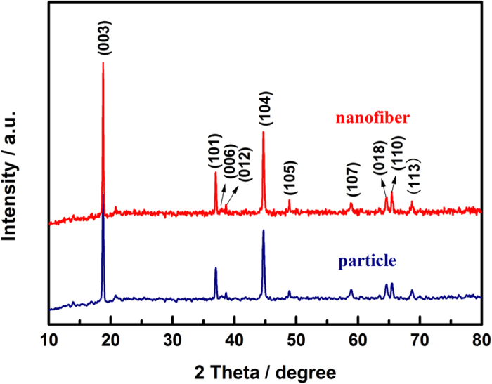
Figure 1 shows the XRD patterns of the pristine particle and nanofiber decorated samples. The pristine particle sample shows the α-NaFeO2 structure without any impurity reflections. The weak superstructure reflections around 2θ = 20°–25° belongs to the ordering of the transition metal and Li ions in the transition metal layer of the lattice25,26. Clear separation of adjacent peaks of (006)/(012) and (108)/(110) indicates that the samples have a well crystalline layered structure27,28,29. For the nanofiber decorated sample, no significant lattice parameter differences were recognized in the main reflections corresponding to the α-NaFeO2 structure, and no additional reflections corresponding to carbon are observed due to its amorphous structure21,22.
Figure 1. XRD patterns of the particle and nanofiber decorated Li1.2Mn0.54Ni0.13Co0.13O2 samples.
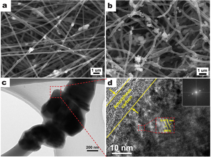
The morphologies and strucutres of the nanofiber decorated sample were shown in Fig. 2. Compared to the pristine particles (Supporting Information, Figure 1S), which showed a universal and slight agglomeration with a uniform particle size ranging between 300 and 500 nm, the carbon nanofiber contained Li1.2Mn0.54Ni0.13Co0.13O2 particles which were evenly dispersed with carbon black, excepting a little agglomeration after electrospinning (Fig. 2a). The morphology of nanofiber decorated Li1.2Mn0.54Ni0.13Co0.13O2 after heat treatment in air was shown in Fig. 2b. The nanofibers shrinked and partly broken, some particles were inevitably squeezed out from the nanofibers and then distributed along the surface or junction of the nanofibers after agglomeration. Figure 2c,d show the HRTEM images of the samples. Result (Fig. 2c) confirms that Li1.2Mn0.54Ni0.13Co0.13O2 particles and carbon black are dispersed inside the carbon nanofiber matrix. The particles exhibit continuous interference fringe spacing of around 0.47 nm corresponding to the (003) lattice fringes of the hexagonal layered structure (Fig. 2d)3,6,28,29. In addition, amorphous carbon layers with a thickness about 10 nm were observed on the surface of the Li1.2Mn0.54Ni0.13Co0.13O2 particles, which may be regarded as protection layer during electrochemical cycling. The selected area electron diffraction (SAED) pattern (inset of Fig. 2d) confirms the single crystallinity for particles.
Figure 2. SEM.
(a,b) and HRTEM (c,d) images of nanofiber decorated Li1.2Mn0.54Ni0.13Co0.13O2 samples before (a) and after (b–d) heat treatment. The inset of d is the SAED pattern.
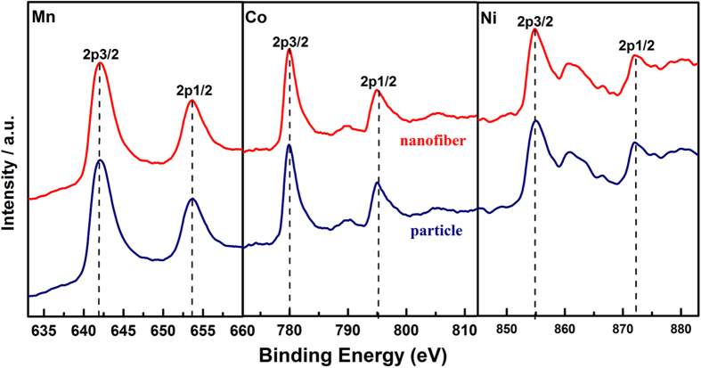
X-ray photoelectron spectroscopy (XPS) measurements were used to investigate the variation in chemical states of Mn, Ni and Co elements. Figure 3 shows typical XPS spectra for Mn2p, Co2p and Ni2p for the pristine and nanofiber decorated samples. For these two samples, the observed binding energies of Mn2p, Co2p and Ni2p remain at 642, 780 and 855 eV, which coincide well with those for Mn4+, Co3+ and Ni2+, respectively30,31. Therefore, the valences of Mn, Co and Ni did not change after the heat treatment with/without electrospinning.
Figure 3. XPS spectra of nanofiber decorated and particle Li1.2Mn0.54Ni0.13Co0.13O2 samples before and after heat treatment.
The first charge-discharge curves during electrochemical cycling of the two cathode materials taken at 0.2 C, are shown in Fig. 4a. During the charging process, both samples exhibit two distinguishable stages, a smoothly sloping voltage profile below 4.5 V corresponding to Li+ deintercalation from LiMn1/3Ni1/3Co1/3O2 component and a long plateau around 4.5 V related to Li+ and O2− extracted from the Li2MnO3 phase and structural rearrangement1,31,32,33. The first discharge capacity of nanofiber decorated Li1.2Mn0.54Ni0.13Co0.13O2/C and pristine sample are 263.7 and 247.2 mAh g−1, with the coulombic efficiency of 83.5% and 75.4%, respectively. The higher coulombic efficiency and discharge capacity may be attributed to the reasons as follow: (a) the amorphous carbon layer prevents the formation of inactive O2 molecules and the side reactions of electrolyte; (b) the better dispersity and less agglomeration of the cathode particles would increase the active surface area and facilitate the electrons and/or ions migration during cycling.
Figure 4. Charge-discharge profiles.
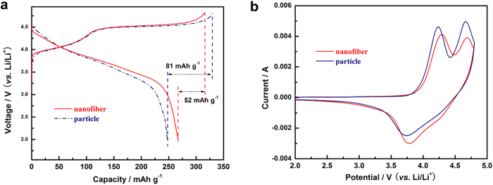
(a) at 0.2 C and CV curves (b) scanned at 1 mV s−1 of particle and nanofiber decorated Li1.2Mn0.54Ni0.13Co0.13O2 electrodes.
Figure 4b shows the CV profiles of the first cycle of the two samples at a sweep rate of 1 mV s−1 between 2.0–4.8 V. As can be seen, the initial anodic peaks of the samples at about 4.2 V are due to the lithium extraction from the Li1.2Mn0.54Ni0.13Co0.13O2 lattice and the oxidation process of Ni2+/Co3+ to higher oxidation states2,30,34,35,36, while the sole cathodic peak occurs at around 3.8 V is attributed to the reduction of Ni2+/Co3+. The second anodic peaks located in the potential of 4.6 V are normally associated with the oxygen release process from Li2MnO3-type component as well as the electrolyte oxidation. Compared with the CV curve of pristine Li1.2Mn0.54Ni0.13Co0.13O2, the decreased intensity and area of anodic peak around 4.6 V of the nanofiber decorated Li1.2Mn0.54Ni0.13Co0.13O2 sample implies that the O2 release was greatly suppressed by the amorphous carbon layers and more available Li+ sites were remained. This agrees with the improved columbic efficiency of the composite sample (Fig. 4a).
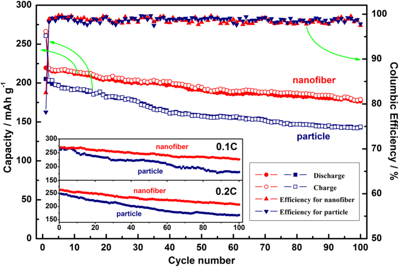
The cyclic performances of both samples at 1 C are shown in Fig. 5. The first discharge capacity of nanofiber decorated Li1.2Mn0.54Ni0.13Co0.13O2 and particle samples are 219.5 and 205 mAh g−1 with a coulombic efficiency of 82.5% and 76.2%, respectively. After 100 cycles, the nanofiber samples still display a discharge capacity of 176.7 mAh g−1 with a capacity retention of 80.5%, while the particle samples only show a discharge capacity of 145.9 mAh g−1 and a capacity retention of 71.1%. Their cyclic stability at low rate of 0.1 and 0.2 C are also shown in the inset of Fig. 5. The two samples deliver a similar discharge capacity in the first several cycles. However, the nanofiber samples still retain a higher discharge capacity than the pristine one both at 0.1 and 0.2 C after 100 cyles. The improved cyclic stability and capacity retention of the nanofiber cathodes may be attributed to the synergistic effect of the larger specific surface area and the protection of coating layer. Comparing with the pristine samples, the larger specific surface area of nanofibers provides more transportation paths for ions and/or electrons, and the coating layer simultaneously prevents the inner oxide from reacting with HF in the electrolyte and twice agglomeration during electrochemical test.
Figure 5. Cyclic performance and coulombic efficiency of particle and nanofiber decorated Li1.2Mn0.54Ni0.13Co0.13O2 samples at 1 C.
The inset is the discharge capacities of two samples at 0.1 and 0.2 C.
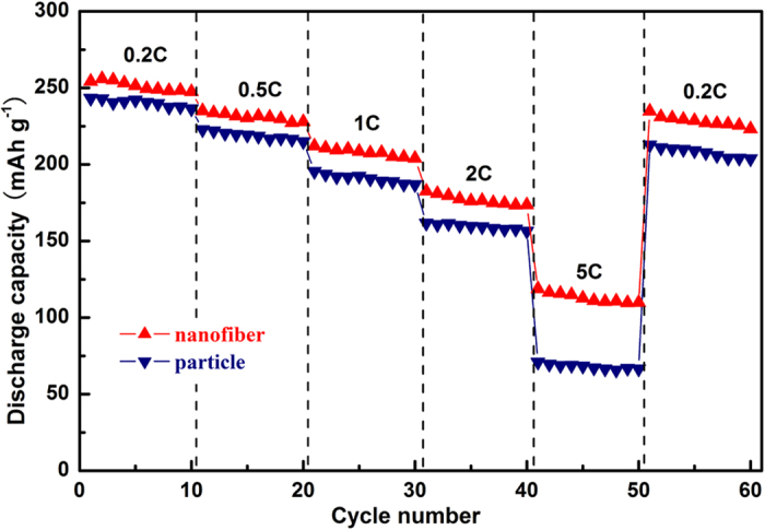
Figure 6 shows a continuous cycling result at incremental rates from 0.2 to 5 C then recovering back to 0.2 C. Similar discharge capacity is observed at lower rate for both samples, but obvious capacity differences increase gradually with the increasing rate. As can be seen, the rate capability for nanofiber decorated Li1.2Mn0.54Ni0.13Co0.13O2 samples has been significantly enhanced compared to the particle. Such contrast observation might be related to the faster ionic and/or electronic diffusion and shorter migration path at high rate, which were benefited from the network structures.
Figure 6. Cycling behavior of particle and nanofiber-decorated Li1.2Mn0.54Ni0.13Co0.13O2 samples at various rates.
To investigate the origin of the improved electrochemical performance of nanofiber decorated Li1.2Mn0.54Ni0.13Co0.13O2 sample, EIS plots of both samples were collected. Figure 7 compares the EIS spectra of the particle and nanofiber samples at different states of cycling, 1st and 10th. An equivalent circuit (inset of Fig. 7)28 was applied to fit the raw data from EIS measurements to obtain the accurate values of different resistances. In this equivalent circuit, RΩ is the resistance of the solution , Rs is the resistance of ion diffusion in the region of the surface layer of particle, Rct represents the charge transfer resistance in the electrode/electrolyte interface and Zw refers to Warburg impedance .The fitting values of Rs and Rct were summarized in Table 1.
Figure 7.
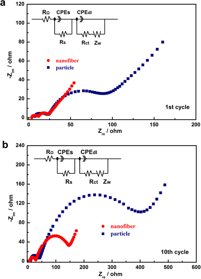
EIS plots of particle and nanofiber decorated Li1.2Mn0.54Ni0.13Co0.13O2 samples after 1st (a) and 10th (b) cycle at a same state of discharge at 3.5 V. The insets of a and b are the equivalent circuits.
Table 1. Fitting values of R s and R ct of the particle and decorated samples at the 1st and 10th cycling.
|
Rs/Ω |
Rct/Ω |
|||
|---|---|---|---|---|
| 1st | 10th | 1st | 10th | |
| Nanofiber | 8.21 | 11.65 | 21.73 | 118.26 |
| Particle | 15.46 | 62.61 | 30.91 | 363.31 |
Compared with the Fig. 7a,b, there was a huge reduction of Rct for the nanofiber sample upon cycling. After 10 cycles, the Rct values of nanofiber and particle samples increased from 21.73 and 30.91 Ω to 118.26 and 363.31 Ω, respectively. The reduction of Rct for nanofiber sample may facilitate lithium transfer on the electrode/electrolyte interface, then leads to improved rate capability. Therefore, the better electrochemical performance of nanofiber sample compared to the particle one can be ascribed to larger surface area and better electrical contact of the particle, which increase the active specific surface area and decrease the polarization effect.
Conclusions
In conclusion, Li1.2Mn0.54Ni0.13Co0.13O2-ecapulated carbon nanofiber network were prepared through a facile electrospinning method following by a heat treatment. XPS spectra demonstrate that the valence of Mn, Ni, Co did not change after the heat treatment. Compared with the pristine Li1.2Mn0.54Ni0.13Co0.13O2 particles, the nanofiber sample has the obviously improved electrochemical performance. The coulombic efficiency of the first charge-discharge process is enhanced from 76.2% to 82.5% with a capacity retention of 80.5% at 1 C after 100 cycles. In addition, the nanofiber sample also exhibits superior rate capability compared to the pristine one. Such improved initial coulombic efficiency, cyclic stability and rate capability can be attributed to the suppression of the side reaction of electrolyte, larger surface area of nanofibers, better dispersity of active particles and more importantly, faster electron and ion transportation, which are benefited from the carbon nanofiber network. Therefore, it is believed that the nanofiber decorated Li1.2Mn0.54Ni0.13Co0.13O2 should be one of ideal cathode candidates and the results also provide a rational direction to synthesize other cathode materials for Li-ion batteries.
Methods
Preparation of Li 1.2 Mn 0.54 Ni 0.13 Co 0.13 O 2 particles
Li1.2Mn0.54Ni0.13Co0.13O2 particles were prepared by a PVP-assisted sol-gel method. Typically, stoichiometric amounts of Li(CH3COO)∙2H2O, Mn(CH3COO)2∙4H2O , Ni(CH3COO)2∙4H2O and Co(CH3COO)2∙4H2O were first dissolved in 10 wt% polyvinylpyrrolidone (PVP)/H2O mixed solution under vigorous stirring for 6 hours to form a homogeneous solution. Then the solution was evaporated at 85 °C until a viscous violet gel emerged. The gel was dried in a vacuum oven at 120 °C for 18 h. The precursor powders were decomposed at 500 °C for 6 h to guarantee the complete decomposition of (PVP). After a grinding for 15 min with a mortar and pestle, the obtained powders were calcined at 950 °C for 8 h in air and ground again for at least 20 min to get the finial products.
Fabrication of Li 1.2 Mn 0.54 Ni 0.13 Co 0.13 O 2 /C composite fibers
After milling for 1 h, the mixture of 30 wt% of Li1.2Mn0.54Ni0.13Co0.13O2 particles and 8 wt% of carbon black were added into the 8 wt% polyacrylonitrile (PAN)/dimethyl formamide (DMF) solution and then stirred with ultrasonic dispersion for at least 24 h. Electrospinning was carried out with 18 KV high voltage, 1 ml/h feeding rate and 20 cm needle-to-collector distance, respectively. The electrospun nanofibers were collected on the aluminum foil and dried in a vacuum oven at 120 °C for 12h. The obtained electrospun nanofibers were firstly heated at 280 °C for 1 h with a heating rate of 5 °C min−1, then at 350 °C for 1 h and finally at 400 °C for 1 h with a heating rate of 2 °C min−1 to form Li1.2Mn0.54Ni0.13Co0.13O2-encapuslated carbon nanofibers. In order to have a fair comparison, the pristine Li1.2Mn0.54Ni0.13Co0.13O2 particles were also having the same heat treatment without electrospinning. The carbon content of nanofiber decorated Li1.2Mn0.54Ni0.13Co0.13O2 was evaluated by elemental analysis to be around 10.57%–10.82%. Additionally, the specific surface areas of the particles and nanofibers were 4.2 m2/g and 28.56 m2/g, respectively, which were tested by using NOVA1200e (Quantachrome).
Structure and morphology characterization
X-ray diffraction (XRD) (Bruker D8 Advance) with Cu Kα radiation operated at 40 KV and 40 mA was employed to identify the crystal structure (2θ = 10°~80°). Scanning electron microscope (SEM) (Hitachi, S-3400N) and high resolution transmission electron microscope (HRTEM) (Tecnai G2 F30) were used to observe the morphologies and structures. X-ray photoelectron spectroscopy (ESCALAB 250Xi) was used to test the chemical state of Mn, Ni and Co of the samples. With regard to the HRTEM samples preparation, a trace of nanofiber decorated Li1.2Mn0.54Ni0.13Co0.13O2 sample were added into ethanol solution with ultrasonic dispersion for 7 h, then the testing liquid was dropped to the micro Cu grid and heated at 60 °C for 1 h.
Electrochemical measurements
Electrochemical tests were made using CR-2032 coin cells, which were assembled in a glove box (MBRAUN,UNILab2000) filled with high purity argon, comprising cathode, Li metal anode and a polymer separator (Celgard 2400) with 1 M LiPF6 in EC:DMC (1:1 in volume ratio) as electrolyte. For fabrication of the cathodes, nanofiber decorated Li1.2Mn0.54Ni0.13Co0.13O2 was mixed with polyvinylidene fluoride (PVDF) with a weight ratio of 95:5 in N-methyl-2-pyrrolidone (NMP), while pristine Li1.2Mn0.54Ni0.13Co0.13O2 particles were mixed with acetylene black and PVDF with a weight ratio of 85:10:5. The obtained slurry was coated onto Al foil and roll-pressed. The electrodes were dried overnight at 120 °C in vacuum oven. Cyclic performance and rate capability tests were conducted on a battery station (LAND, CT2001A) between voltage range of 2.0–4.8 V (1 C = 250 mA g−1). Both of the Cyclic voltammetry (CV) and electrochemical impedance spectroscopy (EIS) (at a frequency range of 10−2–105 Hz) were conducted using an electrochemical workstation (CHI660A), and the CV was applied with a scan rate of 1 mV/s between 2.0–4.8 V. All of the electrochemical tests were carried out at room temperature.
Additional Information
How to cite this article: Ma, D. et al. Li1.2Mn0.54Ni0.13Co0.13O2-Encapsulated Carbon Nanofiber Network Cathodes with Improved Stability and Rate Capability for Li-ion Batteries. Sci. Rep. 5, 11257; doi: 10.1038/srep11257 (2015).
Supplementary Material
Acknowledgments
We are grateful for the financial support provided by the National Natural Science Foundation of China (Grant#51374146, 50874074), Shenzhen Government’s Plan of Science and Technology (Grant #JCYJ20120613173950029, JCYJ20140418095735619), the Special Fund of the Central Finance for the Development of Local Universities (Grant #000022070149).
Footnotes
Author Contributions P.Z. conceived the research. D.M., P.Z., Y.L. and X.R. developed the concept and designed the experiments. D.M. and X.R. developed the experimental setup. D.M., Y.L. and P.Z. prepared the manuscript. All authors discussed the results and commented the manuscript.
References
- Yu H. & Zhou H. High-energy cathode materials (Li2MnO3–LiMO2) for lithium-ion batteries. The J. Phys. Chem. Lett. 4, 1268–1280 (2013). [DOI] [PubMed] [Google Scholar]
- Yu H. et al. High-energy ‘composite’ layered manganese-rich cathode materials via controlling Li2MnO3 phase activation for lithium-ion batteries. Phys. chem. chem. phys.: PCCP 14, 6584–6595 (2012). [DOI] [PubMed] [Google Scholar]
- Thackeray M. M. et al. Li2MnO3-stabilized LiMO2 (M=Mn, Ni, Co) electrodes for lithium-ion batteries. J. Mater. Chem. 17, 3112 (2007). [Google Scholar]
- Gao J., Kim J. & Manthiram A. High capacity Li[Li0.2Mn0.54Ni0.13Co0.13]O2–V2O5 composite cathodes with low irreversible capacity loss for lithium ion batteries. Electrochem. Commun. 11, 84–86 (2009). [Google Scholar]
- Liu J. & Manthiram A. Functional surface modifications of a high capacity layered Li[Li0.2Mn0.54Ni0.13Co0.13]O2 cathode. J. Mater. Chem. 20, 3961 (2010). [Google Scholar]
- Wu C. et al. Surface modification of Li1.2Mn0.54Co0.13Ni0.13O2 with conducting polypyrrole. J. Power Sources 231, 44–49 (2013). [Google Scholar]
- Wang Z. Y. et al. Cycle performance improvement of Li-rich layered cathode material Li[Li0.2Mn0.54Ni0.13Co0.13]O2 by ZrO2 coating. Surf. Coat. Tech. 235, 570–576 (2013). [Google Scholar]
- Kim I. T., Knight J. C., Celio H. & Manthiram A. Enhanced electrochemical performances of Li-rich layered oxides by surface modification with reduced graphene oxide/AlPO4 hybrid coating. J. Mater. Chem. A 2, 8696 (2014). [Google Scholar]
- Li Q. et al. K+-doped Li1.2Mn0.54Co0.13Ni0.13O2: a novel cathode material with an enhanced cycling stability for lithium-ion batteries. ACS appl. mater. interfaces 6, 10330–10341 (2014). [DOI] [PubMed] [Google Scholar]
- Song B., Lai M. O. & Lu L. Influence of Ru substitution on Li-rich 0.55Li2MnO3·0.45LiNi1/3Co1/3Mn1/3O2 cathode for Li-ion batteries. Electrochim. Acta 80, 187–195 (2012). [Google Scholar]
- Song B. et al. Advances in sustain stable voltage of Cr-doped Li-rich layered cathodes for lithium ion batteries. J. Electrochem. Soc. 161, A1723–A1730 (2014). [Google Scholar]
- Wang D., Huang Y., Huo Z. & Chen L. Synthesize and electrochemical characterization of Mg-doped Li-rich layered Li[Li0.2Ni0.2Mn0.6]O2 cathode material. Electrochim. Acta 107, 461–466 (2013). [Google Scholar]
- Deepak P. S., Mulder F. M., Abdelkader A. M. & Wagemaker M. Facile micro templating LiFePO4 electrodes for high performance Li-Ion batteries. Adv. Energy Mater. 3, 572–578 (2013). [Google Scholar]
- Thavasi V., Singh G. & Ramakrishna S. Electrospun nanofibers in energy and environmental applications. Energy Environ. Sci. 1, 205 (2008). [Google Scholar]
- Lu W., Sun J. & Jiang X. Recent advances in electrospinning technology and biomedical applications of electrospun fibers. J. Mater. Chem. B 2, 2369 (2014). [DOI] [PubMed] [Google Scholar]
- Xue L. et al. Si/C composite nanofibers with stable electric conductive network for use as durable lithium-ion battery anode. Nano Energy 2, 361–367 (2013). [Google Scholar]
- Suresh Kumar P. et al. Free-standing electrospun carbon nanofibres—a high performance anode material for lithium-ion batteries. J. Phys. D: Appl. Phys. 45, 265302 (2012). [Google Scholar]
- Ji L. & Zhang X. Fabrication of porous carbon/Si composite nanofibers as high-capacity battery electrodes. Electrochem. Commun. 11, 1146–1149 (2009). [Google Scholar]
- Liu B. et al. Encapsulation of MnO nanocrystals in electrospun carbon nanofibers as high-performance anode materials for lithium-ion batteries. Sci. Rep. 4, 4229 (2014). [DOI] [PMC free article] [PubMed] [Google Scholar]
- Von Hagen R., Lorrmann H., Möller K.-C. & Mathur S. Electrospun LiFe1-yMnyPO4/C nanofiber composites as self-Supporting cathodes in Li-ion batteries. Adv. Energy Mater. 2, 553–559 (2012). [Google Scholar]
- Toprakci O., Ji L., Lin Z., Toprakci H. & Zhang X. Fabrication and electrochemical characteristics of electrospun LiFePO4/carbon composite fibers for lithium-ion batteries. J. Power Sources 196, 7692–7699 (2011). [Google Scholar]
- Zhou H. et al. Fabrication and electrochemical characteristics of electrospun LiMn2O4 nanofiber cathode for Li-ion batteries. Mater. Lett. 117, 175–178 (2014). [Google Scholar]
- Zhang S. et al. Cr-doped Li2MnSiO4/carbon composite nanofibers as high-energy cathodes for Li-ion batteries. J. Mater. Chem. 22, 14661 (2012). [Google Scholar]
- Zhang S. et al. High-capacity Li2Mn0.8Fe0.2SiO4/carbon composite nanofiber cathodes for lithium-ion batteries. J. Power Sources 213, 10–15 (2012). [Google Scholar]
- Chen Y., Chen Z. & Xie K. Effect of annealing on the first-cycle performance and reversible capabilities of lithium-rich layered oxide cathodes. J. Phys. Chem. C 118, 11505–11511 (2014). [Google Scholar]
- Ates M. N. et al. Mitigation of layered to spinel conversion of a Li-rich layered metal oxide cathode material for Li-Ion batteries. J. Electrochem. Soc. 161, A290–A301 (2013). [DOI] [PMC free article] [PubMed] [Google Scholar]
- Ates M. N., Mukerjee S. & Abraham K. M. A Li-rich layered cathode material with enhanced structural stability and rate capability for Li-on batteries. J. Electrochem. Soc. 161, A355–A363 (2014). [Google Scholar]
- Lee E.-S. & Manthiram A. Smart design of lithium-rich layered oxide cathode compositions with suppressed voltage decay. J. Mater. Chem. A 2, 3932–3939 (2014). [Google Scholar]
- Jarvis K. A., Deng Z., Allard L. F., Manthiram A. & Ferreira P. J. Atomic structure of a lithium-rich layered oxide material for lithium-ion batteries: evidence of a solid solution. Chem. Mater. 23, 3614–3621 (2011). [Google Scholar]
- Song B. et al. High rate capability caused by surface cubic spinels in Li-rich layer-structured cathodes for Li-ion batteries. Sci. Rep. 3, 3094 (2013). [DOI] [PMC free article] [PubMed] [Google Scholar]
- Zheng J. et al. The effects of persulfate treatment on the electrochemical properties of Li[Li0.2Mn0.54Ni0.13Co0.13]O2 cathode material. J. Power Sources 221, 108–113 (2013). [Google Scholar]
- Chen D. et al. Porous layered lithium-rich oxide nanorods: Synthesis and performances as cathode of lithium ion battery. Electrochim. Acta 154, 83–93 (2015). [Google Scholar]
- Yabuuchi N., Yoshii K., Myung S. T., Nakai I. & Komaba S. Detailed studies of a high-capacity electrode material for rechargeable batteries, Li2MnO3-LiCo1/3Ni1/3Mn1/3O2. J. Am. Chem. Soc. 133, 4404–4419 (2011). [DOI] [PubMed] [Google Scholar]
- Wang J. et al. Improved rate capability of layered Li-rich cathode for lithium ion battery by electrochemical treatment. ECS Electrochem. Lett. 2, A78–A80 (2013). [Google Scholar]
- Johnson C. S., Li N., Vaughey J. T., Hackney S. A. & Thackeray M. M. Lithium–manganese oxide electrodes with layered–spinel composite structures xLi2MnO3·(1−x)Li1+yMn2−yO4 (0 < x < 1, 0 ≤ y ≤ 0.33) for lithium batteries. Electrochem. Commun. 7, 528–536 (2005). [Google Scholar]
- Wang C. C. & Manthiram A. Influence of cationic substitutions on the first charge and reversible capacities of lithium-rich layered oxide cathodes. J. Mater. Chem. A 1, 10209 (2013). [Google Scholar]
Associated Data
This section collects any data citations, data availability statements, or supplementary materials included in this article.


