Abstract
Introduction
An increasing number of catheter ablations involve the mitral annular region and valve apparatus, increasing the risk of catheter interaction with the mitral valve (MV) complex. We review our experience with catheter ablation-related MV injury resulting in severe mitral regurgitation (MR) to delineate mechanisms of injury and outcomes.
Methods
We searched the Mayo Clinic mitral valve surgical database over a 19-year period (1993–2012) and the electrophysiologic procedures database over a 23-year period (1990–2013) and identified 9 patients with catheter ablation related MV injury requiring clinical intervention.
Results
Indications for ablation included atrial fibrillation (AF) [n=4], ventricular tachycardia (VT) [n =3], and left-sided accessory pathways [n=2]. In all 4 AF patients, a circular mapping catheter entrapped in the MV apparatus was responsible for severe mitral regurgitation. In all 3 VT patients, radiofrequency energy delivery led to direct injury to the MV apparatus. In the 2 patients with accessory pathways, both mechanisms were involved (1 per patient). Six patients required surgical intervention (5 MV repair, 1 catheter removal). One patient developed severe functional MR upon successful endovascular catheter disentanglement that improved spontaneously. Two VT patients with persistent severe post-ablation MR were managed non-surgically, one of whom died 3 months post-procedure.
Conclusion
Circular mapping catheter entrapment and ablation at the mitral annulus are the most common etiologies of MV injury during catheter ablation. Close surveillance of the MV is needed during such procedures and early surgical repair is important for successful salvage if significant injury occurs.
Introduction
Catheter ablation procedures involving the mitral annular region are common and manipulation during procedures increases the risk of catheter interaction with the mitral valve complex. Cases of ablation energy damage to the mitral valve annulus and/or entrapment in the valve apparatus have been reported.1–7 The resultant damage to the mitral valve can be severe and dysfunction of the valve can pose an emergent situation. Patients with severe mitral regurgitation (MR) related to ruptured chordae tendineae or flail leaflets have been shown to have better long term survival and freedom from heart failure when surgical correction is performed promptly following recognition.8
We present the largest series of cases to date that illustrates: 1) two most common causes for mitral valve regurgitation post ablation procedures, 2) necessity for strict surveillance when procedural mitral valve injury is suspected, 3) need for early surgical consultation in order to obtain positive outcomes.
Methods
A retrospective analysis was performed by searching the Mayo Clinic mitral valve surgical database over a 19-year period (January 1, 1993–December 31, 2012) and found that 8400 mitral valve repair or replacements were performed for etiology of mitral valve trauma. We also searched the electrophysiologic (EP) procedures database over a 23-year period (January 4, 1990–December 31, 2013) and found 627 catheter ablations involving mitral valve from the procedure records. We identified 9 patients who had catheter ablation procedures and required strict surveillance and/or surgical intervention for mitral valve damage (Table 1).
Table 1.
Characteristics of patients with catheter ablation and mitral valve damage
| Case | Age | Gender | Indication | Catheter | Complication | Intervention |
|---|---|---|---|---|---|---|
| 1 | 48 | M | Atria fibrillation | Circular mapping | Catheter entrapment | MV repair |
| 2 | 33 | M | Atrial fibrillation | Circular mapping | Catheter entrapment | Surgical removal of catheter |
| 3 | 59 | F | Atrial fibrillation | Circular mapping | Catheter entrapment | Close follow-up |
| 4 | 53 | M | Atrial fibrillation | Circular mapping | Catheter entrapment | MV repair |
| 5 | 23 | M | Accessory pathway (WPW) | Ablation | Catheter entrapment | MV repair |
| 6 | 39 | F | Accessory pathway (WPW) | Ablation | RE energy | MV repair |
| 7 | 48 | M | Ventricular tachycardia | Ablation | RF energy | MV repair |
| 8 | 71 | M | Ventricular tachycardia | Ablation | RF energy | Close follow-up |
| 9 | 79 | M | Ventricular tachycardia | Ablation | RF energy | None (patient death) |
Atrial Fibrillation
A-48-year old male with paroxysmal AF underwent ablation. During transition from the left- to the right-sided PVs, the circular mapping catheter became ensnared in the mitral valve and could not be dislodged with traction. The sheath was advanced in an attempt to disengage the catheter but was not successful, and the catheter fractured. The distal tip was seen on echocardiography to be entrapped in the chordae with severe mitral regurgitation that required surgical repair.
A 33-year-old male with medically refractory AF underwent ablation. Upon removal of the circular mapping catheter from the left inferior pulmonary vein, the catheter flipped into the left ventricle and became entrapped in the valve chordae. Several clockwise and counterclockwise rotations and rotation with sheath support were unsuccessful. The circular mapping catheter was trapped in the anterior mitral valve leaflet and caused mild to moderate mitral regurgitation. This required surgical removal of the catheter with rotation out of the P1 cords.
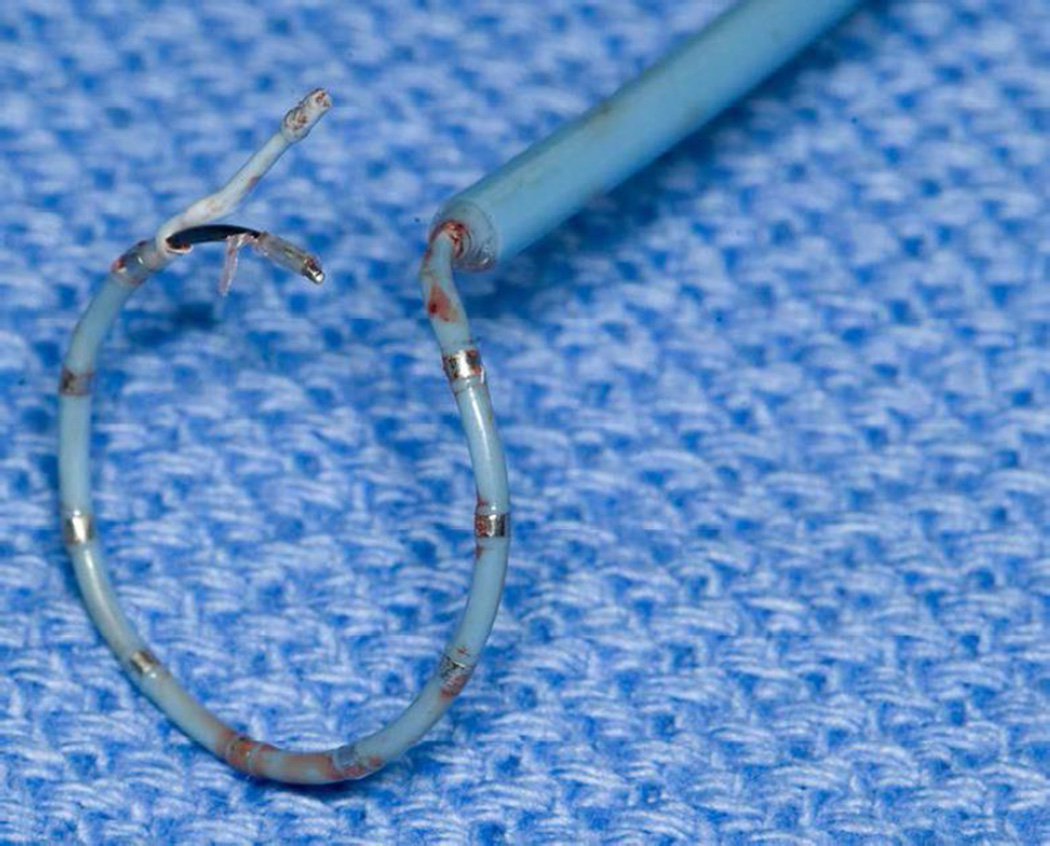
A 59-year-old female underwent ablation for medically refractory AF. The circular mapping catheter became entrapped in the anterior leaflet of the mitral valve while attempting to cannulate the left inferior PV. Intracardiac echocardiography (ICE) confirmed the location of entrapment (Figure 1A and B). The ablation catheter was positioned at the junction point of the trapped catheter and mitral valve, and a 15 to 20 second radiofrequency (RF) pulse with application of countertraction freed the catheter (Figure 2) without the need for surgical intervention.
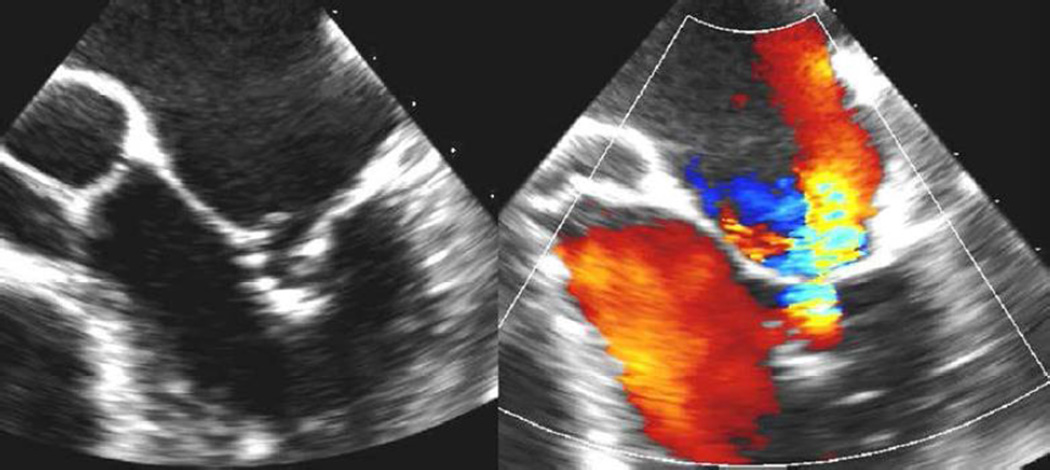
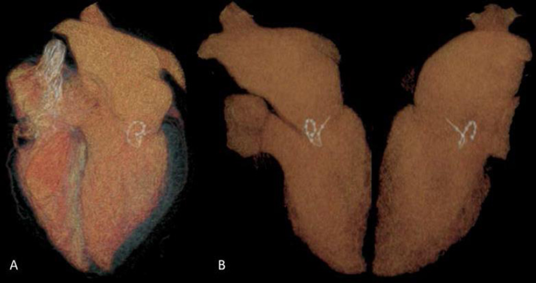
A 53-year-old male presented for surgical intervention post AF ablation. At the time of emergence from the transseptal sheath, the circular mapping catheter tip became embedded in the mitral valve annulus. The tip broke off and became lodged in the valve while attempting to remove the catheter. Transesophageal echocardiogram showed moderate mitral regurgitation, and the catheter tip was interfering with mitral valve closing motion (Figure 3A and B). CT reconstruction scan (Figure 4A and B) showed the circular mapping tip lodged in the posterior mitral subvalvular apparatus, which required surgical intervention.
Figure 1. Mapping catheter entrapment in mitral valve apparatus seen with ICE.
A. ICE imaging of circular mapping catheter entrapped in mitral valve apparatus (white arrow-catheter shaft, yellow arrow- entrapment of catheter in anterior valve leaflet of mitral valve. B. color flow Doppler showing regurgitation due to catheter entrapment.
Figure 2. Circular mapping catheter after rescue from mitral valve apparatus.
Figure 3. Catheter tip interferes with closing motion and leads to moderate regurgitation.
A. Trans-esophageal echocardiographic image of circular mapping catheter entrapped in mitral valve posterior leaflet. B. Color flow Doppler showing mitral regurgitation due to catheter preventing normal mitral valve closure.
Figure 4. Cardiac CT 3D reconstruction imaging showing circular mapping catheter tip enlodged in mitral valve posterior leaflet.
A. Coronal and B. Sagittal views showing circular catheter tip in posterior mitral valve leaflet.
Accessory Pathway Ablation Cases
A 23-year-old male with Wolff-Parkinson-White (WPW) syndrome underwent a left-sided accessory pathway ablation. The ablation catheter became entrapped in the mitral valve chordae, and the posterior leaflet chordae tore when the catheter was retracted, causing a P2 flail segment with moderate to severe mitral regurgitation. He was followed with serial echocardiography but ultimately required valve repair because of worsening valve function and ventricular size.
A 39-year-old female presented for surgical mitral valve repair for severe regurgitation following accessory pathway ablation. Ablation of the left lateral and posterolateral accessory pathways were performed with a total number of 18 ablation lesions at the mitral valve annulus. She developed severe MR with flail segment of the anterior medial scallop, with a torn mitral valve chordae. At surgical intervention for mitral valve repair, the mitral valve was excoriated areas along the posteromedial mitral annulus and posterior mitral leaflet with tissue necrosis.
Ventricular Tachycardia Ablation Cases
A 48-year-old male with symptomatic palpitations underwent VT ablation. There was suspicion that bileaflet mitral valve prolapse was causing a mechanical trigger for papillary muscle ectopy. RF ablation targeted a dominant PVC trigger on the papillary muscle. Postablation, severe mitral regurgitation developed, and surgical intervention was required to repair the valve.
A 71-year-old male presented for repeat VT ablation. RF energy was used to create an ablation line extending from scar to the mitral annulus. An additional ablation was performed 5 days later, and an early pre-potential was found in the anteroseptal region close to the mitral annulus. Postop, the patient developed severe mitral valve regurgitation. The patient was medically managed with aggressive diuresis and continues with serial echocardiography for surveillance.
A 79-year-old male underwent ablation for refractory VT. RF energy applied along the anterior scar border extending to the mitral isthmus for substrate modification. Two days post-procedure, the patient developed worsening mitral regurgitation. The patient was lost to follow up and passed away three months after procedure without any surveillance echocardiography.
Discussion
Catheter ablation is commonly used to treat arrhythmias such as atrial fibrillation, ventricular tachycardia, and supraventricular arrhythmias. Although electrophysiologic techniques, imaging, and tools continue to improve, mitral valve trauma is still a real and relatively underreported complication that can occur during catheter ablation procedures. This can be due to catheter manipulation with direct mechanical disruption of mitral valvular structures or from damage that occurs secondary to ablation energy delivered to the mitral annulus, valve, or subvalvular structures such as the papillary muscle (Figure 5A and B).
Figure 5. Two most common etiologies of damage to mitral valve during catheter ablation.
A. Retrograde Trans-aortic access to the mitral annulus for ablation energy delivery. B. Circular mapping catheter entrapment in the mitral valve apparatus.
It is intriguing that the quoted incidence of mitral valve complications during RF ablation of accessory pathways is only 0.04%.9 In a review published in 2005, only one case of mitral valve damage was registered among more than 8,500 AF ablation procedures.10 However, the actual incidence of mitral complications associated with AF or VT ablation is likely higher, because adverse events during procedures are reported voluntarily, may be under-detected, and are not routinely published in the literature.
There have been few case reports describing ablation procedure-related mitral valve damage, predominantly associated with AF and WPW ablation.3, 4, 7, 11 The first report of catheter entrapment in the mitral valve apparatus was described during radiofrequency ablation of a left-sided accessory pathway.12 Subsequently, ablation involving accessory pathways has been shown to injure the mitral valve, especially when a retrograde aortic approach is utilized.13, 14 In our series, the mechanism of valve injury in both patients undergoing accessory pathway ablation was mechanical chordae rupture secondary to ablation catheter entrapment.
In patients undergoing transcatheter AF ablation, the most common cause of damage to the valve apparatus is mechanical trauma related to the use of circumferential mapping catheters. Since Haïssaguerre et al.15 first described the utilization of circular mapping catheters to identify PV focal triggers or breakthrough sites, loop catheters have become a widely used tool during AF ablation. However, their circular shape, the presence of 10 to 20 electrodes, and a distal knob at the tip that may create friction with tissue contact synergistically increase catheter vulnerability to mitral valve entrapment when it contacts the valve.
Entanglement of the circular spine of the catheter in the mitral valve apparatus has been previously reported.11, 16 Similar with these reports, we identified four patients with mapping catheter entrapment in the valve leaflets or chord apparatus during PV isolation. This complication occurs most frequently during initial deployment through the sheath or during PV mapping, particularly when moving from the left-sided to the right-sided PVs.4 It should be cautioned that any blinded clockwise or rotation of the catheter should be avoided,17 and furthermore, when in the left atrium the only catheter rotational torque that should be performed is in a clockwise fashion.
One strategy to release an entangled catheter is to advance the sheath to the tip of the catheter to straighten it and subsequently retract and release it from the mitral valve.3 Alternatively, if gentle traction of the catheter is ineffective, advancement of the catheter apically towards the papillary muscle (and thus away from the chordal ramifications) may liberate the circular spine without valve damage.2 However, in our cases, both maneuvers were tried without success. At least one report has suggested that if such a situation cannot be resolved with gentle manipulation and moderate traction, open heart surgery may be the best option for removing the catheter.1, 11 In our series, one cases (case #3) had successful disentanglement of the catheter without surgical intervention.
Mitral valve catheter entrapment is an easily recognizable complication during the ablation procedure itself. However, mitral valve damage due to direct RF lesions may not be apparent and presentation delayed until symptoms such as shortness of breath develop, at times fulminantly, as in case #8. In the extreme, this type of injury can be insidious and present only following a marked delay, developing years after the ablation procedure.6, 18
Given the difficulty with retrieving entrapped catheters, avoiding the complication in the first place is ideal. Using the right anterior oblique projection to define the mitral annulus and having coronary sinus electrodes landmark can be helpful. Whenever manipulating circular catheters, the plane of the catheter movement should not cross anterior or ventricular to the annular plane. Using a standard maneuver for pulmonary vein access, for example starting with the catheter near the appendage and using clockwise torque to get into the left-sided pulmonary vein will help the operator to know what torque was used in case inadvertent migration of the catheter has occurred and the opposite torque could potentially be applied as a retrieval technique. When placing the catheter in the right inferior pulmonary vein, once again it is important to be sure the catheter has not crossed near the pulmonary valve before applying the usual significant, and at times marked, torque needed to engage the right lower vein. Given the differences across patient anatomy and the effects of chamber enlargement, no universally applicable torque to avoid this complication altogether is presently available.
Although it has been rarely reported, any type of ablation energy (RF or cryoablation) can cause mitral valve injury during AF, accessory pathway, or VT ablation procedures.5, 14 Our case #6 was previously reported,14 and highlights how RF energy delivery can lead to valve damage. A relatively older case report of damage to the mitral valve using cryoablation5 also has been published. Direct damage from multiple energy deliveries was also seen in case #8 with VT ablation. Because of multiple repeat ablations and the commonality of mapping and/or ablating pathways near the mitral valve apparatus, these cases need to be carefully scrutinized and echocardiographic surveillance is needed.
Six of nine patients in our series required surgical intervention, either for removal of the entangled catheter from the mitral valve apparatus or for repair of the damaged valve. In all the cases the mitral valve was successfully repaired surgically, and repair showed durable results at followup, without the need for valve replacement. This suggests that great care should be taken to avoid manipulation of circular catheters near the mitral valve, and vigilance with early surgical consultation is warranted when ablation near or on valvular structures is performed.
Conclusion
We present the largest case series to date of mitral valve damage as a result of a catheter ablation procedure. This series illustrates that catheter entrapment and/or damage due to ablation lesion application to the mitral annulus can occur and requires the operator to be mindful of this complication regardless of the type of arrhythmia ablation procedure performed. Furthermore, these cases highlight the need to closely follow these patients with serial echocardiography for mitral valve dysfunction if damage or ample ablative energy delivery to the mitral valve annulus occurred. Additionally, early surgical repair is important for successful salvage of mitral valve function.
References
- 1.Je HG, Kim JW, Jung SH, Lee JW. Minimally Invasive Surgical Release of Entrapped Mapping Catheter in the Mitral Valve. Circulation Journal. 2008;72:1378–1380. doi: 10.1253/circj.72.1378. [DOI] [PubMed] [Google Scholar]
- 2.Mansour M, Mela T, Ruskin J, Keane D. Successful release of entrapped circumferential mapping catheters in patients undergoing pulmonary vein isolation for atrial fibrillation. Heart Rhythm. 2004;1:558–561. doi: 10.1016/j.hrthm.2004.07.004. [DOI] [PubMed] [Google Scholar]
- 3.Kesek M, Englund A, Jensen SM, Jensen-Urstad M. Entrapment of circular mapping catheter in the mitral valve. Heart Rhythm. 2007;4:17–19. doi: 10.1016/j.hrthm.2006.09.016. [DOI] [PubMed] [Google Scholar]
- 4.Zeljko HM, Mont L, Sitges M, Tolosana JM, Nadal M, Castella M, Brugada J. Entrapment of the circular mapping catheter in the mitral valve in two patients undergoing atrial fibrillation ablation. Europace. 2011;13:132–133. doi: 10.1093/europace/euq309. [DOI] [PubMed] [Google Scholar]
- 5.Piccione W, Goldin M. Mitral valve dysfunction following papillary muscle cryoablation. Ann Thorac Surg. 1988:347–348. doi: 10.1016/s0003-4975(10)65942-5. [DOI] [PubMed] [Google Scholar]
- 6.Ohtsuka TMY, Hayami N, Kotsuka Y, Takamoto S. Surgically Repaired Delated Mitral Regurgitation After Radiofrequency Catheter Ablation for Wolff-Parkinson-White Syndrome. Pacing and Clinical Electrophysiology. 2002;25:1142–1143. doi: 10.1046/j.1460-9592.2002.01142.x. [DOI] [PubMed] [Google Scholar]
- 7.Mandawat MKTG, el-Sherif N. Catheter Entrapment in the Mitral Valve Apparatus Requiring Surgical Removal: An Unusual Complication of Radiofrequency Ablation. Pacing and Clinical Electrophysiology. 1998;4(Pt 1):772–773. doi: 10.1111/j.1540-8159.1998.tb00138.x. [DOI] [PubMed] [Google Scholar]
- 8.Suri RM, Vanoverschelde J, Grigioni F, et al. ASsociation between early surgical intervention vs watchful waiting and outcomes for mitral regurgitation due to flail mitral valve leaflets. JAMA. 2013;310:609–616. doi: 10.1001/jama.2013.8643. [DOI] [PubMed] [Google Scholar]
- 9.Scheinman MM. History of Wolff-Parkinson-White Syndrome. Pacing and Clinical Electrophysiology. 2005;28:152–156. doi: 10.1111/j.1540-8159.2005.09461.x. [DOI] [PubMed] [Google Scholar]
- 10.Cappato R, Calkins H, Chen SA, Davies W, Iesaka Y, Kalman J, Kim YH, Klein G, Packer D, Skanes A. Worldwide survey on the methods, efficacy, and safety of catheter ablation for human atrial fibrillation. Circulation. 2005;111:1100–1105. doi: 10.1161/01.CIR.0000157153.30978.67. [DOI] [PubMed] [Google Scholar]
- 11.Wu RCBJ, Yuh DD, Berger RD, Calkins HG. Circular Mapping Catheter Entrapment in the Mitral Valve Apparatus: A Previously Unrecognized Complication of Focal Atrial Fibrillation Ablation. Journal of Cardiovascular Electrophysiology. 2002;13:819–821. doi: 10.1046/j.1540-8167.2002.00819.x. [DOI] [PubMed] [Google Scholar]
- 12.Conti JBGE, Curtis AB. Catheter Entrapment in the Mitral Valve Apparatus During Radiofrequency Ablation. Pacing and Clinical Electrophysiology. 1994;17:1681–1685. doi: 10.1111/j.1540-8159.1994.tb02365.x. [DOI] [PubMed] [Google Scholar]
- 13.Minich LLSA, Dick M., 2nd Doppler Detection of Valvular Regurgitation After Radiofrequency Ablation of Accessory Connections. American Journal of Cardiology. 1992;70:116–117. doi: 10.1016/0002-9149(92)91404-r. [DOI] [PubMed] [Google Scholar]
- 14.Penaranda Canal JGE-SM, Asirvatham SJ, Munger TM, Friedman PA, Suri RM. Mitral Valve Injury After Radiofrequency Ablation for Wolff-Parkinson-White Syndrome. Circulation. 2013;127:2551–2552. doi: 10.1161/CIRCULATIONAHA.113.002711. [DOI] [PubMed] [Google Scholar]
- 15.Haïssaguerre M, Shah DC, Jaïs P, Hocini M, Yamane T, Deisenhofer I, Chauvin M, Garrigue S, Clémenty J. Electrophysiological Breakthroughs From the Left Atrium to the Pulmonary Veins. Circulation. 2000;102:2463–2465. doi: 10.1161/01.cir.102.20.2463. [DOI] [PubMed] [Google Scholar]
- 16.Tavernier R, Duytschaever M, Taeymans Y. Fracture of a Circular Mapping Catheter After Entrapment in the Mitral Valve Apparatus During Segmental Pulmonary Vein Isolation. Pacing and Clinical Electrophysiology. 2003;26:1774–1775. doi: 10.1046/j.1460-9592.2003.t01-1-00268.x. [DOI] [PubMed] [Google Scholar]
- 17.Macedo PG, Kapa S, Mears JA, Fratianni AMY, Asirvatham SJ. Correlative Anatomy for the Electrophysiologist: Ablation for Atrial Fibrillation. Part II: Regional Anatomy of the Atria and Relevance to Damage of Adjacent Structures During AF Ablation. Journal of Cardiovascular Electrophysiology. 2010;21:829–836. doi: 10.1111/j.1540-8167.2010.01730.x. [DOI] [PubMed] [Google Scholar]
- 18.Fisch-Thomsen MJJ, Egeblad H, Wierup P, Norgaard BL. Mitral Valve Perforation Appearing Years After Radiofrequency Ablation. Journal of Heart Valve Disease. 2011;20:351–352. [PubMed] [Google Scholar]


