Abstract
Mesenchymal stem cells (MSCs) have the ability to differentiate into a variety of lineages and to renew themselves without malignant changes, and thus hold potential for many clinical applications. However, it has not been well characterized how different the properties of MSCs are depending on the tissue source in which they resided. We previously reported a novel technique for the prospective MSC isolation from bone marrow, and revealed that a combination of cell surface markers (LNGFR and THY-1) allows the isolation of highly enriched MSC populations. In this study, we isolated LNGFR+ THY-1 + MSCs from synovium using flow cytometry. The results show that the synovium tissue contained a significantly larger percentage of LNGFR + THY-1 + MSCs. We examined the colony formation and differentiation abilities of bone marrow-derived MSCs (BM-MSCs) and synovium-derived MSCs (SYN-MSCs) isolated from the same patients. Both types of MSCs exhibited a marked propensity to differentiate into specific lineages. BM-MSCs were preferentially differentiated into bone, while in the SYN-MSC culture, enhanced adipogenic and chondrogenic differentiation was observed. These data suggest that the tissue from which MSCs are isolated should be tailored according to their intended clinical therapeutic application.
Introduction
Mesenchymal stem cells (MSCs) are self-renewing cells that can differentiate into osteoblasts, chondrocytes, and adipocytes [1–4]. These cells are found in various human tissues, including bone marrow (BM), synovium (SYN), placenta, and adipose tissue [3,5–9]. The characterization of MSCs isolated from those various tissues remains relatively unexplored. Because MSCs display no tumorigenicity, therefore they are currently used in clinical applications [10,11]. Number of clinical studies have been performed using MSCs to cure a variety of diseases [12–15]. Because origin tissues and isolation techniques are not unified, these clinical trials led to variable results. It has not been well-characterized how the differentiation potential of MSCs differs according to the tissue from which they are derived.
There have been several reports describing that synovium-derived MSCs (SYN-MSCs) showed a higher colony-forming efficiency than MSCs derived from bone marrow (BM-MSCs) [16,17]. Because SYN-MSCs display a great potential to differentiate into chondrocytes, SYN-MSCs are one of the best candidates for the repair of damaged cartilage [18,19]. A few reports have characterized the surface markers of SYN-MSCs. Cultured SYN-MSCs express such as CD44, CD90 (known as THY-1), CD105, and CD166, which are also found on fibroblasts and stromal lineages, and do not express hematopoietic and endothelial specific markers including CD45, CD253a, and CD31 [16,17]. We recently reported combination of novel markers for prospective MSC isolation and revealed that a combination of cell surface markers (low-affinity nerve growth factor receptor (LNGFR) and THY-1) allows the isolation of highly enriched MSC populations [20]. We also showed that LNGFR+THY-1+ cells, containing MSC-like cells, are present in placenta and adipose tissue [20].
In the current study, we freshly isolated MSCs from synovium and bone using surface markers, LNGFR and THY-1. We show that the MSCs existed high frequency in the synovium tissue, and the pattern of surface marker expression was similar between SYN- and BM- MSCs. BM-MSCs have a preference to differentiate to bone, while SYN-MSCs retains preference to both adipogenic and chondrogenic differentiation. Our results suggest that the tissue from source of MSCs should be tailored according to their intended therapeutic application.
Materials and Methods
Ethics information
Synovium and bone fragments were harvested from donors during total knee arthroplasty surgery at Tokyo Medical and Dental University Hospital. In total, 10 biological samples were used for the experiments. All experiments were approved by the local Institutional Review Board of Tokyo Medical and Dental University (No. 1030) and all study participants provided written informed consent. Tissue sample information and actual value of isolated LNGFR+THY-1+ cells for experiments show in S1 Table.
Tissue preparation
Synovium was digested with 2 mg/mL collagenase (Wako), 3 mg/mL dispase (Wako), and 25 μg/mL deoxyribonuclease I (Sigma-Aldrich) prepared in Dulbecco’s Modified Eagle’s Medium (DMEM, Life Technologies) with shaking at 37°C for 1 hour. Bone fragments were crushed with a pestle, after which the crushed bones were washed gently once in phosphate buffered saline (PBS) (for remove the marrow cells). Bone and bone fragments were incubated for 1 hour at 37°C in DMEM in the presence of 2 mg/mL collagenase (Wako Chemicals) and 25 μg/mL deoxyribonuclease I (Sigma-Aldrich). The cell suspensions (synovium and bone fragments) were filtered through a cell strainer (Falcon, 70 μm) to remove debris. After lyse the red blood cells, these cells were re-suspended in calcium- and magnesium-free Hank's Balanced Salt Solution (HBSS) (Gibco) supplemented with 2% FBS, 10 mM HEPES and 1% penicillin/streptomycin.
Flow cytometric analysis and cell sorting
Digested cells were suspended in HBSS at a density of 1–5 × 10⁷ cells/mL and stained for 30 minutes on ice with THY-1-APC (BD Pharmingen) and LNGFR-PE (Miltenyi Biotec) antibodies for sorting. Cultured cells (at three and five passages) were harvested using cell-dissociation buffer (Gibco). Cells (1.0 × 10⁵) were suspended in ice-cold HBSS and stained for 30 minutes on ice with the monoclonal antibodies CD45-PE-Cy7 (Tonbo Biosciences), CD29-PE, CD31-PE-Cy7, CD44-PE, CD105-PE and CD166-PE (BioLegend) for cell surface analysis. Flow cytometric analysis and cell sorting were performed on a triple-laser Moflo system (Beckman Coulter) and data were analyzed using Flowjo software (Tree Star).
Colony-forming unit-fibroblast (CFU-F) assay
The CFU-F assay was performed by culturing sorted 2,000 cells in a 100-mm dish for 14 days in culture medium, which consisted of DMEM supplemented with 20% fetal bovine serum (Hyclone), 1% penicillin/streptomycin (Gibco), and 5 ng/mL basic fibroblast growth factor (Repro Cell). The medium was changed every 3–4 days. Colonies containing more than 50 cells were counted.
Cell differentiation
Cultured MSCs at three passages were harvested using Trypsin-EDTA (Gibco). For adipogenic differentiation, 1.0 × 10⁴ cells were transferred to a 24-well plate and cultured overnight in culture medium. Adherent cells were cultured in adipogenic induction medium (Lonza), which was changed every 3–4 days. After 14 days, oil red O staining (Muto Pure Chemicals) confirmed the differentiation of these cells into adipocytes. For osteogenic differentiation, 7.0 × 10³ cells were transferred to a 24-well plate and cultured overnight in culture medium. Adherent cells were cultured in osteogenic induction medium (Lonza), which was changed every 3–4 days. After 14 days, the differentiation of these cells into osteoblasts was assessed by alizarin red staining (Millipore). For chondrogenic differentiation, 5.0 × 10⁵ cells were transferred to a 15-mL tube and cultured in chondrogenic induction medium (Lonza) containing 10 ng/mL transforming growth factor-β3 (Lonza) and 500 ng/mL bone morphogenetic protein 6 (R&D Systems), which was changed every 3–4 days. After 21 days, chondrogenic differentiated cells were was analyzed by Toluidine blue (Wako) and Safranin O staining (TCI) staining.
Real-time RT-PCR
After cell differentiation, total RNA was prepared with TRI reagent (Sigma-Aldrich) as described previously [21,22]. Complementary DNA was amplified using the StepOne Real-Time PCR System (Life Technologies) and normalized against β-actin expression (each gene were determined between 20–35 cycles). The experiments were performed averaged duplicates value as a data and using more than three independent biological samples. The probes used to identify gene expression of the key markers were confirmed to be human-specific sequence (TaqMan gene Expression Assays). All analyzed genes are listed in S2 Table. The PCR data using FAST SYBR Green showed in S1 Fig and S3 Table.
Statistical analysis
Quantitative data are presented as means ± standard deviation of at least three independent experiments. For statistical analysis, data were evaluated using Paired T-test. In all cases, p-values of < 0.05 were considered significant.
Results
The LNGFR+THY-1+ population of synovium-derived cells is enriched in clonogenic cells
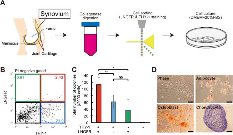
In human BM, LNGFR and THY-1 surface makers allow the isolation of highly enriched MSCs [20]. To isolate MSCs from synovium, we digested synovium with collagenase and collagenase-treated cells were stained with antibodies against LNGFR and THY-1 (Fig 1A). We identified four populations, corresponding with these markers expression (Fig 1B). To analyze the clonogenic potential of four populations, we performed the CFU-F assay for each population cells. After 2 weeks of culture, we counted the number of colonies containing more than 50 cells. Cells that were negative for both LNGFR and THY-1 (black) did not form colonies, whereas cells that were positive for both markers formed many colonies (Fig 1C). In the LNGFR+THY-1- (green) and LNGFR-THY-1+ (blue) populations, cells formed few colonies and had a flat morphology (data not shown). Cells in the LNGFR+THY-1+ (red) population were spindle-shaped and had a robust ability to differentiate into adipocytes, chondrocytes, and osteoblasts (Fig 1D).
Fig 1. Isolation of LNGFR+THY-1+ MSCs from SYN.
(A) Synovium (SYN) was harvested from patients undergoing total knee arthroplasty surgery and digested with collagenase and dispase. Mesenchymal stem cells (MSCs) were isolated using flow cytometry. DMEM, Dulbecco’s Modified Eagle’s Medium; FBS, fetal bovine serum. (B) Representative flow cytometric profile of SYN-derived cells stained for LNGFR and THY-1. Each population shows in LNGFR+/THY-1+(red), LNGFR+/THY- (green), LNGFR-/THY+ (blue) and LNGFR-/THY- (black) gates. PI, propidium iodide. (C) Number of colonies formed after 2 weeks of culturing 2,000 of the following cells: LNGFR+/THY-1+, LNGFR+/THY-, LNGFR-/THY+, and LNGFR-/THY- (mean ± standard deviation, n = 4 per group; *p<0.05; **p < 0.01; ns, not significant). (D) Phase contrast micrographs of LNGFR+THY-1+ cells showing their potential to differentiate into adipocytes, osteoblasts, and chondrocytes (scale bar = 100 μm).
SYN-MSCs have a high colony-forming ability and proliferation potential compared with BM-MSCs
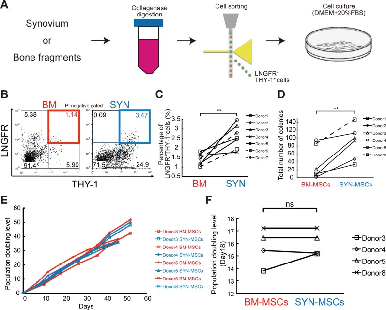
We digested human synovum and bone fragments and stained the obtained cells with antibodies against LNGFR and THY-1, and then performed flow cytometric analysis (Fig 2A). Synovium tissue contained a significantly larger percentage of LNGFR+THY-1+ cells than BM (Fig 2B and 2C). We performed the CFU-F assay using freshly sorted cells from BM and SYN to evaluate the clonogenic potential of LNGFR+THY-1+ cells. SYN-MSCs had high clonogenic ability after 2 weeks of culture (Fig 2D). The population doubling level (Day 18) and growth kinetic analyses revealed that SYN-MSCs could be cultured for longer than 7 weeks in a similar fashion to BM-MSCs (Fig 2E and 2F).
Fig 2. Colony-forming potential of SYN-MSCs and BM-MSCs.
(A) MSCs were isolated from synovium and bone fragments. (B) Representative flow cytometric profiles of cells derived from bone marrow (BM, left) and synovium (SYN, right) of the same donor and stained for LNGFR and THY-1. PI, propidium iodide. (C) Flow cytometric analysis of the percentage of LNGFR+THY-1+ cells among BM- and SYN-derived cells (n = 7, **p < 0.01). (D) Number of colonies formed by BM-MSCs and SYN-MSCs on day 14 (n = 6, *p<0.05; **p < 0.01). (E) Representative population doubling level of BM-MSCs and SYN-MSCs. (F) Population doubling level of SYN-MSCs and BM-MSCs at day 18 (n = 4; ns, not significant).
Expression of THY-1 correlate with cell proliferation in BM-MSCs and SYN-MSCs
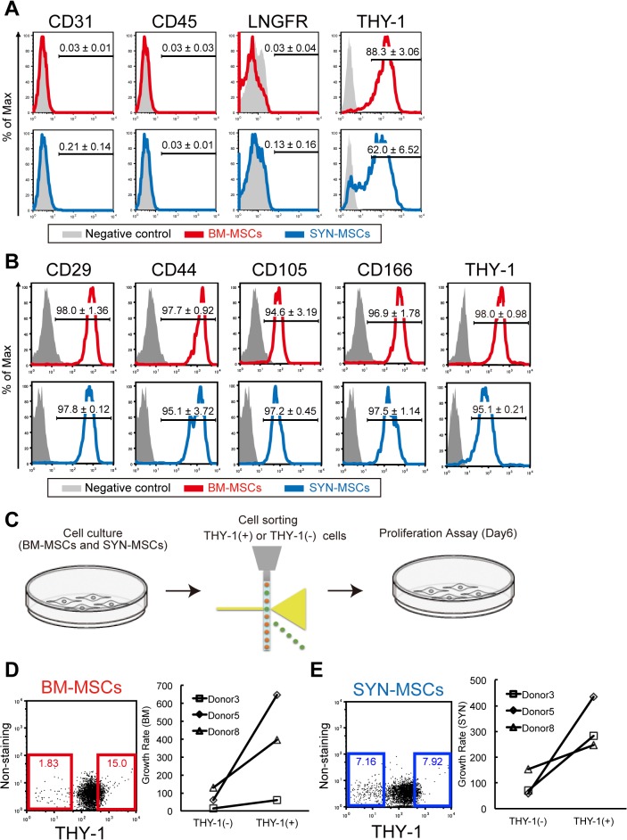
We examined the surface epitopes of MSCs by flow cytometry. At passage 3, there were no differences in the expression of CD31 (endothelial marker), CD45 (hematopoietic marker), LNGFR and THY-1 between SYN-MSCs and BM-MSCs (Fig 3A). After passage 5, we analyze the mesenchymal markers (CD29, CD44, CD105 and CD166) (Fig 3B). The cell surface antigen of SYN-MSCs and BM-MSCs isolated from the same donor were similar profiles (n = 3). We previously reported that murine cultured BM-MSC clones with stem cell potential kept highly expressing of THY-1, whereas clones with a limited differentiation potential turned to THY-1- [23]. Therefore, we isolated THY-1+ and THY-1- cells from BM and SYN, and analyzed the cell proliferation ability of each sorted fraction (Fig 3C). We found that THY-1+ cells had a high proliferation ability compared with THY-1- cells (Fig 3D and 3E). These data suggest that cell surface marker THY-1 was suitable for isolating MSCs in human BM but also in human SYN.
Fig 3. Comparison of cell surface proteins between SYN-MSCs and BM-MSCs.
(A) Representative flow cytometric profiles of cells at passage 3, showing the percentage of cells expressing the antigen (red or blue line) versus the isotype control (gray). Markers: CD31, CD45, LNGFR, and THY-1. (B) The analysis of mesenchymal cell markers at passage 5, (C) Proliferation assay of cultured THY-1+ or THY-1- cells derived from the BM-MSCs and SYN-MSCs after passage 5. (D) Flow cytometric profiles and growth rate in cultured BM-MSCs. (E) Flow cytometric profiles and growth rate in cultured SYN-MSCs.
BM-MSCs preferentially differentiate into bone, while SYN-MSCs differentiate better into adipocytes
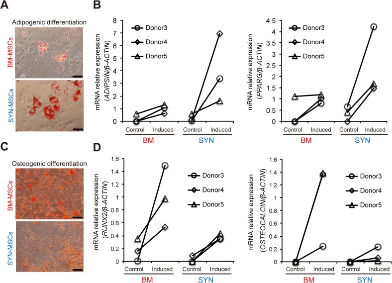
We examined whether the differentiation potential of SYN-MSCs differs from that of BM-MSCs. First, we performed adipogenic differentiation of these MSCs. Although both types of MSCs effectively differentiated into adipocytes after 2 weeks, as determined by oil red O staining (Fig 4A). We analyzed the key gene expression into adipocyte lineage using TaqMan probes to examine the differentiation propensity. The expression of peroxisome proliferator-activated receptor γ (PPARG) and ADIPSIN was higher in SYN-MSCs than in BM-MSCs (more than two-fold) (Fig 4B and S1 Fig). Next, SYN-MSCs and BM-MSCs underwent osteogenic differentiation for 2 weeks (Fig 4C). The expression ratio of RUNX2 and OCN to β-actin was lower in SYN-MSCs than in BM-MSCs (less than three-fold) (Fig 4D and S1 Fig).
Fig 4. Comparison of the adipogenic and osteogenic differentiation abilities of SYN-MSCs and BM-MSCs.
(A) Representative micrographs of adipocytes differentiated BM-MSCs (top) and SYN-MSCs (bottom) stained with oil red O (scale bar = 50 μm). (B) Expression ratio of ADIPSIN and PPARG mRNA to β-ACTIN mRNA following adipogenic differentiation of BM-MSCs and SYN-MSCs (n = 3). Control: non-induced MSCs, Induced: induced MSCs for adipogenic lineage. (C) Representative micrographs of osteoblasts differentiated from BM-MSCs (top) and SYN-MSCs (bottom) stained with alizarin red (scale bar = 200 μm). (D) Expression ratio of RUNX2 and OSTEOCALCIN mRNA to β-ACTIN mRNA following osteogenic differentiation of BM-MSCs and SYN-MSCs (n = 3). Control: non-induced MSCs, Induced: induced MSCs for osteogenic lineage.
Chondrogenic differentiation propensity markedly differs between SYN-MSCs and BM-MSCs
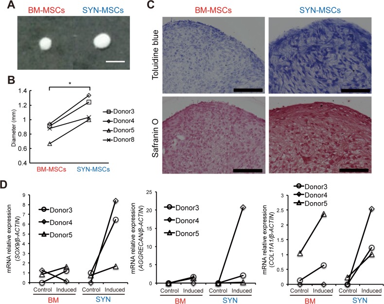
We performed chondrogenic differentiation of SYN-MSCs and BM-MSCs for 3 weeks. Both types of MSCs could undergo chondrogenic differentiation; however, the pellet of SYN-MSC-derived chondrocytes was 1.2-fold larger than that of BM-MSC-derived chondrocytes (Fig 5A and 5B). We also evaluated the level of chondrogenesis by staining cell pellets with Toluidine blue and Safranin O after chondrogenic differentiation. Strong staining was observed in cell pellets generated from SYN-MSCs (Fig 5C). To further verify chondrogenesis, quantitative RT-PCR was performed of the chondrogenic markers SOX9, AGGRECAN (ACAN), and COL11A1. After chondrogenic induction, expression of these markers was markedly higher in SYN-MSCs than in BM-MSCs (SOX9: 5.5-fold, ACAN: 7.6-fold, COL11A1: 1.5-fold) (Fig 5D and S1 Fig). These results suggest that SYN-MSCs have a higher chondrogenic differentiation potential than BM-MSCs.
Fig 5. High chondrogenic differentiation ability of SYN-MSCs.
(A) Representative images of chondrocyte pellets on day 21 derived from BM-MSCs and SYN-MSCs (scale bar = 1 mm). (B) Comparison of chondrocyte pellet size between BM-MSCs and SYN-MSCs (n = 4, *p < 0.05). (C) Micrographs of chondrocyte pellets derived from BM-MSCs and SYN-MSCs on day 21 stained with Toluidine blue and Safranin O (scale bar = 200 μm). (D) Expression ratio of SOX9, AGGRECAN, and COL11A1 mRNA to β-ACTIN mRNA in chondrocyte pellets (n = 3). Control: non-induced MSCs, Induced: induced MSCs for chondrogenic lineage.
Discussion
The combination of cell surface marker LNGFR and THY-1 which are originally characterized as BM-MSCs were also valuable to purify MSCs from synovium. The colony-forming ability of the LNGFR+THY-1+ population was higher than the other sub-fraction. LNGFR+THY-1+ cells exhibited a higher colony-forming potential in the CFU-F assay than that of BM-derived cells. LNGFR and THY-1 are expressed in a variety of other human tissues including placenta, adipose tissue, heart, brain, and liver [20,24–27]. Among human cells, LNGFR is predominantly expressed in melanoma-initiating cells and transit-amplifying cells [28–30]. LNGFR expression may have a common function in rapidly proliferating cells. Analyses of sorted cells revealed that the LNGFR and THY-1 markers could be used to isolate MSCs from human tissues.
We compared the surface marker expression and differentiation potential of MSCs according to the tissue from which they were derived. The osteogenic differentiation capacity of SYN-MSCs was lower than that of BM-MSCs. On the other hand, the chondrogenic differentiation capacity of SYN-MSCs was high. MSCs are shown to specify lineage and commit to phenotypes with extreme sensitivity to in vivo microenvironment and tissue-level elasticity [31]. The synovium supports adjacent cartilage and protects bone tissue from mechanical stresses. The characteristics of SYN-MSCs may be affected by the extracellular matrix and physical stress. THY-1 is a glycophosphatidylinositol anchored conserved cell surface protein and is used to identify MSCs. In murine MSCs, THY-1 is a key marker of multipotency [23], and besides, the expression significantly differs between human perichondrocytes and chondrocytes; its expression level is low in the latter cells [32]. The proliferation assay confirmed that THY-1+ cells had a high proliferation ability compared with THY-1- cells in cultured SYN-MSCs and BM-MSCs. THY-1 can probably be used as a negative marker to identify MSCs which committed to chondrocytes. Taken together, SYN-MSCs may have a high chondrogenic differentiation potency because of the external environment, and immature SYN-MSCs could be isolated with THY-1 marker.
Adipose tissue-derived MSCs have a higher adipogenic differentiation potential and lower osteogenic and chondrogenic differentiation potentials than BM-MSCs [18,33]. Adipose tissue contained the MSCs that express LNGFR and THY-1 markers [20]. Synovium specimens obtained during operations contain adipose tissues; therefore, the adipogenic potential of SYN-MSCs may have been enhanced by adipose tissue-derived MSCs in this study. We used BM cells that were enzymatically isolated from bone fragments, not a BM aspirate. It is difficult to obtain MSCs from BM aspirates owing to the quality of samples. It was reported that human trabecular bone-derived cells obtained by collagenase digestion are similar to BM aspirate-derived MSCs in terms of their proliferation ability and chondrogenic, adipogenic, and osteogenic differentiation potentials [34]. These contents would not significantly change the results regarding the properties of SYN-MSCs. Therefore, this study has a relevance to the true properties of tissue-derived stem cells.
Because of many aged patients suffer from osteoarthritis (OA), regenerative therapy using MSCs has large hope in the world. When the MSCs are used in autograft, the cell isolation efficiency from tissue is an important problem. MSCs that express the same surface antigens differ in terms of their differentiation potential according to the tissue from which they are derived. Therefore, the origin of MSCs must be carefully considered before they are transplanted. Recently reports showed that the degree of inflammation was correlated with MSC isolation efficiency [35]. Because there are millions of OA patients, the synovium from OA patients are large source to isolate MSCs, similarly to cartilage injury and meniscal lesion [36]. It is an essential mission to isolate the MSC stably from SYN for facilitating regenerative medicine.
Conclusion
The LNGFR+THY-1+ cell population of synovium forms many colonies in the CFU-F assay. LNGFR+THY-1+ SYN-MSCs have a high chondrogenic differentiation ability. SYN-MSCs may be a suitable cell source for cartilage regeneration
Supporting Information
Expression ratio of tergeted mRNA to β-ACTIN mRNA following key gene. (A) PPARγ for adipogenic differentiation. (B) bone sialoprotein (BSP) for osteogenic differentiation. (C) AGGRECAN, (D) COL2A1, and (E) COL10A1 for chondrogenic differentiation.
(TIF)
(DOCX)
(DOCX)
(DOCX)
Acknowledgments
We thank M. Mizuno (Tokyo Medical and Dental University) for discussions, D. D. Houlihan for critically reading this manuscript, and laboratory members for technical supports.
Data Availability
All relevant data are within the paper.
Funding Statement
The authors have no support or funding to report.
References
- 1. Pittenger MF, Mackay AM, Beck SC, Jaiswal RK, Douglas R, Mosca JD, et al. (1999) Multilineage potential of adult human mesenchymal stem cells. Science 284: 143–147. [DOI] [PubMed] [Google Scholar]
- 2. Piersma AH, Brockbank KG, Ploemacher RE, van Vliet E, Brakel-van Peer KM, Visser PJ (1985) Characterization of fibroblastic stromal cells from murine bone marrow. Exp Hematol 13: 237–243. [PubMed] [Google Scholar]
- 3. Conget PA, Minguell JJ (1999) Phenotypical and functional properties of human bone marrow mesenchymal progenitor cells. J Cell Physiol 181: 67–73. [DOI] [PubMed] [Google Scholar]
- 4. Simmons PJ, Torok-Storb B (1991) Identification of stromal cell precursors in human bone marrow by a novel monoclonal antibody, STRO-1. Blood 78: 55–62. [PubMed] [Google Scholar]
- 5. Prockop DJ, Sekiya I, Colter DC (2001) Isolation and characterization of rapidly self-renewing stem cells from cultures of human marrow stromal cells. Cytotherapy 3: 393–396. [DOI] [PubMed] [Google Scholar]
- 6. De Bari C, Dell'Accio F, Vandenabeele F, Vermeesch JR, Raymackers JM, Luyten FP (2003) Skeletal muscle repair by adult human mesenchymal stem cells from synovial membrane. J Cell Biol 160: 909–918. [DOI] [PMC free article] [PubMed] [Google Scholar]
- 7. Yen BL, Huang HI, Chien CC, Jui HY, Ko BS, Yao M, et al. (2005) Isolation of multipotent cells from human term placenta. Stem Cells 23: 3–9. [DOI] [PubMed] [Google Scholar]
- 8. Battula VL, Bareiss PM, Treml S, Conrad S, Albert I, Hojak S, et al. (2007) Human placenta and bone marrow derived MSC cultured in serum-free, b-FGF-containing medium express cell surface frizzled-9 and SSEA-4 and give rise to multilineage differentiation. Differentiation 75: 279–291. [DOI] [PubMed] [Google Scholar]
- 9. Liu TM, Martina M, Hutmacher DW, Hui JH, Lee EH, Lim B (2007) Identification of common pathways mediating differentiation of bone marrow- and adipose tissue-derived human mesenchymal stem cells into three mesenchymal lineages. Stem Cells 25: 750–760. [DOI] [PubMed] [Google Scholar]
- 10. Liu Y, Wang L, Kikuiri T, Akiyama K, Chen C, Xu X, et al. (2011) Mesenchymal stem cell-based tissue regeneration is governed by recipient T lymphocytes via IFN-gamma and TNF-alpha. Nat Med 17: 1594–1601. 10.1038/nm.2542 [DOI] [PMC free article] [PubMed] [Google Scholar]
- 11. Bianco P, Cao X, Frenette PS, Mao JJ, Robey PG, Simmons PJ, et al. (2013) The meaning, the sense and the significance: translating the science of mesenchymal stem cells into medicine. Nat Med 19: 35–42. 10.1038/nm.3028 [DOI] [PMC free article] [PubMed] [Google Scholar]
- 12. Horwitz EM, Prockop DJ, Fitzpatrick LA, Koo WW, Gordon PL, Neel M, et al. (1999) Transplantability and therapeutic effects of bone marrow-derived mesenchymal cells in children with osteogenesis imperfecta. Nat Med 5: 309–313. [DOI] [PubMed] [Google Scholar]
- 13. Hanley PJ, Mei Z, da Graca Cabreira-Hansen M, Klis M, Li W, Zhao Y, et al. (2013) Manufacturing mesenchymal stromal cells for phase I clinical trials. Cytotherapy 15: 416–422. 10.1016/j.jcyt.2012.09.007 [DOI] [PMC free article] [PubMed] [Google Scholar]
- 14. Jackson WM, Nesti LJ, Tuan RS (2012) Concise review: clinical translation of wound healing therapies based on mesenchymal stem cells. Stem Cells Transl Med 1: 44–50. 10.5966/sctm.2011-0024 [DOI] [PMC free article] [PubMed] [Google Scholar]
- 15. Wang Y, Han ZB, Song YP, Han ZC (2012) Safety of mesenchymal stem cells for clinical application. Stem Cells Int 2012: 652034 10.1155/2012/652034 [DOI] [PMC free article] [PubMed] [Google Scholar]
- 16. Nimura A, Muneta T, Koga H, Mochizuki T, Suzuki K, Makino H, et al. (2008) Increased proliferation of human synovial mesenchymal stem cells with autologous human serum: comparisons with bone marrow mesenchymal stem cells and with fetal bovine serum. Arthritis Rheum 58: 501–510. 10.1002/art.23219 [DOI] [PubMed] [Google Scholar]
- 17. Sakaguchi Y, Sekiya I, Yagishita K, Muneta T (2005) Comparison of human stem cells derived from various mesenchymal tissues: superiority of synovium as a cell source. Arthritis Rheum 52: 2521–2529. [DOI] [PubMed] [Google Scholar]
- 18. Yoshimura H, Muneta T, Nimura A, Yokoyama A, Koga H, Sekiya I (2007) Comparison of rat mesenchymal stem cells derived from bone marrow, synovium, periosteum, adipose tissue, and muscle. Cell Tissue Res 327: 449–462. [DOI] [PubMed] [Google Scholar]
- 19. Koga H, Shimaya M, Muneta T, Nimura A, Morito T, Hayashi M, et al. (2008) Local adherent technique for transplanting mesenchymal stem cells as a potential treatment of cartilage defect. Arthritis Res Ther 10: R84 10.1186/ar2460 [DOI] [PMC free article] [PubMed] [Google Scholar]
- 20. Mabuchi Y, Morikawa S, Harada S, Niibe K, Suzuki S, Renault-Mihara F, et al. (2013) LNGFR(+)THY-1(+)VCAM-1(hi+) Cells Reveal Functionally Distinct Subpopulations in Mesenchymal Stem Cells. Stem Cell Reports 1: 152–165. 10.1016/j.stemcr.2013.06.001 [DOI] [PMC free article] [PubMed] [Google Scholar]
- 21. Suzuki N, Fukushi M, Kosaki K, Doyle AD, de Vega S, Yoshizaki K, et al. (2012) Teneurin-4 is a novel regulator of oligodendrocyte differentiation and myelination of small-diameter axons in the CNS. J Neurosci 32: 11586–11599. 10.1523/JNEUROSCI.2045-11.2012 [DOI] [PMC free article] [PubMed] [Google Scholar]
- 22. Suzuki N, Mizuniwa C, Ishii K, Nakagawa Y, Tsuji K, Muneta T, et al. (2014) Teneurin-4, a transmembrane protein, is a novel regulator that suppresses chondrogenic differentiation. J Orthop Res 32: 915–922. 10.1002/jor.22616 [DOI] [PubMed] [Google Scholar]
- 23. Morikawa S, Mabuchi Y, Kubota Y, Nagai Y, Niibe K, Hiratsu E, et al. (2009) Prospective identification, isolation, and systemic transplantation of multipotent mesenchymal stem cells in murine bone marrow. J Exp Med 206: 2483–2496. 10.1084/jem.20091046 [DOI] [PMC free article] [PubMed] [Google Scholar]
- 24. Quirici N, Soligo D, Bossolasco P, Servida F, Lumini C, Deliliers GL (2002) Isolation of bone marrow mesenchymal stem cells by anti-nerve growth factor receptor antibodies. Exp Hematol 30: 783–791. [DOI] [PubMed] [Google Scholar]
- 25. Gago-Lopez N, Awaji O, Zhang Y, Ko C, Nsair A, Liem D, et al. (2014) THY-1 receptor expression differentiates cardiosphere-derived cells with divergent cardiogenic differentiation potential. Stem Cell Reports 2: 576–591. 10.1016/j.stemcr.2014.03.003 [DOI] [PMC free article] [PubMed] [Google Scholar]
- 26. van Strien ME, Sluijs JA, Reynolds BA, Steindler DA, Aronica E, Hol EM (2014) Isolation of neural progenitor cells from the human adult subventricular zone based on expression of the cell surface marker CD271. Stem Cells Transl Med 3: 470–480. 10.5966/sctm.2013-0038 [DOI] [PMC free article] [PubMed] [Google Scholar]
- 27. Patil PB, Joshi M, Kuna VK, Xu B, Johannesson L, Olausson M, et al. (2014) CD271 identifies functional human hepatic stellate cells, which localize in peri-sinusoidal and portal areas in liver after partial hepatectomy. Cytotherapy 16: 990–999. 10.1016/j.jcyt.2014.03.001 [DOI] [PubMed] [Google Scholar]
- 28. Boiko AD, Razorenova OV, van de Rijn M, Swetter SM, Johnson DL, Ly DP, et al. (2010) Human melanoma-initiating cells express neural crest nerve growth factor receptor CD271. Nature 466: 133–137. 10.1038/nature09161 [DOI] [PMC free article] [PubMed] [Google Scholar]
- 29. Civenni G, Walter A, Kobert N, Mihic-Probst D, Zipser M, Belloni B, et al. (2011) Human CD271-positive melanoma stem cells associated with metastasis establish tumor heterogeneity and long-term growth. Cancer Res 71: 3098–3109. 10.1158/0008-5472.CAN-10-3997 [DOI] [PubMed] [Google Scholar]
- 30. Truzzi F, Saltari A, Palazzo E, Lotti R, Petrachi T, Dallaglio K, et al. (2014) CD271 Mediates Stem Cells to Early Progeny Transition in Human Epidermis. J Invest Dermatol. [DOI] [PubMed] [Google Scholar]
- 31. Engler AJ, Sen S, Sweeney HL, Discher DE (2006) Matrix elasticity directs stem cell lineage specification. Cell 126: 677–689. [DOI] [PubMed] [Google Scholar]
- 32. Kobayashi S, Takebe T, Inui M, Iwai S, Kan H, Zheng YW, et al. (2011) Reconstruction of human elastic cartilage by a CD44+ CD90+ stem cell in the ear perichondrium. Proc Natl Acad Sci U S A 108: 14479–14484. 10.1073/pnas.1109767108 [DOI] [PMC free article] [PubMed] [Google Scholar]
- 33. Zimmerlin L, Donnenberg VS, Pfeifer ME, Meyer EM, Peault B, Rubin JP, et al. (2010) Stromal vascular progenitors in adult human adipose tissue. Cytometry A 77: 22–30. 10.1002/cyto.a.20813 [DOI] [PMC free article] [PubMed] [Google Scholar]
- 34. Sakaguchi Y, Sekiya I, Yagishita K, Ichinose S, Shinomiya K, Muneta T (2004) Suspended cells from trabecular bone by collagenase digestion become virtually identical to mesenchymal stem cells obtained from marrow aspirates. Blood 104: 2728–2735. [DOI] [PubMed] [Google Scholar]
- 35. Matsukura Y, Muneta T, Tsuji K, Miyatake K, Yamada J, Abula K, et al. (2015) Mouse synovial mesenchymal stem cells increase in yield with knee inflammation. J Orthop Res 33: 246–253. 10.1002/jor.22753 [DOI] [PMC free article] [PubMed] [Google Scholar]
- 36. Berenbaum F (2013) Osteoarthritis as an inflammatory disease (osteoarthritis is not osteoarthrosis!). Osteoarthritis Cartilage 21: 16–21. 10.1016/j.joca.2012.11.012 [DOI] [PubMed] [Google Scholar]
Associated Data
This section collects any data citations, data availability statements, or supplementary materials included in this article.
Supplementary Materials
Expression ratio of tergeted mRNA to β-ACTIN mRNA following key gene. (A) PPARγ for adipogenic differentiation. (B) bone sialoprotein (BSP) for osteogenic differentiation. (C) AGGRECAN, (D) COL2A1, and (E) COL10A1 for chondrogenic differentiation.
(TIF)
(DOCX)
(DOCX)
(DOCX)
Data Availability Statement
All relevant data are within the paper.


