Abstract
The Drosophila stomatogastric nervous system (SNS) is a compact collection of neurons that arises from the migration of neural precursors. Here we describe genetic tools allowing functional analysis of the SNS during the migratory phase of development. We constructed GAL4 lines driven by fragments of the Ret promoter, which yielded expression in a subset of migrating neural SNS precursors and also included a distinct set of midgut associated cells. Screening of additional GAL4 lines driven by fragments of the Gfrl/Munin, forkhead, twist and goosecoid (Gsc) promoters identified a Gsc fragment with expression from initial selection of SNS precursors until the end of embryogenesis. Inhibition of EGFR signaling using three identified lines disrupted the correct patterning of the frontal and recurrent nerves. To manipulate the environment traveled by SNS precursors, a FasII-GAL4 line with strong expression throughout the entire intestinal tract was identified. The transgenic lines described offer the ability to specifically manipulate the migration of SNS precursors and will allow the modeling and in-depth analysis of neuronal migration in ENS disorders such as Hirschsprung’s disease.
Introduction
The invertebrate stomatogastric nervous system (SNS) has provided a wealth of information on the functioning of simple neural networks [1]. In Drosophila, all aspects of the adult gut including the enteric nervous system (ENS) have received intense attention in recent years [2]. After initial characterization of the embryonic development of the SNS primarily by the Hartenstein and Jäckle groups [3–9], the early SNS has received relatively little consideration. This is surprising as the SNS is a simple developmental system and likely to be of clinical relevance to vertebrate ENS disorders.
The SNS begins as three epithelial pouches in the primitive mouth (stomatogastric) that delaminate and migrate along the developing foregut as coherent clusters (referred to as invaginating SNS precursors or iSNSPs; reviewed in [4]). An additional group of cells (dSNSPs) delaminate in front of the iSNSPs [5]. The SNS anlage is located within the roof epithelium of the stomodeum, the primitive mouth of the embryo. Within the anlage, three single cells, called tip cells (tSNSPs), are selected by the action of the proneural (achate-scute), neurogenic (Notch) and wingless (wg) genes [8]. The tip cells secrete an Epidermal Growth Factor (EGF), Spitz, which induces EGF receptor (EGFR) signaling in the surrounding cells, inducing them to delaminate from the epithelium and form migratory vesicles [3, 9]. These three clusters of cells migrate along the foregut and then start to produce daughter cells that separate and migrate both anteriorly and posteriorly to form discrete ganglia [5]. In anterior to posterior order, the ganglia are: the frontal ganglion which lies on top of the pharynx anterior to the brain commissure, two sets of esophageal ganglia which lie alongside the esophagus, and the proventricular ganglion which innervates the crop-like proventriculus that forms at the junction of the foregut and midgut [10]. Cells from each iSNSP cluster contribute to each of the ganglia, whereas the dSNSPs contribute only to the frontal ganglion [5].
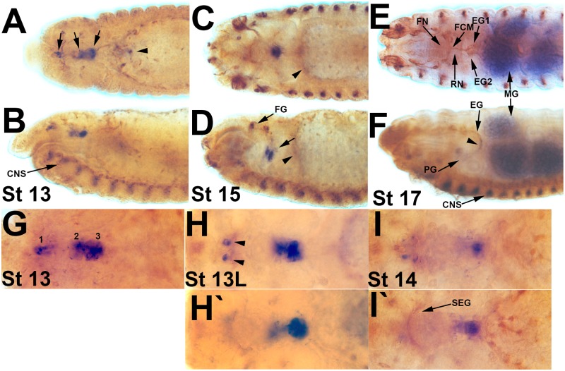
The fly SNS has strong parallels with the vertebrate neural crest as epithelial cells delaminate and migrate to their final destinations. In vertebrates, the RET receptor tyrosine kinase has a critical role in the migration of enteric neuron precursors and mutations are a key cause of Hirschsprung’s disease in which the colon and rectum have severely decreased innervation [11–13]. Intriguingly the fly Ret gene is expressed in the migrating SNS precursors (Fig 1) suggesting there may be a shared evolutionary origin [14]. Drosophila Ret mutants affect dendrite growth but have not yet been examined for SNS defects [15]. We wished to generate transgenic reagents specific to the developing SNS as many developmental genes affect multiple stages and tissues during development, which can hinder phenotypic analysis. Some of the reagents may allow functional assays of feeding and peristalsis to be conducted in larvae. We constructed fragments of the Ret promoter to the GAL4 gene and also screened additional GAL4 lines. Three specific GAL4 lines, GscG-GAL4, Gfrla-GAL4 and RetP-GAL4, were identified that allow the manipulation of SNS precursors and these will be made available to the research community.
Fig 1. Ret expression in the developing SNS.
Drosophila embryos with an in situ hybridization for the Ret gene (dark blue) and antibody staining with the 22c10 antibody (brown) to reveal the SNS, the PNS and elements of the CNS. A, C, E, G, H and I are dorsal views, B, D and F are lateral views. (A, B) Stage 13 embryo with expression in the migrating SNS clusters (arrows). Limited expression can also be seen in a discrete set of cells of the anterior midgut (arrowhead) and in the CNS midline at the bottom of panel B (CNS). (C, D) Stage 15 embryo in which the esophagus has started to loop. The three SNS clusters are immediately adjacent to one another within the loop and all express Ret (arrow). Additional Ret staining occurs in the developing frontal ganglion (FG). Faint expression can be seen in the anterior midgut (arrowheads), the ventral midline and PNS cells towards the anterior of the embryo. (E, F) Stage 17 embryo with Ret expression in some cells of the esophageal ganglion (EG) and proventricular ganglion (PG). Significant Ret expression is observed in the midgut (MG) and the CNS midline (CNS). (G) Expression of Ret in the three migrating SNS clusters in a stage 13 embryo. (H, H’) Two different focal planes of late stage 13 embryo. Ret is expressed in the SNS clusters which are clustered in the looping esophagus (compare to D), and in CNS cells that project through the subesophageal ganglion (arrows). (I, I’) Stage 14 embryo with diminishing Ret expression. Some axons of the subesophageal ganglion (SEG) are labeled by 22c10.
Materials and Methods
Molecular Biology
A 527 base pair fragment upstream of the Ret transcription start site was amplified with Phusion high fidelity DNA polymerase from genomic DNA derived from an Exelixis isogenic stock [16] with CCAGGTAAACCCTTTTATCG (forward) and CCGCGGAAATACTTTTTGG (reverse) primers (written from 5’ to 3’), cloned into pCR8/GW/TOPO (Life Technologies Inc.) and subcloned into the StuI and EcoRI sites of pPTGAL (Addgene; [17]). P-element injections were performed by Genetic Services, Inc. (Sudbury, MA) and one transformant was recovered. The same fragment was cloned into pENTR/D-TOPO (Life Technologies Inc.) and subcloned into pBPGUw (Addgene; [18] using LR Clonase II (Life Technologies Inc.). Two additional fragments were amplified (Fig 2) and cloned the same way using the forward primer above and GTATGACTGCTAATTATT (reverse), and GTCGTATGTTATTAGCAT and CGGATATTTAGACCACGAAC primers. Sequencing of constructs was performed by the Nevada Genomics Center. Injection using phiC31 integrase into the attP2 landing site (Bloomington #25710 nos-phiC31-int.NLS, attP2) was performed by Rainbow Transgenics (Camarillo, CA) and Genetic Services Inc. Six additional transformants were recovered.
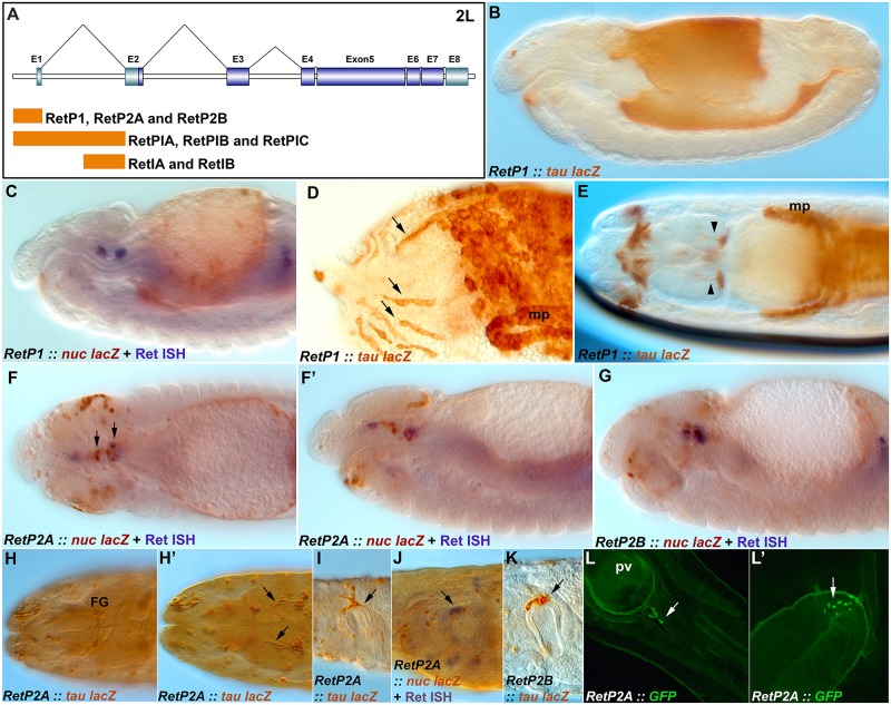
Fig 2. Expression of Ret-GAL4 transgenes.
Embryos and larvae with Ret-GAL4 lines driving expression of either nuclear lacZ (nuc-lacZ) or tau-lacZ reporters (both brown) with select counterstaining with an in situ hybridization probe for Ret (blue). (A) Diagram of the Drosophila Ret gene showing the location of fragments used to construct GAL4 lines. (B) Expression pattern of RetP1-GAL4 driving tau-lacZ showing broad expression throughout the inner lining of the gut. (C) RetP1-GAL4 driving expression of nuc-lacZ showing expression in discrete gut cells. (D) RetP1-GAL4 driving expression of tau-lacZ with individual cells in the midgut sometimes aligning into linear arrays (arrows). (E) RetP1-GAL4 and tau-lacZ with expression in brain neurons (arrowheads), elements of the CNS and PNS at the anterior of the embryo (left), malphigian tubules (mp) and hindgut. (F) Dorsal view of an embryo with RetP2-GAL4 driving nuc-lacZ in a subset of migrating SNS precursors (arrows and blue stain) and the gut. (F’) Lateral view of the same embryo as F with SNS precursors and gut staining visible. (G) An independently recovered line of RetP2 showing similar staining as F. (H, H’) Stage 17 embryo showing persistence of RetP2 expression in the neurons of the frontal ganglion (FG) and brain neurons (arrows). (I) First instar larvae with RetP2 and tau-lacZ displaying prominent staining in the proventricular ganglion (arrow). (J) Late stage 17 embryo showing overlap of RetP2, nuc-lacZ and Ret mRNA expression in the proventricular ganglion(arrow). (K) First instar larva with RetP2-GAL4 and tau-lacZ expression on the proventricular ganglion (arrow). (L,L’) Expression of UAS-CD8-GFP under control of RetP2A-GAL4 in a second instar larva showing expression (arrows) in cells adjacent to and downstream of the proventriculus (pv).
Immunohistochemistry
Antibody staining was performed as described in [19], and in situ hybridizations per [20]. Ret probe was generated by transcription of a 3 kilobase genomic fragment cloned in pBluescript. We generally use 70% glycerol in PBS or 0.1M Tris pH 8.0 as clearing agents. However, the lipid rich midgut can be hard to resolve with microscopy, so we tested several clearing protocols including ClearT [21]. We found that the best results were obtained with either Focus Clear and Rapid Clear (Cedarlane; [22]); both reagents were also useful for imaging late stage 17 embryos.
Drosophila Genetics
Janelia Farm GAL4 lines were all obtained through the Bloomington Drosophila Stock Center (BDSC). Gsc-GAL4 lines are: 46772, 48376, 46773, 48377, 40381, 40382, and 40383. Gfrl-GAL4 lines are: 47237, 47238, 47239, 47275, 40663, 40664, 40665, 40666 and 40667. fkh-GAL4 lines are: 47326, 48746, 48764, and 48795. twi-GAL4 lines are: 46150, 48725, 48729, and 48760. Please note some of these lines are no longer available but we are happy to supply them on request. The stock number for FasII-GAL4 is 46123, UAS-EGFR RNAi on III is 36770 and UAS-EGFR-DN on II and III 5364. UAS-nuclear-lacZ and UAS-CD8-GFP were obtained from the BDSC. UAS-tau-lacZ was obtained from M. Fujioka. Several of the GAL4 lines are no longer available from Bloomington and we are more than willing to supply them upon request. The w1118 stock was the most reliable wild type stock as other reference stocks do not consistently display the wild type neuroanatomy described in previous publications.
Statistics
For each genotype, stage 17 embryos were collected at random and scored for the presence, absence or thinning of the frontal nerve, and for defasciculation defects in the recurrent nerve. At least ten embryos were collected for each genotype. The 95% confidence interval and the Fisher exact test with two tails for the phenotypes was calculated using the GraphPad website (www.graphpad.com/quickcalcs). Statistical significance was assessed using the Bonferroni correction.
Results
Ret expression in the developing SNS
Expression of the Ret gene has been thoroughly documented in the Drosophila embryo [14]. We confirmed expression in the migrating SNS precursors (Fig 1A–1D and 1G–1I). Ret expression is dynamic, with expression reduced in SNS cells that have completed migration (Fig 1E and 1F). We also noted expression in the anterior midgut, which is present throughout the midgut by the end of embryogenesis (Fig 1E and 1F). Gut expression is robust but appears weaker than expression in other tissues. We additionally noted expression in a paired set of CNS neurons at the level of the subesophageal ganglia (Fig 1H) along with an ordered row of midline cells in the ventral nerve cord.
Generation of Ret-GAL4 lines
Traditional pan-neural promoters do not express during SNS precursor migration and previously identified promoter elements either have broad or highly limited expression [5, 9]. We chose to place fragments of the Ret promoter in front of the GAL4 gene with the goal of generating more SNS specific reagents. Ret is distinguished by a short promoter region upstream of the transcriptional start and three large introns (Fig 2A). We cloned the promoter region into the pPTGAL vector [17] and generated transformants using P element transposase (RetP1-GAL4). We also placed the promoter into the pBPGUw vector (RetP2-GAL4) [18], as well as the promoter fused to the first intron (RetPI-GAL4), and the second half of the first intron (RetI-GAL4; Fig 2A); transformants were generated using the PhiC31 integrase. Transformant recovery proved especially difficult for all transgenes and even identical constructs integrated into the same site yielded differences in expression (Table 1), suggesting there may be negative selective pressure towards the Ret control regions when fused to GAL4. The promoter constructs yielded broad expression particularly in the epithelial lining of the midgut (Fig 2B). Expression occurs after migration of the endodermal cells [5] and often appears continuous, but then becomes restricted to a large number of discrete cells, sometimes in linear arrangements (Fig 2C and 2D). Midgut expression is most pronounced in the RetP1-GAL4 construct. Expression was also seen in the brain (Fig 2E), in what may be a single lineage for either the mushroom bodies or a more basal lateral cluster [23]. The pBPGUw insertions (RetP2A,B-GAL4) displayed expression in a subset of the migrating SNS precursors as defined by Ret expression (Fig 2F and 2G). Additional isolated cells throughout the head region express Ret as has been seen for Ret mRNA [14], including a subset of cells projecting through the subesophageal commissure. Expression of Ret-P2 persists to the end of embryogenesis and was found to label a subset of cells in the frontal and esophageal ganglia (Fig 2H). Strong expression of reporters was observed in SNS cells at the proventriculus and additional labeled cells further along the gut (Fig 2I, 2K and 2L). Finally expression was also observed in larval midgut and body wall neurons (Fig 2L’). Additionally, we cloned the entire first intron into pBPGUw yielding strong expression in the midgut and hindgut, but not in the SNS (Table 1). The second half of the first intron produced expression in gut related tissues but not the SNS (Table 1). The RetP2 transgene appeared the most useful for SNS manipulation even though expression was only observed in a subset of cells, because expression persists into larval stages primarily in the proventricular ganglion.
Table 1. Summary of GAL4 line embryonic expression patterns.
| Gal 4 Driver Line | Expression Time | Expression Place | Primary Expression Feature | Embryonic SNS expression (stage) | Larval Expression (mCD8 GFP) |
|---|---|---|---|---|---|
| Gsc A 46772 | embryo 11–17 | Surrounding brain lobes and weak midline (15–17) | Exterior brain lobes | - | - |
| Gsc B 48376 | embryo 11–17 | Broad brain lobe expression (11–17); anterior sensory neurons (11–17); lining of esophagus (13–16) | Full brain lobes | - | - |
| Gsc C 46773 | embryo 13–17 | Weak CNS expression (11–17) | Weak CNS | - | - |
| Gsc D 48377 | embryo 12–17 | Strong CNS expression (12–17) | Complete CNS | - | - |
| Gsc E 40381 | embryo 11–17 | Weak CNS expression (12–17) | Weak CNS | - | - |
| Gsc F 40382 | embryo 12–16 | Esophagus and foregut/esophageal ganglion (12–16); mild outer brain lobe (12–16); small hindgut segment (12–16) | SNS | st 12–16 | Esophagus (pharyngeal muscles?); hindgut |
| Gsc G 40383 | embryo 11–17 | Pre-migrating SNS (11); early esophagus and foregut (11–13); proventriculus/foregut (16/17); frontal ganglion and FNJ (17); posterior brain lobe cluster (15–17) | Foregut/SNS | st 11–17 | - |
| Mun α 47237 | embryo 10–17 | Brain lobes (12–17); SNS precursors (10/11); anterior end of midline (12–16) | Very early SNS | st 11–14 | Anterior midgut cell bodies; hindgut |
| Mun β 47238 | embryo 11–17 | Anterior tip of esophagus (13–15); esophageal ganglion (13); brain lobes (15–17); large posterior brain lobe cluster (16–17); anterior receptor cell clusters (11–17) | Mid-stages SNS marker | st 11–13 | Anterior midgut cell bodies |
| Mun γ 47239 | embryo 11–17 | Lining developing esophagus (12–16); optic lobe precursors (11–12); weak brain lobe (15–17); receptor cells in anterior end of embryo (16–17) | Early SNS/anterior sensory neurons | st 11–15 | - |
| Mun δ 47275 | embryo 11, 15–17 | SNS precursors (11); anterior sensory receptors (16–17) | Mild anterior sensory neurons | st 11 | - |
| Mun ε 40663 | embryo 13–17 | Dorsal closure (13–17) | Dorsal vessel | - | - |
| Mun Ζ 40664 | embryo 11–17 | Midline precursors (13–17); presumptive foregut/hindgut (9–11); lining of developing esophagus (13–15); developing brain lobes (13–17) | Developing CNS | st 13 | Similar expression as Mun I |
| Mun Η 40665 | embryo 13–17 | Broad expression in small punctate 13–17 | Nonspecific | - | - |
| Mun Θ 40666 | embryo 11–17 | Pre-migrating SNS and early esophagus (11); sporadic and nonspecific esophageal tissue (16–17); receptor cells in anterior end of embryo (13–17) | Anterior dorsal sensory neurons | st 11 | None observed |
| Mun Ι 40667 | embryo 11–17 | Presumptive hindgut (11); spread along developing esophagus (13–17); weak midline glia expression (13–17); brain lobes (14–17) | Early foregut/hindgut; midline glia | st 15–17 | Posterior to PV; anterior midgut cell bodies; midbrain?; IMR? |
| Fkh 1 47326 | embryo 11–17 | Hindgut lining (11–17); light expression in whole midgut (11–17) | Hindgut lining | - | Hindgut |
| Fkh 2 48746 | embryo 11–17 | Weak CNS expression (11–17) | Non specific | - | - |
| Fkh 3 48764 | embryo 9–17 | Foregut/hindgut (9–12); complete Intestinal tract (13–17); Malpighian tubules (13–16); mild CNS expression (13–17) | CNS/Intestinal Anatomy outline | - | - |
| Fkh 4 48795 | embryo 11–17 | Similar to Fkh 3; intestinal tract expression (11–17); Malpighian tubules (15/16) | Intestinal anatomy outline | st 11–15 | - |
| Twi 1 46150 | embryo 13–17 | SNS cluster (13); esophageal/pharyngeal muscles, dorsal side of esophagus/EG? (15–17) | Esophageal/pharyngeal clusters | st 13–17 | Anterior sensory neurons |
| Twi 2 48725 | embryo 13–17 | Mild CNS expression (13–17) | Weak CNS | - | - |
| Twi 3 48729 | embryo 11–17 | Developing anterior Sensory receptors (11–17); mild CNS (13–17); | Anterior sensory neurons | - | - |
| Twi 4 48760 | embryo 11–17 | Developing CNS (11–17) | CNS | st 13,17 | - |
| RetP1 (Herna#1 in pPTGAL) | embryo 12–17 | Distinct expression in cells of the dorsal fold (14); putative adult midgut precursors and other endoderm/endoderm adjacent cells (14–17); specific subset of brain cells (15–16); periventricular ganglion, dorsal pharyngeal muscles, Malpighian tubules (17) | CNS; SNS; foregut/midgut/hindgut; brain lobes | st 13–17 | Brain; posterior to PV; anterior midgut cell bodies and hindgut |
| RetP2A (Herna#2 in pBPGUw) | embryo 12–17 | Pre-migrating SNS clusters (11–12); migrating SNS clusters (13–16); specific subset of brain cells, putative adult midgut precursors and other endoderm/endoderm adjacent cells and Malpighian tubules (15–17) | CNS; SNS subset; foregut/midgut/hindgut; brain lobes | st 12–17 | SNS; posterior to PV; anterior midgut cell bodies |
| RetP2B (Herna#3 pBPGUw) | embryo 12–17 | Distinct expression in cells in the esophageal clusters (13–14); esophageal and periventricular ganglion, specific subset of brain cells, putative adult midgut precursors and other endoderm/endoderm adjacent cells (15–17) | CNS; SNS subset; foregut/midgut/hindgut; brain lobes | st 12–17 | SNS; posterior to PV; anterior midgut cell bodies |
| RetPIA (Herna#4 pBPGUw) | embryo 13–17 | Tracheal/peripheral (ventral) expression (13–14); midgut lining (endoderm/endoderm adjacent cells) (14–17 and later) | PNS (ventral); midgut | - | Midgut and hindgut |
| RetPIB (Herna#5 pBPGUw) | embryo 13–17 | Proventriculus (13–14); minimal midgut/hindgut lining (endoderm/endoderm adjacent cells) (15–17); cephalopharyngeal ganglia/pharyngeal muscles (17) | Proventriculus; midgut; hindgut lining | st 13–14 (Proventriculus) | - |
| RetPIC (Herna#6 pBPGUw) | embryo 12–17 | (Anterior) midgut lining (endoderm/endoderm adjacent cells) (14–17); hindgut lining (16–17) | Anterior midgut; hindgut | - | Midgut and hindgut |
| RetIA (Herna#7) pBPGUw) | embryo 12–17 | CNS, broad PNS expression, trachea (12–16); proventriculus (16–17) | CNS/PNS; proventriculus | st 16–17 (Proventriculus) | - |
| RetIB (Herna#8 pBPGUw) | embryo 11–17 | Developing CNS (11); distinct expression in cells in the esophagus (12–17); proventriculus (16–17); (anterior) midgut lining (endoderm/endoderm adjacent cells) (15–17) | Proventriculus; anterior midgut | st 12–17 (Proventriculus) | - |
Identification of Additional SNS Specific GAL4 Lines
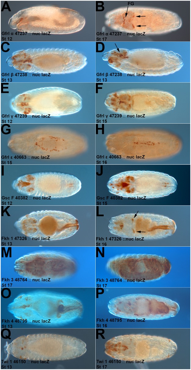
The limited SNS expression and additional expression of the Ret promoter fragments prompted us to look for additional reagents. We examined the Janelia Farm Fly Light GAL4 lines [18, 24] for driver fragments derived from genes with known SNS expression. Ret has an evolutionarily conserved co-receptor known as Gfrl or Munin in flies [25], and we tested nine Gfrl lines for SNS expression (Table 1). One line (#47237) had highly specific SNS expression from initial delamination of the SNS precursors until the end of embryogenesis (Fig 3A and 3B). This line (which we will refer to as Gfrl-GAL4) also displays brain lobe expression strongly resembling that of the RetP1 construct. Two additional lines (#47238, #47239) had broader expression in the esophagus and likely the SNS too (Fig 3C–3F). #47239 also has brain lobe expression. A fourth line expresses in cells at the leading edge of dorsal closure (Fig 3G and 3H). These lines have expression that appears significantly more restricted to the SNS than the Ret lines, with Gfrla-GAL4 having the greatest potential for SNS manipulation. We also tested fragments of the forkhead (fkh), Goosecoid (Gsc) and twist (twi) genes as their expression has been reported in the SNS [8, 14, 26, 27]. Two Gsc lines were of interest, #40382 with esophageal and likely SNS expression (Fig 3I and 3J) and #40383 with strong specific SNS expression (see below). Three fkh lines had midgut and hindgut expression (Fig 3K–3P), with #47326 having brain lobe expression like Ret-P1, #48764 having very broad expression and #48795 strongly resembling Ret-P1 in the overall expression pattern. One twi line #46150 has potential expression in a very small subset of the SNS. However Gsc line #40383 stood out for its striking SNS specificity and duration of expression so we chose to characterize it further.
Fig 3. Expression of Gfrl, Gsc and Fkh GAL4 lines.
Expression of nuc-lacZ or tau-lacZ (brown) by selected Janelia Farm GAL4 lines. The driver, reporter and embryo stage are noted on the Fig panel. Please also refer to Table 1. (A) Gfrl fragment driving expression in the roof of the stomatodeum in presumptive SNS precursor clusters and a few additional cells. (B) Gfrl fragment with expression in the frontal ganglion (FG) and brain lobe clusters (arrows). (C, D) Dorsal and lateral views of a Gfrl fragment driving expression in esophageal and SNS cells (arrow), cells presumed to be the subesophageal ganglion and additional cells. (E) Esophageal, SNS and brain lobe expression of a Gfrl fragment. (F) Esophageal, SNS and brain lobe expression with additional CNS and PNS cells. (G, H) Expression in cells of the leading edge during dorsal closure. (I, J) Expression of a Gsc-GAL4 line in foregut, esophageal, SNS and brain cells. (K, L) Expression of a Fkh-GAL4 line in the midgut, hindgut, brain lobe cells (arrows) and additional cells near the anterior of the embryo (left). (M, N) Expression of a Fkh-GAL4 line throughout the gut and CNS in a late stage embryo. (O, P) Fkh-GAL4 expression in the esophagus, SNS, brain lobes and gut cells in a pattern that resembles RetP1. (Q) Twi-GAL4 expression in a subset of SNS cells. (R) Twi-GAL4 expression in the pharynx and additional unidentified cells that likely include parts of the SNS and PNS.
Maternal Effect of the attP2 Integration Site Chromosome
The Fly Light lines are integrated into a third chromosome site, attP2, using the phiC31 site-specific integrase system [28]. We noticed that several GAL4 lines, but especially GscG-GAL4 produced shorter embryos. This effect was regardless of reporter used and was only observed when the GscG-GAL4 was the mother. The embryos themselves appear completely normal when assessed with 22c10 or 1D4 staining, just compressed along the anterior-posterior axis. A large number of embryos fail to hatch and the lines were quite difficult to maintain as homozygotes. Balancing in combination with a dominant male sterile mutation helped significantly. For all crosses, we used the GAL4 line as the male parent.
Characterization of a Gsc-GAL4 line
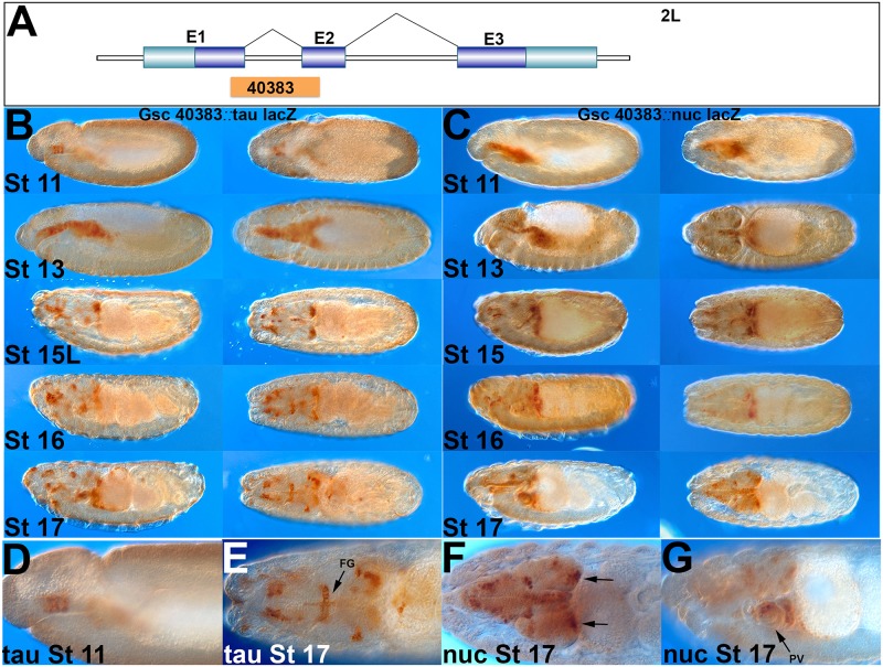
A transposable element, SNS1-GAL4, inserted in the Goosecoid (Gsc) gene had been previously used to drive early SNS expression [9], which led us to test Gsc promoter fragments. Gsc #40383 expresses in all three SNS clusters from the start of SNS delamination until the end of embryogenesis (we will refer to this line as GscG-GAL4). This element contains parts of exon coding regions from the first and second exons of Gsc (Fig 4A). Early stage 11 expression of GscG-GAL4 may be restricted to the three delaminating clusters but rapidly broadens to include cells in the underlying esophagus (Fig 4B and 4C). Expression in stage 16/17 is subsequently restricted to the migrating SNS precursors as well as the cluster of brain cells seen in RetP1-GAL4 and the Gfrl-GAL4 lines (Figs 2E, 3B,3F and 3L). Additional cells are seen in the head region as seen for Ret mRNA. At the end of embryogenesis the line has strong expression in the frontal nerve, frontal commissure, recurrent nerve, and proventricular ganglion (Fig 4E–4G). This line has the most complete expression in the anterior elements of the SNS, while RetP1-GAL4 appears to express in more proventricular cells and for longer.
Fig 4. Characterization of the GscG-GAL4 line.
Developmental series of embryos expressing tau-lacZ (left hand columns) or nuc-lacZ (right hand columns) under control of the GscG-GAL4 line. (A) Schematic of the position of the promoter fragment of GscG-GAL4 inside the Gsc gene containing parts of exon 1 and 2. (B) Nuclear lacZ reporter displaying expression of GscG-GAL4 in the initial SNS precursor clusters in the stomatodeum (stage 11, see also panel D), expanding to include most esophageal cells (stage 13), and resolving into SNS, brain lobe, subesophageal ganglion and PNS specific staining in later stages (15–17; see also panels E-G). (C) Tau-lacZ reporter in a similar developmental series to panel B. All SNS cells are labeled by the end of embryogenesis including the frontal ganglion, esophageal ganglia and proventriculus. (D-E) Higher magnification views showing the initial three delaminating SNS clusters at stage 11 and the entire frontal ganglion and nerve at stage 17 (FG). The nuclear staining shows the brain lobe clusters (arrows) as well as the proventriculus (PV).
Manipulation of SNS Migration using Gfrl-GAL4 and RetP2-GAL4
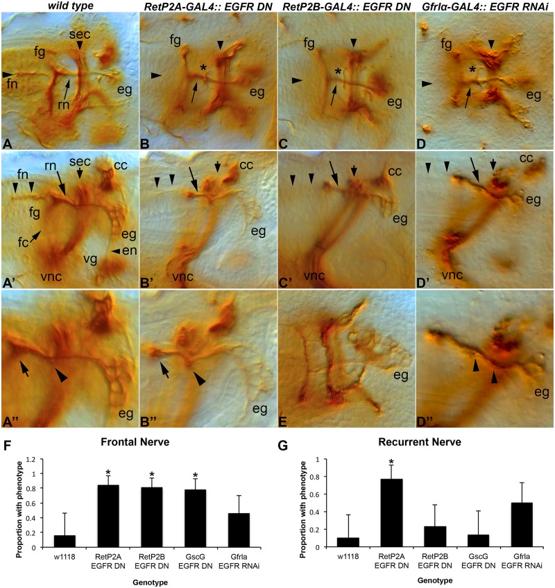
To test the utility of the identified GAL4 lines we chose to manipulate EGFR function. As noted above EGFR plays an important role in delamination of the SNS precursors and this early phenotype would preclude later phenotypes from being observed. We used RetP2A- and RetP2B-GAL4 to drive an EGFR dominant negative construct [29, 30]. In each case we observed frequent loss or reduction of the frontal nerve and disruption of the recurrent nerve (Fig 5B, 5B', 5B'', 5C, 5C', 5E, 5F and 5G). We observed the same phenotype with Gfrla-GAL4 driving EGFR RNAi (Fig 5D, 5D' and 5D''). Obtaining the same phenotype with three different GAL4 lines and two different constructs validates the lines identified. Similar results were obtained with GscG-GAL4 and both dominant negative and RNAi transgenes. The observed phenotypes indicate that EGFR signaling is likely required for axon growth/guidance of the frontal and recurrent nerves. Based on the appearance of the esophageal ganglia (Fig B'', C'') cell number is not disrupted by EGFR inhibition. We could not accurately determine whether cells in the frontal ganglia are lost as anti-FasII staining does not reveal the cell bodies of the frontal ganglia in later stages (when the frontal nerve has developed). Most of the recurrent nerve axons project from the esophageal ganglia and project anteriorly, but a few appear to originate in the frontal ganglia and project posteriorly [10]. The recurrent nerve phenotype appears to arise from esophageal ganglion axons projecting anteriorly and defasciculating from the nerve (Fig 5D''). The axons can either change direction after passing under the brain commissure, or may fail to join the recurrent nerve in the first place projecting around the commissural surface of the brain lobe. Confocal microscopy could not further distinguish the origins of the phenotype (S1 Fig). Similar results were observed with Gsc-GAL4 and both EGFR RNAi and the dominant negative transgene (Fig 5F and 5G and S2 Fig). With our current level of analysis, we cannot rule out alternative explanations including non-cell autonomous effects on brain neurons although we think this unlikely. We note that similar recurrent nerve phenotypes were observed for the misshapen/Ste20/l(3)6683 kinase that regulates MAPK signaling [7]. The Gsc expression pattern suggests there could be a cell type boundary at or near the defasciculation point at the pharynx-esophageal junction [26], perhaps indicating that secreted cues change at this position and that there could be choice point for growing axons.
Fig 5. SNS manipulation using the RetP2-GAL4 and Gfrl-GAL4 lines.
Anti-Fasciclin II staining (monoclonal antibody 1D4) revealing the mature embryonic SNS. We use 1D4 as it reliably stains the frontal nerve (fn) whereas 22c10 frequently fails to stain this nerve in wild type. (A) Dorsal view of a stage 17 wild type embryo with elements of the SNS labeled. The recurrent nerve (rn, arrow) runs from the esophageal ganglion (eg) along the esophagus underneath the supraesophageal commissure (sec, also known as the brain commissure) to the frontal ganglion (fg). The frontal nerve (fn, arrowhead) projects anteriorly from the frontal ganglion (fg). (A') Lateral view of the same embryo. The frontal connective (fc) which links the frontal ganglion to the brain, and the esophageal nerve (en) which links the esophageal ganglia to the ventricular ganglion (vg) can be seen. The corpora cardiaca (cc) neuroendocrine organ is visible close to the dorsal surface. (A'') Close-up of the frontal ganglion (arrow), recurrent nerve (arrowhead) and the esophageal ganglia (EG). A slight expansion of the recurrent nerve (arrowhead) can be seen, marking a location where defasciculated axons are often seen in transgenic manipulations. (B) RetP2A-GAL4 driving a dominant negative EGFR transgene. The frontal nerve (arrowhead) is absent and the recurrent nerve (arrow) exhibits a defasciculated axon or axons (asterisk). (B') Lateral view of the embryo in B. The frontal nerve is clearly missing (arrowheads). (B'') Close-up of the same embryo. The recurrent nerve displays a kink and expansion underneath the supraesophageal commissure (arrowhead). The frontal ganglion (arrow) is also visible. The number of cells in the esophageal ganglia appear comparable to wild type. (C) RetP2B-GAL4 driving the dominant negative EGFR transgene. The frontal nerve is absent (arrowhead) and a defasciculated axon is crossing the recurrent nerve (asterisk). The latter axon may originate at the esophageal ganglia, but we have been unable to conclusively determine this for any examples studied. (C') Lateral view of the same embryo showing the absence of the frontal nerve (arrowheads) and axon defasciculation from the recurrent nerve (arrow). From this angle, at least one defasciculated axon appears to originate from the recurrent nerve itself. (D) Gfrlα-GAL4 driving transgenic RNAi for EGFR. The frontal nerve is absent (arrowhead) and the recurrent nerve (arrow) is defasciculated (asterisk). (D') Lateral view of the same embryo showing the absence of the frontal nerve (arrowheads) and a slight swelling of the recurrent nerve (arrow) at the point that defasciculation occurs. (D'') Higher magnification view showing a defasciculated axon growing alongside the recurrent nerve from the esophageal ganglia to the point of defasciculation. (E) Late stage 17 embryo with the EGFR dominant negative transgene driven by RetP2B-GAL4 showing that the number of cells within the esophageal ganglia appears unaffected by EGFR inhibition. (F, G) Quantification of frontal nerve defects (reduced or absent; F) or recurrent nerve (defasciculation; G) defects in the genotypes examined. The error bars represent the 95% confidence interval. Statistical significance (*) relative to the wild type control (w1118) was assessed using the Fisher exact test with two tails and the Bonferroni correction.
A GAL4 Line for Gut Specific Expression
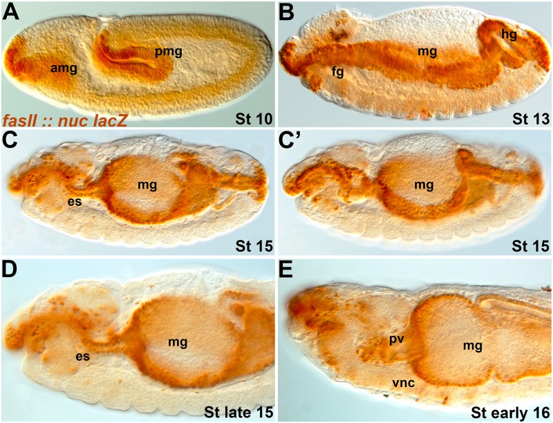
We anticipate wanting to be able to manipulate the esophagus and gut tissue over which the SNS precursors migrate and differentiate. We identified a GAL4 line with a fragment of the FasII promoter (#46123) that expresses strongly from the earlier stages of gut formation until the point at which SNS precursors stop migrating (Fig 6). The line is striking as it expresses simultaneously in the foregut, midgut and hindgut. As the gut is derived from different cell populations and germ layers [31], GAL4 lines typically express in a subsection of the gut rather than the entire tissue [32]. The FasII line identified expresses in the intestinal epithelium as opposed to the visceral muscle that surrounds the gut. Over-expression of the EGFR ligand Spitz led to extremely disrupted embryos as would be expected from high expression of a potent growth factor.
Fig 6. A FasII-GAL4 line drives in all parts of the developing gut.
Lateral views of embryos with a nuclear lacZ reporter (brown) driven by FasII-GAL4 (#46123). (A) Stage 11 embryo in which the anterior (amg) and posterior (pmg) midgut are stained. (B) Stage 13 embryo displaying continuous staining throughout the gut tissue, including the foregut (fg), midgut (mg) and hindgut (hg). (C, C’) Early stage 15 embryos in which the esophagus (es) is starting to loop. By this stage the visceral mesoderm that forms the visceral muscles surrounding the gut has migrated to the epithelial lining, but we see no evidence that the FasII line expresses in this tissue. (D) Late stage 15 embryo with persistent staining throughout the gut. (E) Early stage 16 embryo in which the proventriculus (pv) has started to form. Expression is widespread and continues to stage 17/early larval stages but at weaker levels.
Discussion
Lines identified in this study
Our analysis has identified three GAL4 lines that have embryonic specific SNS expression with limited expression in other tissues. The RetP2-GAL4 line expresses in a subset of SNS cells with strong persistent expression in the proventricular ganglion in the first instar larva. The Gfrl-GAL4 line expresses in most SNS cells until the end of embryogenesis. The Gsc-GAL4 line expresses in likely all SNS cells, and based on reporter gene expression is the strongest driver we identified in the embryo, although SNS expression does not persist into the larvae. All three lines have utility in SNS manipulation as they produce EGFR phenotypes that occur after EGFR’s role in the specification of SNS precursor clusters. As SNS delamination was unaffected by any of the GAL4 lines used, these lines may not be used to manipulate the earliest stages of SNS migration. The strength of reporter genes suggests the GAL4 lines may have relatively low expression. We are currently building stocks to amplify or permanently switch on expression to assess whether the lines will be useful in larval analysis.
Relationships between identified gene fragments
Striking similarities are observed between the expression patterns of the promoter fragments analyzed. RetP1-GAL4, RetP2A-GAL4, RetP2B-GAL4, Gfrl-GAL4 (2 different lines), fkh-GAL4 and Gsc-GAL4 all express in cells near the back of the brain lobes, in the migrating SNS precursors and/or the underlying esophageal cells, suggesting that these genes may be functionally linked. It has been suggested that Ret and Gfrl did not function as a cis receptor-coreceptor pair before the emergence of GDNF family ligands in vertebrate lineages [25]. The shared expression patterns of Ret and Gfrl regulatory elements suggest a functional relationship exists in flies and likely other invertebrates. We note that Ret and Gfrl share expression both in the SNS and in the malphigian tubules [14, 25], the fly homologue of the vertebrate kidney where Ret also plays a role [33, 34]. We are currently generating Ret mutants in the fly to establish the precise role it plays in SNS formation.
The Ret gene is one of the key markers for neural crest cells migrating into the gut [35] and plays a key role in enteric nervous system formation [12]. Neural crest cells acquire their identities through the expression of neural crest specifier genes such as the Snail, FoxD3 and SoxE genes. The fly Sox10 ortholog is not expressed in the SNS and appears to have been co-opted into neural crest development during the course of evolution through altered expression patterns [36]. It is therefore interesting to note that although the fly FoxD3 homologue is not expressed in the SNS [37], the related fkh gene is required for SNS formation [38]. Similarly, Gsc homologues have roles in neural crest development [39, 40]. Identifying regulators of SNS development in the fly and their functional relationships has the potential to shed light on vertebrate neural crest formation. Other insects display a similar migration of enteric precursors [41–43], suggesting that SNS precursor migration is an ancient developmental program. It will be interesting to see whether there is an evolutionarily conserved regulatory network driving Ret expression. Such information will likely be useful in unraveling the genetic complexity of Hirschsprung’s disease in humans.
Future directions
Identification of three independent SNS specific drivers with different expression characteristics provides an opportunity to investigate the development of the fly SNS. Relatively few SNS developmental components have been identified and fewer have been characterized to date, particularly in the later stages of embryonic development. Reporter genes can be used as markers to dissect phenotypes in greater detail. The ability to complement loss of function data with gain of function data is an important tool in analyzing function, as is the ability to rescue phenotypes. Some of the lines such as Ret-P2 may be useful in larval feeding assays that can provide comprehensive functional readouts [44–47]. Hirschsprung’s disease occurs one hundred times more frequently in Down Syndrome patients and overexpression of the Dscam gene is the leading candidate gene [48]. The SNS drivers will facilitate modeling of Hirschsprung's disease in a simple organism.
Supporting Information
Anti-Fasciclin II staining (monoclonal antibody 1D4) revealing the mature SNS in lateral views. (A) Wild type (w1118) embryo showing the frontal nerve (fn), frontal ganglion (fg), recurrent nerve (rn), supraesophageal or brain commissure (sec), corpora cardiaca (cc), frontal connective (fc), esophageal ganglia (EG), esophageal nerve (en), ventricular ganglion (vg) and ventral nerve cord (vnc). (B) RetP2A-GAL4 driving the EGFR dominant negative transgene. The frontal nerve is absent (arrowheads) and the recurrent nerve is possibly less tightly bundled (arrow). (C) RetP2B-GAL4 driving the EGFR dominant negative transgene. The frontal nerve is absent (arrowheads) whereas the recurrent nerve looks normal.
(TIF)
Anti-Fasciclin II staining (monoclonal antibody 1D4) revealing the mature SNS in ventral views. (A) Stage 17 wild type embryo with elements of the SNS labeled. The recurrent nerve (rn, arrow) runs from the esophageal ganglion (EG) along the esophagus underneath the brain commissure (bc) to the esophageal gangion (EG). (B) GscG-GAL4 driving transgenic RNAi for EGFR. The recurrent nerve (arrow) is clearly disrupted. (C) GscG-GAL4 driving an EGFR dominant negative transgene. The frontal nerve is missing (arrowhead) and the recurrent nerve is disrupted.
(TIF)
Acknowledgments
We thank C. Abrescia, C. Ibáñez, I. Miguel-Aliaga, P. Soba, R. Reed, J. Kallijarvi, R. Cagan, A. Teague, M. Fujioka, S. Sprecher for advice and reagents, M. Lamanuzzi for drawing our attention to the FasII-GAL4 stock and help with dissections/analysis, J. Hogan and C. Loosbroock for cloning the Ret genomic fragment, C. Jenkins, S. Hamilton-Jones, M. Contreras and members of the Kidd laboratory for technical assistance. We thank S. Ward, C. von Bartheld and D. Mathew and unidentified reviewers for comments on the manuscript. Antibodies were obtained from the Developmental Studies Hybridoma Bank (DHSB) developed under the auspices of the NICHD and maintained by the University of Iowa. KH was supported by the Michael (Mick) J. M. Hitchcock, Ph.D. Graduate Student Research Fund.
Data Availability
All relevant data are within the paper and its Supporting Information files.
Funding Statement
This project was supported by a grant from the National Institutes of Health (R15NS075918) to T.K. This study also received funding from the NIH National Institute of General Medical Sciences grants P20 GM103650, P20 GM103554, P20 GM103440. Additionally, Michael (Mick) J. M. Hitchcock, Ph.D. Graduate Student Research Fund to KH. The funders had no role in study design, data collection and analysis, decision to publish, or preparation of the manuscript.
References
- 1. Marder E, Bucher D. Understanding circuit dynamics using the stomatogastric nervous system of lobsters and crabs. Annual review of physiology. 2007;69:291–316. Epub 2006/10/03. 10.1146/annurev.physiol.69.031905.161516 . [DOI] [PubMed] [Google Scholar]
- 2. Lemaitre B, Miguel-Aliaga I. The digestive tract of Drosophila melanogaster. Annual review of genetics. 2013;47:377–404. Epub 2013/09/11. 10.1146/annurev-genet-111212-133343 . [DOI] [PubMed] [Google Scholar]
- 3. Dumstrei K, Nassif C, Abboud G, Aryai A, Aryai A, Hartenstein V. EGFR signaling is required for the differentiation and maintenance of neural progenitors along the dorsal midline of the Drosophila embryonic head. Development (Cambridge, England). 1998;125(17):3417–26. Epub 1998/08/07. . [DOI] [PubMed] [Google Scholar]
- 4. Hartenstein V. Development of the insect stomatogastric nervous system. Trends in neurosciences. 1997;20(9):421–7. Epub 1997/09/18. . [DOI] [PubMed] [Google Scholar]
- 5. Hartenstein V, Tepass U, Gruszynski-Defeo E. Embryonic development of the stomatogastric nervous system in Drosophila. The Journal of comparative neurology. 1994;350(3):367–81. Epub 1994/12/15. 10.1002/cne.903500304 . [DOI] [PubMed] [Google Scholar]
- 6. Hartenstein V, Tepass U, Gruszynski-deFeo E. Proneural and neurogenic genes control specification and Morphogenesis of stomatogastric nerve cell precursors in Drosophila. Developmental biology. 1996;173(1):213–27. Epub 1996/01/10. 10.1006/dbio.1996.0018 . [DOI] [PubMed] [Google Scholar]
- 7. Forjanic JP, Chen CK, Jackle H, Gonzalez Gaitan M. Genetic analysis of stomatogastric nervous system development in Drosophila using enhancer trap lines. Developmental biology. 1997;186(2):139–54. Epub 1997/06/15. . [DOI] [PubMed] [Google Scholar]
- 8. Gonzalez-Gaitan M, Jackle H. Invagination centers within the Drosophila stomatogastric nervous system anlage are positioned by Notch-mediated signaling which is spatially controlled through wingless. Development (Cambridge, England). 1995;121(8):2313–25. Epub 1995/08/01. . [DOI] [PubMed] [Google Scholar]
- 9. Gonzalez-Gaitan M, Jackle H. Tip cell-derived RTK signaling initiates cell movements in the Drosophila stomatogastric nervous system anlage. EMBO reports. 2000;1(4):366–71. Epub 2001/03/28. 10.1093/embo-reports/kvd064 ; PubMed Central PMCID: PMCPmc1083741. [DOI] [PMC free article] [PubMed] [Google Scholar]
- 10. Spiess R, Schoofs A, Heinzel HG. Anatomy of the stomatogastric nervous system associated with the foregut in Drosophila melanogaster and Calliphora vicina third instar larvae. Journal of morphology. 2008;269(3):272–82. Epub 2007/10/27. 10.1002/jmor.10581 . [DOI] [PubMed] [Google Scholar]
- 11. Lake JI, Heuckeroth RO. Enteric nervous system development: migration, differentiation, and disease. American journal of physiology Gastrointestinal and liver physiology. 2013;305(1):G1–24. Epub 2013/05/04. 10.1152/ajpgi.00452.2012 ; PubMed Central PMCID: PMCPmc3725693. [DOI] [PMC free article] [PubMed] [Google Scholar]
- 12. McKeown SJ, Stamp L, Hao MM, Young HM. Hirschsprung disease: a developmental disorder of the enteric nervous system. Wiley interdisciplinary reviews Developmental biology. 2013;2(1):113–29. Epub 2013/06/27. 10.1002/wdev.57 . [DOI] [PubMed] [Google Scholar]
- 13. Ibanez CF. Structure and physiology of the RET receptor tyrosine kinase. Cold Spring Harbor perspectives in biology. 2013;5(2). Epub 2013/02/05. 10.1101/cshperspect.a009134 . [DOI] [PMC free article] [PubMed] [Google Scholar]
- 14. Hahn M, Bishop J. Expression pattern of Drosophila ret suggests a common ancestral origin between the metamorphosis precursors in insect endoderm and the vertebrate enteric neurons. Proceedings of the National Academy of Sciences of the United States of America. 2001;98(3):1053–8. Epub 2001/02/07. 10.1073/pnas.021558598 ; PubMed Central PMCID: PMCPmc14707. [DOI] [PMC free article] [PubMed] [Google Scholar]
- 15. Soba P, Han C, Zheng Y, Perea D, Miguel-Aliaga I, Jan LY, et al. The Ret receptor regulates sensory neuron dendrite growth and integrin mediated adhesion. Elife. 2015;4 Epub 2015/03/13. 10.7554/eLife.05491 [DOI] [PMC free article] [PubMed] [Google Scholar]
- 16. Thibault ST, Singer MA, Miyazaki WY, Milash B, Dompe NA, Singh CM, et al. A complementary transposon tool kit for Drosophila melanogaster using P and piggyBac. Nature genetics. 2004;36(3):283–7. Epub 2004/02/26. 10.1038/ng1314 . [DOI] [PubMed] [Google Scholar]
- 17. Sharma Y, Cheung U, Larsen EW, Eberl DF. PPTGAL, a convenient Gal4 P-element vector for testing expression of enhancer fragments in drosophila. Genesis (New York, NY: 2000). 2002;34(1–2):115–8. Epub 2002/09/27. 10.1002/gene.10127 ; PubMed Central PMCID: PMCPmc1805626. [DOI] [PMC free article] [PubMed] [Google Scholar]
- 18. Pfeiffer BD, Jenett A, Hammonds AS, Ngo TT, Misra S, Murphy C, et al. Tools for neuroanatomy and neurogenetics in Drosophila. Proceedings of the National Academy of Sciences of the United States of America. 2008;105(28):9715–20. Epub 2008/07/16. 10.1073/pnas.0803697105 ; PubMed Central PMCID: PMCPmc2447866. [DOI] [PMC free article] [PubMed] [Google Scholar]
- 19. Patel NH. Imaging neuronal subsets and other cell types in whole-mount Drosophila embryos and larvae using antibody probes. Methods in cell biology. 1994;44:445–87. Epub 1994/01/01. . [DOI] [PubMed] [Google Scholar]
- 20. Kopczynski CC, Davis GW, Goodman CS. A neural tetraspanin, encoded by late bloomer, that facilitates synapse formation. Science (New York, NY). 1996;271(5257):1867–70. Epub 1996/03/29. . [DOI] [PubMed] [Google Scholar]
- 21. Kuwajima T, Sitko AA, Bhansali P, Jurgens C, Guido W, Mason C. ClearT: a detergent- and solvent-free clearing method for neuronal and non-neuronal tissue. Development (Cambridge, England). 2013;140(6):1364–8. Epub 2013/02/28. 10.1242/dev.091844 ; PubMed Central PMCID: PMCPmc3912244. [DOI] [PMC free article] [PubMed] [Google Scholar]
- 22. Liu YC, Chiang AS. High-resolution confocal imaging and three-dimensional rendering. Methods (San Diego, Calif). 2003;30(1):86–93. Epub 2003/04/16. . [DOI] [PubMed] [Google Scholar]
- 23. Sprecher SG, Reichert H, Hartenstein V. Gene expression patterns in primary neuronal clusters of the Drosophila embryonic brain. Gene expression patterns: GEP. 2007;7(5):584–95. Epub 2007/02/16. 10.1016/j.modgep.2007.01.004 ; PubMed Central PMCID: PMCPmc3928073. [DOI] [PMC free article] [PubMed] [Google Scholar]
- 24. Jenett A, Rubin GM, Ngo TT, Shepherd D, Murphy C, Dionne H, et al. A GAL4-driver line resource for Drosophila neurobiology. Cell reports. 2012;2(4):991–1001. Epub 2012/10/16. 10.1016/j.celrep.2012.09.011 ; PubMed Central PMCID: PMCPmc3515021. [DOI] [PMC free article] [PubMed] [Google Scholar]
- 25. Kallijarvi J, Stratoulias V, Virtanen K, Hietakangas V, Heino TI, Saarma M. Characterization of Drosophila GDNF receptor-like and evidence for its evolutionarily conserved interaction with neural cell adhesion molecule (NCAM)/FasII. PloS one. 2012;7(12):e51997 Epub 2013/01/04. 10.1371/journal.pone.0051997 ; PubMed Central PMCID: PMCPmc3527400. [DOI] [PMC free article] [PubMed] [Google Scholar]
- 26. Hahn M, Jackle H. Drosophila goosecoid participates in neural development but not in body axis formation. The EMBO journal. 1996;15(12):3077–84. Epub 1996/06/17. ; PubMed Central PMCID: PMCPmc450248. [PMC free article] [PubMed] [Google Scholar]
- 27. Hammonds AS, Bristow CA, Fisher WW, Weiszmann R, Wu S, Hartenstein V, et al. Spatial expression of transcription factors in Drosophila embryonic organ development. Genome biology. 2013;14(12):R140 Epub 2013/12/24. 10.1186/gb-2013-14-12-r140 ; PubMed Central PMCID: PMCPmc4053779. [DOI] [PMC free article] [PubMed] [Google Scholar]
- 28. Groth AC, Fish M, Nusse R, Calos MP. Construction of transgenic Drosophila by using the site-specific integrase from phage phiC31. Genetics. 2004;166(4):1775–82. Epub 2004/05/06. ; PubMed Central PMCID: PMCPmc1470814. [DOI] [PMC free article] [PubMed] [Google Scholar]
- 29. Ni JQ, Liu LP, Binari R, Hardy R, Shim HS, Cavallaro A, et al. A Drosophila resource of transgenic RNAi lines for neurogenetics. Genetics. 2009;182(4):1089–100. Epub 2009/06/03. 10.1534/genetics.109.103630 ; PubMed Central PMCID: PMCPmc2728850. [DOI] [PMC free article] [PubMed] [Google Scholar]
- 30. Buff E, Carmena A, Gisselbrecht S, Jimenez F, Michelson AM. Signalling by the Drosophila epidermal growth factor receptor is required for the specification and diversification of embryonic muscle progenitors. Development (Cambridge, England). 1998;125(11):2075–86. Epub 1998/06/24. . [DOI] [PubMed] [Google Scholar]
- 31. Tepass U, Hartenstein V. Epithelium formation in the Drosophila midgut depends on the interaction of endoderm and mesoderm. Development (Cambridge, England). 1994;120(3):579–90. Epub 1994/03/01. . [DOI] [PubMed] [Google Scholar]
- 32. Ward EJ, Thaipisuttikul I, Terayama M, French RL, Jackson SM, Cosand KA, et al. GAL4 enhancer trap patterns during Drosophila development. Genesis (New York, NY: 2000). 2002;34(1–2):46–50. Epub 2002/09/27. 10.1002/gene.10138 . [DOI] [PubMed] [Google Scholar]
- 33. Pachnis V, Mankoo B, Costantini F. Expression of the c-ret proto-oncogene during mouse embryogenesis. Development (Cambridge, England). 1993;119(4):1005–17. Epub 1993/12/01. . [DOI] [PubMed] [Google Scholar]
- 34. Schuchardt A, D'Agati V, Larsson-Blomberg L, Costantini F, Pachnis V. Defects in the kidney and enteric nervous system of mice lacking the tyrosine kinase receptor Ret. Nature. 1994;367(6461):380–3. Epub 1994/01/27. 10.1038/367380a0 . [DOI] [PubMed] [Google Scholar]
- 35. Sauka-Spengler T, Bronner-Fraser M. A gene regulatory network orchestrates neural crest formation. Nature reviews Molecular cell biology. 2008;9(7):557–68. Epub 2008/06/05. 10.1038/nrm2428 . [DOI] [PubMed] [Google Scholar]
- 36. Cossais F, Sock E, Hornig J, Schreiner S, Kellerer S, Bosl MR, et al. Replacement of mouse Sox10 by the Drosophila ortholog Sox100B provides evidence for co-option of SoxE proteins into vertebrate-specific gene-regulatory networks through altered expression. Developmental biology. 2010;341(1):267–81. Epub 2010/02/11. 10.1016/j.ydbio.2010.01.038 . [DOI] [PubMed] [Google Scholar]
- 37. Hacker U, Grossniklaus U, Gehring WJ, Jackle H. Developmentally regulated Drosophila gene family encoding the fork head domain. Proceedings of the National Academy of Sciences of the United States of America. 1992;89(18):8754–8. Epub 1992/09/15. ; PubMed Central PMCID: PMCPmc49999. [DOI] [PMC free article] [PubMed] [Google Scholar]
- 38. De Velasco B, Shen J, Go S, Hartenstein V. Embryonic development of the Drosophila corpus cardiacum, a neuroendocrine gland with similarity to the vertebrate pituitary, is controlled by sine oculis and glass. Developmental biology. 2004;274(2):280–94. Epub 2004/09/24. 10.1016/j.ydbio.2004.07.015 . [DOI] [PubMed] [Google Scholar]
- 39. Gottlieb S, Hanes SD, Golden JA, Oakey RJ, Budarf ML. Goosecoid-like, a gene deleted in DiGeorge and velocardiofacial syndromes, recognizes DNA with a bicoid-like specificity and is expressed in the developing mouse brain. Human molecular genetics. 1998;7(9):1497–505. Epub 1998/08/13. . [DOI] [PubMed] [Google Scholar]
- 40. Parry DA, Logan CV, Stegmann AP, Abdelhamed ZA, Calder A, Khan S, et al. SAMS, a syndrome of short stature, auditory-canal atresia, mandibular hypoplasia, and skeletal abnormalities is a unique neurocristopathy caused by mutations in Goosecoid. American journal of human genetics. 2013;93(6):1135–42. Epub 2013/12/03. 10.1016/j.ajhg.2013.10.027 ; PubMed Central PMCID: PMCPmc3853132. [DOI] [PMC free article] [PubMed] [Google Scholar]
- 41. Copenhaver PF, Taghert PH. Origins of the insect enteric nervous system: differentiation of the enteric ganglia from a neurogenic epithelium. Development (Cambridge, England). 1991;113(4):1115–32. Epub 1991/12/01. . [DOI] [PubMed] [Google Scholar]
- 42. Copenhaver PF, Taghert PH. Neurogenesis in the insect enteric nervous system: generation of premigratory neurons from an epithelial placode. Development (Cambridge, England). 1990;109(1):17–28. Epub 1990/05/01. . [DOI] [PubMed] [Google Scholar]
- 43. Knipp S, Bicker G. A developmental study of enteric neuron migration in the grasshopper using immunological probes. Developmental dynamics: an official publication of the American Association of Anatomists. 2009;238(11):2837–49. Epub 2009/10/21. 10.1002/dvdy.22115 . [DOI] [PubMed] [Google Scholar]
- 44. Gasque G, Conway S, Huang J, Rao Y, Vosshall LB. Small molecule drug screening in Drosophila identifies the 5HT2A receptor as a feeding modulation target. Scientific reports. 2013;3:srep02120 Epub 2013/07/03. 10.1038/srep02120 ; PubMed Central PMCID: PMCPmc3698492. [DOI] [PMC free article] [PubMed] [Google Scholar]
- 45. Melcher C, Pankratz MJ. Candidate gustatory interneurons modulating feeding behavior in the Drosophila brain. PLoS biology. 2005;3(9):e305 Epub 2005/08/27. 10.1371/journal.pbio.0030305 ; PubMed Central PMCID: PMCPmc1193519. [DOI] [PMC free article] [PubMed] [Google Scholar]
- 46. Neckameyer WS, Bhatt P. Neurotrophic actions of dopamine on the development of a serotonergic feeding circuit in Drosophila melanogaster. BMC neuroscience. 2012;13:26 Epub 2012/03/15. 10.1186/1471-2202-13-26 ; PubMed Central PMCID: PMCPmc3364880. [DOI] [PMC free article] [PubMed] [Google Scholar]
- 47. Osborne KA, Robichon A, Burgess E, Butland S, Shaw RA, Coulthard A, et al. Natural behavior polymorphism due to a cGMP-dependent protein kinase of Drosophila. Science (New York, NY). 1997;277(5327):834–6. Epub 1997/08/08. . [DOI] [PubMed] [Google Scholar]
- 48. Jannot AS, Pelet A, Henrion-Caude A, Chaoui A, Masse-Morel M, Arnold S, et al. Chromosome 21 scan in Down syndrome reveals DSCAM as a predisposing locus in Hirschsprung disease. PloS one. 2013;8(5):e62519 Epub 2013/05/15. 10.1371/journal.pone.0062519 ; PubMed Central PMCID: PMCPmc3646051. [DOI] [PMC free article] [PubMed] [Google Scholar]
Associated Data
This section collects any data citations, data availability statements, or supplementary materials included in this article.
Supplementary Materials
Anti-Fasciclin II staining (monoclonal antibody 1D4) revealing the mature SNS in lateral views. (A) Wild type (w1118) embryo showing the frontal nerve (fn), frontal ganglion (fg), recurrent nerve (rn), supraesophageal or brain commissure (sec), corpora cardiaca (cc), frontal connective (fc), esophageal ganglia (EG), esophageal nerve (en), ventricular ganglion (vg) and ventral nerve cord (vnc). (B) RetP2A-GAL4 driving the EGFR dominant negative transgene. The frontal nerve is absent (arrowheads) and the recurrent nerve is possibly less tightly bundled (arrow). (C) RetP2B-GAL4 driving the EGFR dominant negative transgene. The frontal nerve is absent (arrowheads) whereas the recurrent nerve looks normal.
(TIF)
Anti-Fasciclin II staining (monoclonal antibody 1D4) revealing the mature SNS in ventral views. (A) Stage 17 wild type embryo with elements of the SNS labeled. The recurrent nerve (rn, arrow) runs from the esophageal ganglion (EG) along the esophagus underneath the brain commissure (bc) to the esophageal gangion (EG). (B) GscG-GAL4 driving transgenic RNAi for EGFR. The recurrent nerve (arrow) is clearly disrupted. (C) GscG-GAL4 driving an EGFR dominant negative transgene. The frontal nerve is missing (arrowhead) and the recurrent nerve is disrupted.
(TIF)
Data Availability Statement
All relevant data are within the paper and its Supporting Information files.


