Description
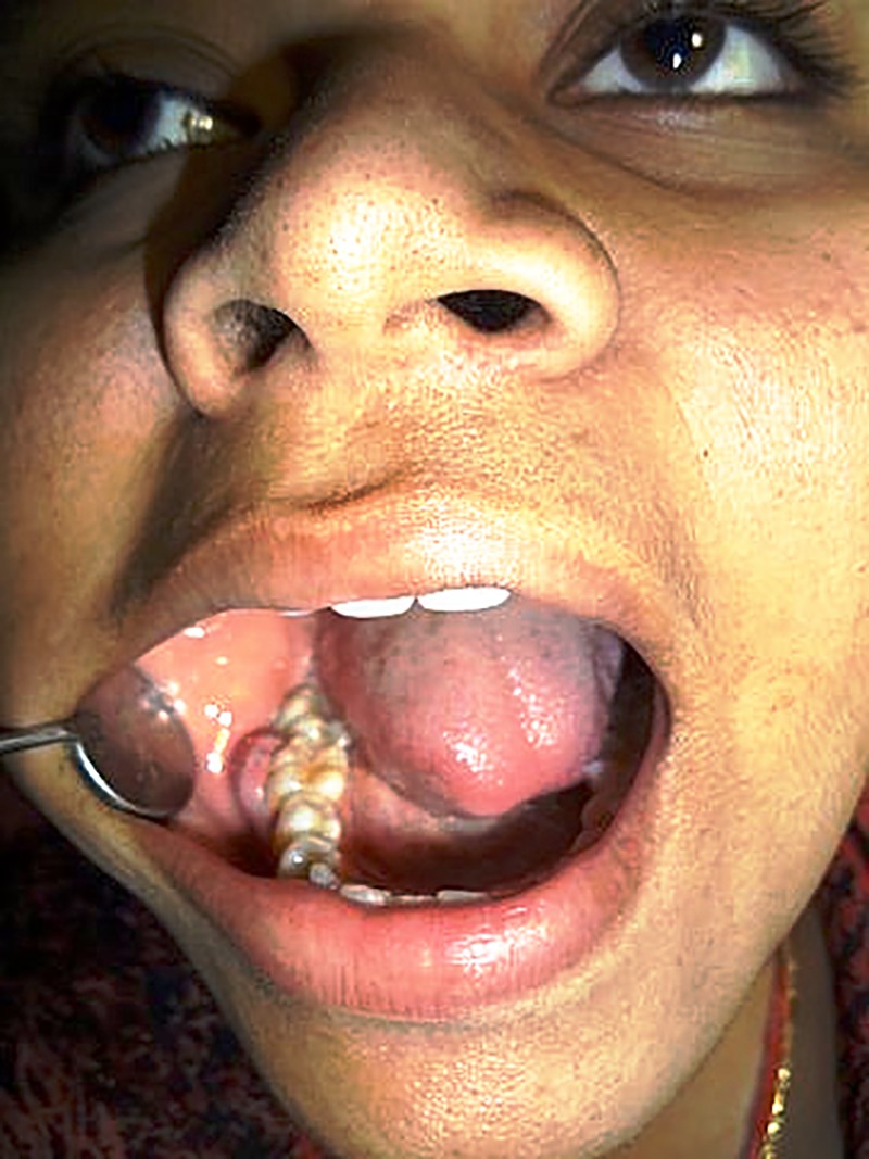
A 25-year-old female patient presented to the dental clinic with a painless gingival swelling in the right mandibular first molar (46) region (figure 1). The swelling had been present for the past 4 months. The medical and surgical histories were unremarkable.
Figure 1.

Preoperative clinical photograph showing gingival swelling in relation to right mandibular first molar.
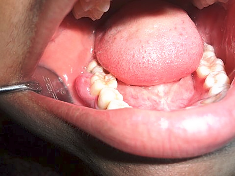
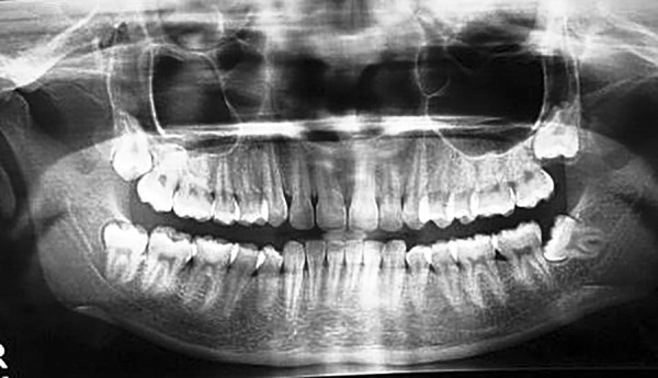
Intraoral examination revealed a reddish, sessile, firm, well-defined mass measuring 1.0 cm at its greatest dimension (figure 2). Panoramic radiographic examination showed no alterations in the underlying bone or in the adjacent teeth (figure 3). The first clinical impression at examination was that of pyogenic granuloma, irrational fibroma. Surgical excision of the lesion under local anaesthesia was planned. After administrating inferior alveolar nerve block on the right side, complete excision of the lesion was performed with a scalpel number 15. Minimal bleeding was encountered, which was controlled with a pressure pack. The surgical wound was sutured with 3-0 vicryl.
Figure 2.

Intraoral examination shows a sessile, firm, well-defined mass in relation to 46 region.
Figure 3.

Panoramic radiographic examination shows no alterations in the underlying bone or in the adjacent teeth.
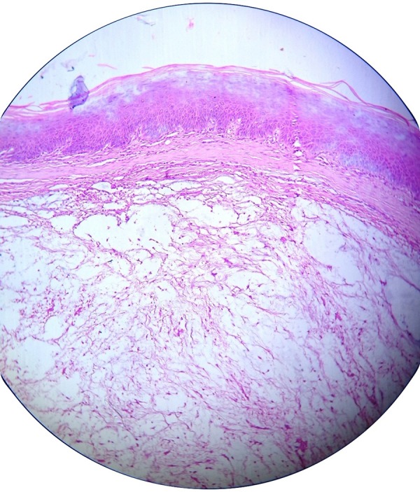
The excised lesion was sent for histopathological examination. The H&E stained microscopic slide revealed parakeratinised stratified surface squamous epithelium in association with a nodule of myxomatous tissue associated with spindle cells (figure 4). Based on the microscopic findings, the final diagnosis was oral focal mucinosis (OFM).
Figure 4.

Photomicrograph showing parakeratinised stratified surface squamous epithelium with myxomatous stroma.
OFM is an uncommon clinicopathological condition that is considered to be the oral counterpart of cutaneous focal mucinosis (CFM).1 OFM has no distinctive clinical features and is most often clinically thought to be fibroma, pyogenic granuloma, mucocele or similar lesions.2
The lesions are difficult to clinically diagnose, as there are no clinically distinctive features. The histological features are always the basis for diagnosis.1 OFM is treated with surgical excision, after which it rarely recurs.2
In this case, the diagnosis of OFM was based on histological examination. After 1 year follow-up, the patient showed no signs of recurrence.
Learning points.
Oral focal mucinosis (OFM) is a rare disease of unknown aetiology, and most commonly occurs in adult women; it has a slight predilection for the keratinised mucosa directly overlying the bone.
OFM has no characteristic features and is most commonly diagnosed clinically as irritational fibroma, pyogenic granuloma, or mucocele. The diagnosis is based on histopathological examination only.
Footnotes
Contributors: SKM and HKM contributed to diagnosis of the patient, acquisition of the data, and conceptualising, drafting and revision of the manuscript. VS contributed to diagnosis of the patient, acquisition of data, and conceptualising and revision of the manuscript. SHK contributed to diagnosis of the patient, and conceptualising, drafting and revision of the manuscript. All the authors gave their approval for the final version of the manuscript.
Competing interests: None declared.
Patient consent: Obtained.
Provenance and peer review: Not commissioned; externally peer reviewed.
References
- 1.Bharti V, Singh J. Oral focal mucinosis of palatal mucosa: a rare case report. Contemp Clin Dent 2012;3:S214–18. doi:10.4103/0976-237X.101098 [DOI] [PMC free article] [PubMed] [Google Scholar]
- 2.Ena S, Nadellamanjari, Chatterjeeanirban, et al. Oral focal mucinosis: a rare case report of two cases. Ethiop J Health Sci 2013;23:178–82. [PMC free article] [PubMed] [Google Scholar]


