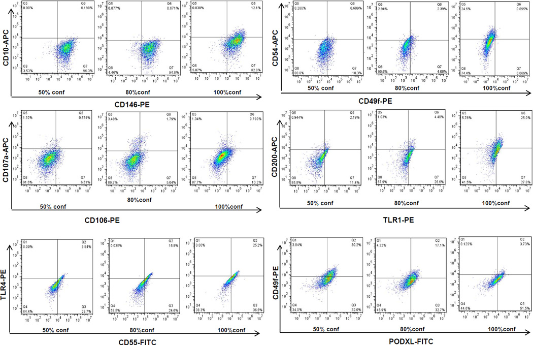
Figure 3. Changes in BMSC surface marker expression at 50%, 80% and 100% confluence.
BMSCs from 2 donors were cultured to 50%, 80% and 100% confluence and the expression of surface markers were measured by flow cytometry. Representative plots from one donor were shown. The markers were indicated by x-axis, and y-axis.

