Abstract
We report a case of blood cyst of the anterior mitral leaflet leading to severe mitral regurgitation and heart failure in a 70-year-old woman with no other factors that could explain the severe mitral regurgitation.
Blood cysts of the cardiac valves are rare. They are relatively common in newborns, but disappear spontaneously during infancy in most cases. Finding a blood cyst on the mitral valve in an older adult prompted this case report.
CASE REPORT
A 70-year-old woman was admitted for progressive effort dyspnea and palpitation of 5 years' duration and orthopnea and paroxysmal dyspnea for 6 months. She had no history of rheumatic fever. She had acute loss of vision in her left eye, diagnosed as retinal artery occlusion 3 weeks prior to the present admission. On examination, she was dyspneic and tachypneic, with a heart rate of 98 beats per minute. Her jugular venous pressure was 8 cm above the sternal angle. Both ankles were edematous, and her blood pressure was 100/80 mm Hg. Her heart was enlarged, and a systolic thrill and left parasternal heave were palpated. The first heart sound was soft, the second heart sound was widely split with an accentuated pulmonary component, and the left ventricular third heart sound was heard over the apex. There was a grade 4/6 pansystolic murmur at the apex, and it radiated to the back. An electrocardiogram showed sinus rhythm with a right axis deviation and left ventricular volume overload pattern. A chest radiograph showed cardiomegaly with pulmonary venous and arterial enlargement.
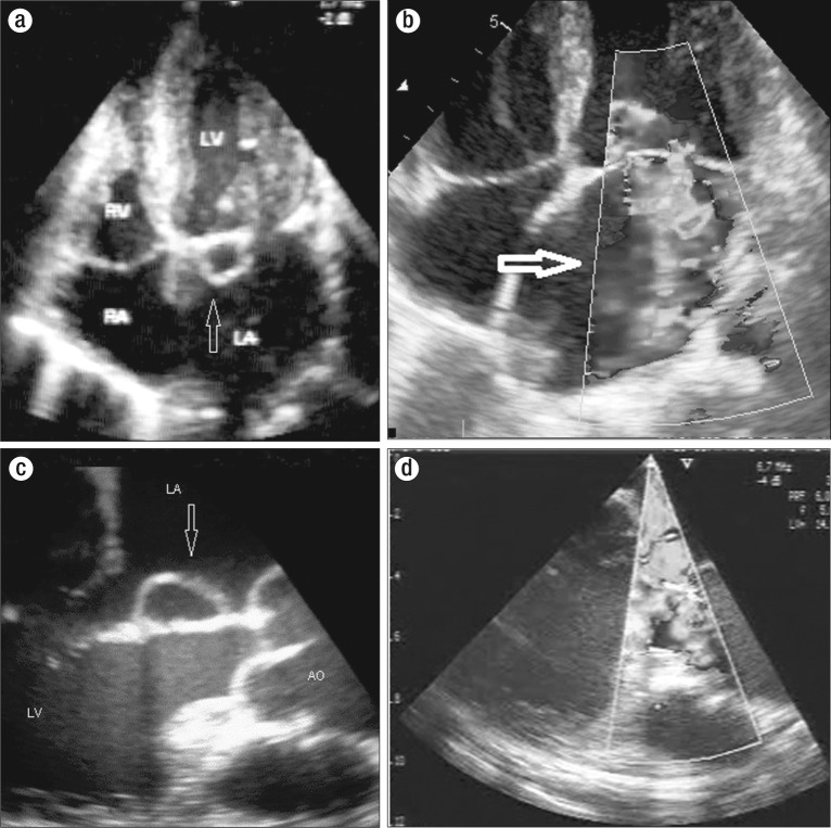
A transthoracic echocardiogram showed a dilated left ventricle and a left atrium with an ejection fraction of 55%. A cystic swelling was attached to the atrial aspect of the anterior mitral leaflet, present during both systole and diastole (Figure 1a). The chordae and papillary muscles were normal. Severe mitral regurgitation was observed (Figure 1b). There was no regional wall motion abnormality or mitral annular calcium. Pulmonary arterial hypertension was estimated to be 64 mm Hg. Transesophageal examination confirmed the cystic swelling in the anterior mitral leaflet (Figure 1c) with severe mitral regurgitation (Figure 1d), with no other morphological changes in the mitral valve apparatus (Video supplement). The coronary arteries were normal. The patient was treated with diuretics, digoxin, and vasodilators. Intraoperatively, surgeons noted a 16 mm round, bluish cystic swelling with a broad base attached to the atrial aspect of the anterior mitral leaflet. The patient subsequently underwent successful mitral valve replacement with a TTK Chitra tilting disc prosthesis. Histopathology revealed blood-filled space lined with a single layer of endothelium consistent with a blood cyst. At 1-year follow up, the patient was clinically stable.
Figure 1.
Apical four-chamber echocardiography showing (a) a blood cyst of the anterior mitral leaflet and (b) severe mitral regurgitation. Transesophageal echocardiography showing (c) a blood cyst of the anterior mitral leaflet and (d) severe mitral regurgitation.
DISCUSSION
Intracardiac blood cysts are rare. Blood cysts are lined by flattened endothelial cells and filled with nonorganized blood (1). Generally blood cysts are small and round, although giant cysts have been reported. Their etiology is thought to be congenital or acquired. There are various theories regarding the origin of intracardiac blood cysts. Boyd's theory states that during valvular development, blood is pressed into the crevices on the valvular surfaces of the cusps, which later gets sealed off (2). Kantelip's theory proposes dilatation of the normal invagination of the valve cusp as the mechanism of genesis of the blood cyst (3). Blood cysts may be derived from ectatic blood vessels or angiomas. Inflammation, vagal stimulation, anoxia, and hemorrhagic diathesis could lead to sudden occlusion of small vascular channels and lead to subsequent hematoma formation in the subvalvular region.
Congenital blood cysts of the heart valves are mostly seen on tricuspid and mitral valves of fetuses and infants. Autopsy reports of fetuses and infants have shown a higher occurrence of cardiac blood cysts (4), indicating that severe hypoxia or inflammation may be an etiology for the appearance of this entity. Blood cysts are often found in neonates dying of various causes and probably have no clinical significance. Blood cysts may persist and enlarge to form giant cysts of heart valves.
Blood cysts are usually asymptomatic in adults and have often been discovered incidentally during routine echocardiographic evaluation. They can cause inflow and outflow obstruction and valvular regurgitation due to incomplete coaptation (5). Mitral regurgitation can also occur due to the mitral paraannular location of the cyst (6). Cysts may be a potential source of cerebrovascular embolism (7). There is not necessarily a correlation between the size of the cyst and hemodynamic consequences; even giant blood cysts can be asymptomatic and may be an incidental finding during echocardiography (8).
There is no consensus regarding the management of blood cysts. Pelikan et al suggested that asymptomatic cysts, because of their benign character, can be monitored with echocardiography, and resection should be reserved for cysts that interfere with normal cardiac function (9). Paşaoğlu et al advised surgical excision of all cystic tumors of the heart, especially in a valvular location, because the precise diagnosis can be made only by intraoperative examination (10).
Video supplement
Transesophageal echocardiography at 130° showing the cyst in the atrial aspect of the anterior mitral valve leaflet (see www.BaylorHealth.edu/Proceedings/Documents/BUMC%20Proceedings/2015%20Vol%2028/No_3/28_3_Jayaprakash.mp4).
References
- 1.Burke A, Virmani R. Tumors of the Heart and Great Vessels (Atlas of Tumor Pathology, Third Series, Vol. 15) Washington, DC: American Registry of Pathology; 1996. pp. 171–177. [Google Scholar]
- 2.Boyd TA. Blood cysts on the heart valves of infants. Am J Pathol. 1949;25(4):757–759. [PMC free article] [PubMed] [Google Scholar]
- 3.Kantelip B, Satge D, Camilleri L, Chenard MP, De Riberolles C. Valvular cyst and atrioventricular canal in a child with trisomy 21. Ann Pathol. 1994;14(2):101–107. [PubMed] [Google Scholar]
- 4.Zimmerman KG, Paplanus SH, Dong S, Nagle RB. Congenital blood cysts of the heart valves. Hum Pathol. 1983;14(8):699–703. doi: 10.1016/s0046-8177(83)80142-7. [DOI] [PubMed] [Google Scholar]
- 5.Xie SW, Lu OL, Picard MH. Blood cyst of the mitral valve: detection by transthoracic and transesophageal echocardiography. J Am Soc Echocardiogr. 1992;5(5):547–550. doi: 10.1016/s0894-7317(14)80047-3. [DOI] [PubMed] [Google Scholar]
- 6.Sekine S, Abe T, Kuribayashi R, Aida H, Seki K, Shibata Y. Mitral regurgitation caused by mitral paraannular cyst. J Heart Valve Dis. 1997;6(1):67–68. [PubMed] [Google Scholar]
- 7.Kuvin J, Saha P, Rastegar H, Salomon RN, Pandian N, Denofrio D. Blood cyst of the mitral valve apparatus in a woman with a history of orthotopic liver transplantation. J Am Soc Echocardiogr. 2004;17(5):480–482. doi: 10.1016/j.echo.2004.01.013. [DOI] [PubMed] [Google Scholar]
- 8.Abreu A, Galrinho A, Sá EP, Ramos S, Martins AP, Fragata J, Ferreira R. Hamartoma of the mitral valve with blood cysts: a rare tumor detected by echocardiography. J Am Soc Echocardiogr. 1998;11(8):832–836. doi: 10.1016/s0894-7317(98)70060-4. [DOI] [PubMed] [Google Scholar]
- 9.Pelikan HM, Tsang TS, Seward JB. Giant blood cyst of the mitral valve. J Am Soc Echocardiogr. 1999;12(11):1005–1007. doi: 10.1016/s0894-7317(99)70157-4. [DOI] [PubMed] [Google Scholar]
- 10.Paşaoğlu I, Doğan R, Nazli N, Güngen Y, Bozer AY. Blood cyst originating from tricuspid septal leaflet. J Cardiovasc Surg (Torino) 1991;32(5):589–591. [PubMed] [Google Scholar]


