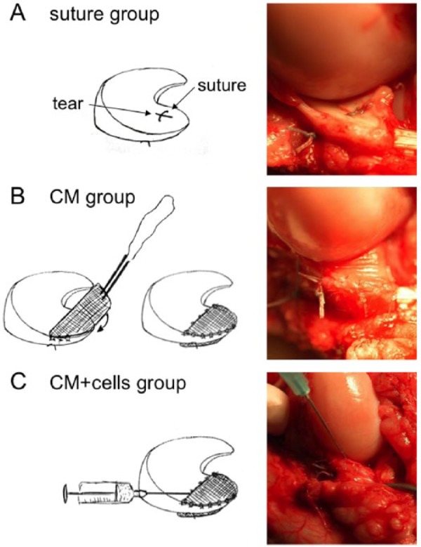
Figure 1.

Treatments of meniscal tears. In the control group (A), a single horizontal suture was applied to hold tear margins in tight contact. In the CM group (B), upon suturing, the collagen membrane was wrapped around the meniscus and secured on both surfaces. In the CM+cells group (C), upon suturing the tear and securing the collagen membrane, expanded autologous chondrocytes were injected into the tear and under the collagen membrane.
