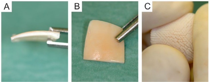
Figure 3.

Macroscopic appearance of the multiply incised pure cartilage allograft. (A) A side view of the graft showing the cartilage graft without subchondral bone (1.5-2 mm in thickness). (B) The superficial (articular) side of the graft; please note that this surface of the graft remains intact providing the graft with good tensile strength. (C) The “hedgehog” appearance of the deep zone of the graft after folding it to a concave shape. The picture demonstrates how the incisions of the deep zone render the sample to an easily pliable implant with increased basal surface.
