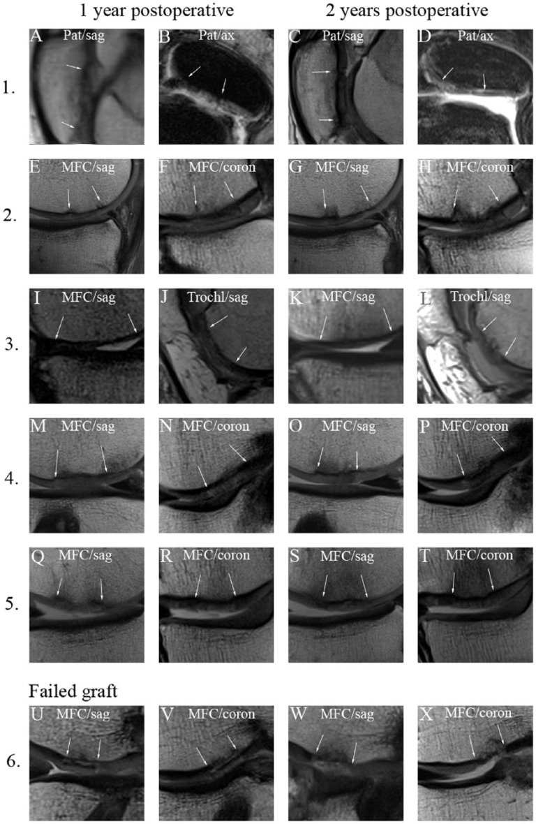
Figure 6.
Postoperative proton density–weighted images of the cartilage lesions repaired with multiply incised pure cartilage allograft. The repair sites are shown 1 year (1-2. columns) and 2 years (3-4. columns) postoperatively. The white arrows mark the edge of the graft. Numbers for each row correspond to the case numbers in Table 1. Case 3 shows 2 distinct location of the same knee joint. While the first 5 cases show acceptable integration case 6 represents a failed graft. Pat = patella; MFC = medial femoral condyle; Trochl = trochlea; sag = sagittal plane; ax = axial plane; coron = coronal plane.

