Abstract
Objective
Report initial experience with the Frozen Elephant Trunk technique.
Methods
From July 2009 to October 2013, Frozen Elephant Trunk technique was performed in 21 patients (66% male, mean age 56 ±11 years). They had type A aortic dissection (acute 9.6%, chronic 57.3%), type B (14.3%, all chronic) and complex aneurysms (19%). It was 9.5% of reoperations and 38% of associated procedures (25.3% miocardial revascularization, 25.3% replacement of aortic valve and 49.4% aortic valved graft). Aortic remodeling was evaluated comparing preoperative and most recent computed tomography scans. One hundred per cent of complete follow-up, mean time of 28 months.
Results
In-hospital mortality of 14.2%, being 50% in acute type A aortic dissection, 8.3% in chronic type A aortic dissection, 33.3% in chronic type B aortic dissection and 0% in complex aneurysms. Mean times of cardiopulmonary bypass (152±24min), myocardial ischemia (115±31min) and selective cerebral perfusion (60±15min). Main complications were bleeding (14.2%), spinal cord injury (9.5%), stroke (4.7%), prolonged mechanical ventilation (4.7%) and acute renal failure (4.7%). The need for second-stage operation was 19%. False-lumen thrombosis was obtained in 80%.
Conclusion
Frozen Elephant Trunk is a feasible technique and should be considered. The severity of the underlying disease justifies high mortality rates. The learning curve is a reality. This approach allows treatment of more than two segments at once. Nonetheless, if a second stage is made necessary, it is facilitated.
Keywords: Aortic Diseases; Aorta, Thoracic; Cardiovascular Surgical Procedures; Aortic Aneurysm, Thoracic; Aneurysm, Dissecting; Endovascular Procedures
Abstract
Objetivo
Relatar experiência inicial com a técnica "Frozen Elephant Trunk".
Métodos
Entre julho de 2009 e outubro de 2013, 21 pacientes, 66% homens, média de idade de 56±11 anos, 66,7% portadores de dissecção da aorta tipo A de Stanford (9,6% agudas e 57,1% crônicas), tipo B (14,3%, todas crônicas) e aneurismas complexos (19%), foram operados pela técnica Frozen Elephant Trunk. Foram 9,5% de reoperações e 38% com procedimentos associados (25,3% revascularizações do miocárdio, 25,3% troca da valva aórtica e 49,4% tubos valvulados). Remodelamento da aorta foi avaliado com a comparação de angiotomografia pré-operatória e pós-operatória mais recente. Seguimento 100% dos pacientes, tempo médio de 28 meses.
Resultados
Mortalidade hospitalar de 14,2%, sendo 50% nas dissecções do tipo A agudas, 8,3% nas tipo A crônicas, 33,3% nas tipo B crônicas e 0% nos aneurismas complexos. Tempos médios de CEC (152±24min), isquemia miocárdica (115±31min) e perfusão cerebral seletiva (60±15min). Principais complicações pós-operatórias foram sangramento (14,2%), acidente vascular encefálico (4,7%), paraplegia (9,5%), intubação>72h (4,7%) e insuficiência renal aguda (4,7%). Houve necessidade de complementação do tratamento (distal ao stent) em 19%. Houve trombose da falsa luz em 80%.
Conclusão
Frozen Elephant Trunk é opção técnica a ser utilizada. A gravidade e extensão da doença justificam mortalidade mais elevada. A curva de aprendizado é uma realidade. Esta abordagem permite abordar mais de dois segmentos de aorta em um estágio, mas se necessário segundo estágio, este é facilitado.
| Abbreviations, acronyms & symbols | |
|---|---|
| CPB | Cardiopulmonary bypass |
| CT | Computed tomography |
| FET | Frozen elephant trunk |
INTRODUCTION
The treatment of complex diseases of the aorta that involve the aortic arch using a hybrid procedure aims at reconstructing several segments within a single intervention. The "Frozen Elephant Trunk" (FET) surgical technique has been used for the treatment of complex aneurysms and chronic dissections of the thoracic aorta since 1996[1]. The prostheses used in these procedures consist of a proximal segment made of polyester (used to reconstruct the ascending aorta and aortic arch segment) distally attached to a self-expanding endovascular prosthesis (used to treat the proximal segment of the descending thoracic aorta). The main purpose of this technique is to potentially avoid a second procedure required after the classic elephant trunk technique[2]. Over time, indications for the use of the FET have extended to patients with acute aortic dissections. This surgical approach was established to expand aortic treatment as well as to favor thrombosis of the false lumen or the aneurysm, since the false lumen that remains patent (and, therefore, pressurized) is a risk factor for dilation and rupture of the aorta and subsequent need for a new surgical treatment[3-5].
The aim of this study is to report an initial experience with the FET technique to treat complex diseases of the thoracic aorta with involvement of the aortic arch.
METHODS
Between July 2009 and October 2013, 21 patients underwent surgery with the FET technique, all with the E-vita Open® prosthesis (Jotec GmbH, Hechingen, Germany). Consequently, all patients underwent antegrade implantation of the endovascular prosthesis through the aortic arch.
The choice of prosthesis was due to it being the only one available in the Brazilian market. A series of previously published cases using this prosthesis have shown satisfactory perioperative and mid-term results in terms of the extent of the aneurysm, false lumen thrombosis, and aortic remodeling[6,7].
The patients who were treated had complex diseases of the thoracic aorta involving the aortic arch. A complex disease of the aorta was defined as simultaneous involvement of three or more aortic segments.
Operations performed in the distal aorta were defined as new surgical approaches involving treatment of any segment distal to the stent in order to supplement the primary treatment or because of the evolution of the disease.
Cardiopulmonary bypass was maintained with arterial cannulation of the right subclavian artery in 5 patients and innominate # 16 patients. Venous drainage was performed with double-stage cannula through the right atrium.
Brain protection was performed by antegrade cerebral perfusion by both carotid arteries, with monitoring of the right upper limb perfusion pressure (invasive blood pressure in the right radial artery), associated with moderate hypothermia (25ºC), hypothermia with ice and sodium thiopental (spinal cord protection exclusively by 25ºC hypothermia associated with systemic circulatory arrest).
The reconstruction of the supra-aortic vessels was made from reimplantation in island anastomosed to the polyester tube for all cases.
All operations were performed in conventional operating room, without use of scopes or guidewire. The introduction of the prosthesis was performed through direct identification of the true light for patients with aortic dissection.
The FET technique was used in type A acute or chronic dissections with inlet port located in the distal aortic arch or descending thoracic aorta, and patients with these aorta segments greater than than four centimeters.
During the study period, all patients with involvement of the ascending aorta, aortic arch and proximal descending aorta underwent this technique.
There were 21 patients, 66% male, with a mean age of 56±11 years (ranging from 27 to 70 years-old).
Out of the 21 patients, 67% had Stanford type A aortic dissections (10% acute and 57% chronic), 14% had Stanford type B chronic aortic dissections, and 19% had complex aneurysms.
In 9.5% of the patients, there was a reoperation (all had previous surgery in the aortic root and ascending aorta), and 38% required associated procedures (50% underwent aortic root reconstruction with composite-graft valve replacement, 25% underwent aortic valve replacement, and 25% underwent myocardial revascularization).
In addition, 28% were urgency or emergency surgeries, 10% of which were Stanford type A acute dissections.
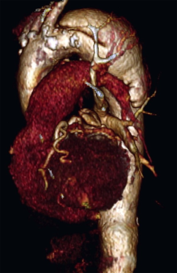
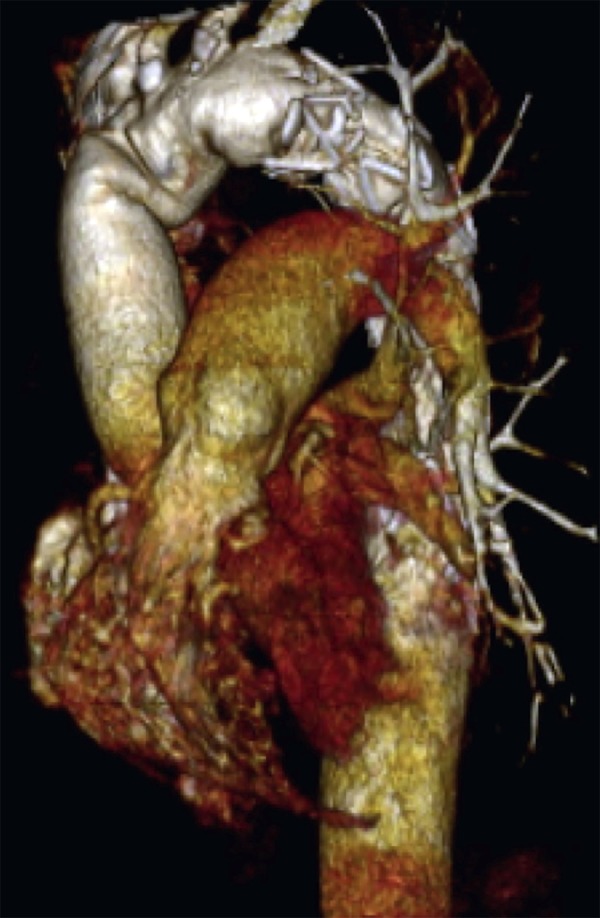
Radiological assessment was carried out taking into consideration preoperative angiotomographies of the aorta and comparing it to the last postoperative exam (Figures 1A and 1B).
Fig. 1A.

Tridimensional reconstruction of angiotomography of the aorta: preoperative.
Fig. 1B.

Tridimensional reconstruction of angiotomography of the aorta: postoperative.
Patient data were retrospectively analyzed from a database prospectively built. Full clinical follow-up was done up to November 30, 2013. Follow-up was performed at the institution's outpatient facility or via telephone.
This study was approved by the Research Ethics Committee of the institution (No. 837) for specific database creation of the aforementioned patients and the consent form is not required by the retrospective feature of the research.
RESULTS
Hospital mortality was 14.2%. According to etiology, mortality rates for Stanford type A acute dissections, Stanford type A chronic dissections, and Stanford type B chronic dissections were 50%, 8.3%, and 33.3%, respectively. There was no mortality in patients with complex thoracic aortic aneurysms.
Mean CPB time was 152 min±24 min, mean myocardial ischemia time was 115 min±31 min, and mean time of selective cerebral perfusion at 25ºC was 60 min±15 min.
The main complications observed were reoperation due to bleeding in three patients, stroke in one patient, paraplegia in two patients, prolonged intubation (longer than 72 hours) in one patient, and acute renal failure requiring dialysis in one patient.
Four patients (19%) required a second surgical time for intervention in the descending thoracic aorta. Most of them (three patients) underwent implantation of vascular endoprosthesis in the segment distal to the one first treated during the same hospital stay whereas one patient, while recovering from the first stage and awaiting thoracoabdominal reconstruction, was admitted to another facility four months after hospital discharge and underwent open surgery, leading to death.
Angiotomographies for postoperative control of patients diagnosed with dissection showed false lumen thrombosis in 80% of the aortic segments with vascular endoprosthesis and in 60% of the aortic segments without endoprosthesis. All of the patients with aneurysm had thrombosis around the stent.
DISCUSSION
The use of a hybrid approach to complex diseases of the thoracic aorta allows for the treatment of long segments of the aorta in a single stage.
Some authors have questioned the possibility of treating complex aortic diseases in a single stage through median sternotomy using the FET technique[12], giving preference to other approaches, such as bithoracotomy[13,14]. However, the FET technique has shown fewer pulmonary complications as well as similar survival rates in 5-year follow-ups[11,14].
Shimamura et al.[8] and Uchida et al.[9] demonstrated survival free from reintervention in the distal segment of the aorta of 75% and 92%, respectively, in up to 10 years of follow-up, and with mortality ranging from 46% to 25%.
The first mid-term results obtained with the use of the E-vita Open® prosthesis showed 5-year survival rates of 79%, where 96% of the patients were free from thoracoabdominal aorta open reintervention and 84% were free from additional endovascular treatments[10].
Our experience has shown that, in less than a 3-year follow-up period, there was 20% of reinterventions, all of them as a consequence of the inability to fully approach the disease of the aorta through the FET procedure. None of the reinterventions was due to progression of the initial disease or to the appearance of a new disease in the distal segment of the aorta.
The percentage of reoperations, the high number of associated procedures required as well as procedures characterized as urgency or emergency attest the high surgical complexity and high surgical risk of patients included in this study.
This study states the initial experience of the Heart Institute of the University of São Paulo Medical School with this operation. Follow-up time is still short (average of 28 months, ranging from 1 to 54 months), with hospital mortality compatible with that reported in the international literature[11].
In the last 10 years, the use of the FET technique has spread to include the treatment of acute aortic dissections and mega-aorta syndrome. As a result, the need for second-stage surgery is real, since the approach of the aorta has to be extended distally to the segment associated with the stent. Thus, the FET stent facilitates the second stage of surgery while complementing the treatment, in cases of open thorax or endovascular operations, and providing a better region for new endoprosthesis anchoring, especially when compared to the classic elephant trunk technique or with the diseased native aorta initially untreated[11,15,16].
Likewise, in patients with chronic aortic dissection, a second stage is likely to be required since the possibility of excluding false lumen is lower. In this disease, the delaminated membrane is thicker than in acute dissections and true lumen is lower, which increases the risk of leakage and progression of aneurysmatic dilation. Consequently, a large number of publications have shown high percentage of false lumen thrombosis around the endoprosthesis, but inferior outcomes in the distal segments of the aorta without stent and no improvement throughout follow-up[9,17,10]. Therefore, we believe that the need for second stage surgery is not related to the failure of the prosthesis used in the FET, but rather to the characteristics of the primary disease.
Some authors have shown that the prevalence of aortic reinterventions distally to the original surgery is higher in patients with chronic aortic dissection when compared to patients with acute dissection[11]. Conversely, in acute dissections, the FET technique prevented late dilation of the proximal segment of the descending aorta, stabilized the dissected membrane, and promoted true lumen expansion, even in segments distal to the stent[11]. As a result, knowing that in these patients the distal portion of the dissected aorta tends to dilate over time when only a conduit interposition in the ascending aorta is performed, especially the proximal segment of the descending aorta[3-5], the FET technique has become the technique of choice for the treatment of Stanford type A acute dissections in some centers[11]. This technique has been used even in patients with connective tissue diseases[18].
Lastly, when compared to other series of cases[19,20], the prevalence of spinal injury in this study was equal or lower to what has been reported in the international literature and it was present only in the first cases, in particular, when systemic circulatory arrest time was longer due to initial difficulties to perform the procedure.
Limitations of the study
Main limitations of the study are the reduced number of patients and short, though complete, follow-up period.
CONCLUSION
The FET technique has been shown to be an option in the surgical treatment of complex diseases of the thoracic aorta. High mortality is warranted in view of the severity of primary diseases and the complexity of the patients.
The learning curve is a reality in these operations.
This technique can treat complex diseases of the thoracic aorta in a single stage. When required, FET simplifies reintervention in the distal segments, providing a more appropriate region both for surgical manipulation and for new endoprosthesis anchoring.
| Authors' roles & responsibilities | |
|---|---|
| RRD | Analysis and/or interpretation of data, statistical analysis, final approval of the manuscript, conception and study design study, carried out procedures and/or experiments, writing of the manuscript or review of its content |
| JAD | Analysis and/or interpretation of data, carried out procedures and/or experiments, writing of the manuscript or review of its content |
| DSV | Analysis and/or interpretation of data, carried out procedures and/or experiments, writing of the manuscript or review of its content |
| LBF | Analysis and/or interpretation of data, statistical analysis, carried out procedures and/or experiments |
| FF | Conception and study design |
| FJAR | Conception and study design |
| CM | Conception and study design |
| FBJ | Conception and study design |
Footnotes
This study was carried out at the Heart Institute of the University of São Paulo Medical School, São Paulo, SP, Brazil.
No financial support.
REFERENCES
- 1.Kato M, Ohnishi K, Kaneko M, Ueda T, Kishi D, Mizushima T, et al. New graft-implanting method for thoracic aortic aneurysm or dissection with a stented graft. Circulation. 1996;94(9) Suppl I:II188–II193. [PubMed] [Google Scholar]
- 2.Borst HG, Walterbusch G, Schaps D. Extensive aortic replacement using 'elephant trunk' prosthesis. Thorac Cardiovasc Surg. 1983;31(1):37–40. doi: 10.1055/s-2007-1020290. [DOI] [PubMed] [Google Scholar]
- 3.Halstead JC, Meier M, Etz C, Spielvogel D, Bodian C, Wurm M, et al. The fate of the distal aorta after repair of acute type A aortic dissection. J Thorac Cardiovasc Surg. 2007;133(1):127–135. doi: 10.1016/j.jtcvs.2006.07.043. [DOI] [PubMed] [Google Scholar]
- 4.Song SW, Chang BC, Cho BK, Yi G, Youn YN, Lee S, et al. Effects of partial thrombosis on distal aorta after repair of acute DeBakey type I aortic dissection. J Thorac Cardiovasc Surg. 2010;139(4):841–847. doi: 10.1016/j.jtcvs.2009.12.007. [DOI] [PubMed] [Google Scholar]
- 5.Kim JB, Lee CH, Lee TY, Jung SH, Choo SJ, Lee JW, et al. Descending aortic aneurysmal changes following surgery for acute DeBakey type I aortic dissection. Eur J Cardiothorac Surg. 2012;42(5):851–856. doi: 10.1093/ejcts/ezs157. [DOI] [PubMed] [Google Scholar]
- 6.Di Bartolomeo R, Di Marco L, Armaro A, Marsilli D, Leone A, Pilato E, et al. Treatment of complex disease of the thoracic aorta: the frozen elephant trunk technique with the E-vita open prosthesis. Eur J Cardiothorac Surg. 2009;35(4):671–675. doi: 10.1016/j.ejcts.2008.12.010. [DOI] [PubMed] [Google Scholar]
- 7.Gorlitzer M, Weiss G, Moidl R, Folkmann S, Waldenberger F, Czerny M, et al. Repair of stent graft-induced retrograde type A aortic dissection using the E-vita open prosthesis. Eur J Cardiothorac Surg. 2012;42(3):566–570. doi: 10.1093/ejcts/ezs041. [DOI] [PubMed] [Google Scholar]
- 8.Shimamura K, Kuratani T, Matsumiya G, Kato M, Shirakawa Y, Takano H, et al. Long-term results of the open stent-grafting technique for extended aortic arch disease. J Thorac Cardiovasc Surg. 2008;135(6):1261–1269. doi: 10.1016/j.jtcvs.2007.10.056. [DOI] [PubMed] [Google Scholar]
- 9.Uchida N, Katayama A, Tamura K, Sutoh M, Kuraoka M, Murao N, et al. Long-term results of the frozen elephant trunk technique for extended aortic arch disease. Eur J Cardiothorac Surg. 2010;37(6):1338–1345. doi: 10.1016/j.ejcts.2010.01.007. [DOI] [PubMed] [Google Scholar]
- 10.Jakob H, Dohle DS, Piotrowski J, Benedik J, Thielmann M, Marggraf G, et al. Six-year experience with a hybrid stent graft prosthesis for extensive thoracic aortic disease: an interim balance. Eur J Cardiothorac Surg. 2012;42(6):1018–1025. doi: 10.1093/ejcts/ezs201. [DOI] [PubMed] [Google Scholar]
- 11.Ius F, Fleissner F, Pichlmaier M, Karck M, Martens A, Haverich A, et al. Total aortic arch replacement with the frozen elephant trunk technique: 10-year follow-up single centre experience. Eur J Cardiothorac Surg. 2013;44(5):949–957. doi: 10.1093/ejcts/ezt229. [DOI] [PubMed] [Google Scholar]
- 12.Kouchoukos NT. Frozen elephant trunk technique for extensive chronic thoracic aortic dissection: is the final answer? Ann Thorac Surg. 2011;92(5):1557–1558. doi: 10.1016/j.athoracsur.2011.08.005. [DOI] [PubMed] [Google Scholar]
- 13.Beaver TM, Martin TD. Single-stage transmediastinal replacement of the ascending, arch, and descending thoracic aorta. Ann Thorac Surg. 2001;72(4):1232–1238. doi: 10.1016/s0003-4975(01)02998-8. [DOI] [PubMed] [Google Scholar]
- 14.Kouchoukos NT, Masetti P, Mauney MC, Murphy MC, Castner CF. One-stage repair of extensive chronic aortic dissection using the arch-first technique and bilateral anterior thoracotomy. Ann Thorac Surg. 2008;86(5):1502–1509. doi: 10.1016/j.athoracsur.2008.07.059. [DOI] [PubMed] [Google Scholar]
- 15.Pichlmaier MA, Teebken OE, Khaladj N, Weidemann J, Galanski M, Haverich A. Distal aortic surgery following arch replacement with a frozen elephant trunk. Eur J Cardiothorac Surg. 2008;34(3):600–604. doi: 10.1016/j.ejcts.2008.05.038. [DOI] [PubMed] [Google Scholar]
- 16.Uchida N, Kodama H, Katayama K, Takasaki T, Katayama A, Takahashi S, et al. Endovascular aortic repair as second-stage surgery after hybrid open arch repair by the frozen elephant trunk technique for extended thoracic aneurysm. Ann Thorac Cardiovasc Surg. 2013;19(3):257–261. doi: 10.5761/atcs.nm.12.01918. [DOI] [PubMed] [Google Scholar]
- 17.Sun LZ, Qi RD, Chang Q, Zhu JM, Liu YM, Yu CT, et al. Surgery for acute type A dissection using total arch replacement combined with stented elephant trunk implantation: experience with 107 patients. J Thorac Cardiovasc Surg. 2009;138(6):1358–1362. doi: 10.1016/j.jtcvs.2009.04.017. [DOI] [PubMed] [Google Scholar]
- 18.Sun L, Li M, Zhu J, Chang Q, Zheng J, Qi R. Surgery for patients with Marfan syndrome with type A dissection involving the aortic arch using total arch replacement combined with stented elephant trunk implantation: the acute versus the chronic. J Thorac Cardiovasc Surg. 2011;142(3):e85–e91. doi: 10.1016/j.jtcvs.2011.01.038. [DOI] [PubMed] [Google Scholar]
- 19.Flores J, Kunihara T, Shiiya N, Yoshimoto K, Matsuzaki K, Yasuda K. Extensive deployment of the stented elephant trunk is associated with an increased risk of spinal cord injury. J Thorac Cardiovasc Surg. 2006;131(2):336–342. doi: 10.1016/j.jtcvs.2005.09.050. [DOI] [PubMed] [Google Scholar]
- 20.Pacini D, Tsagakis K, Jakob H, Mestres CA, Armaro A, Weiss G, et al. The frozen elephant trunk for the treatment of chronic dissection of the thoracic aorta: a multicenter experience. Ann Thorac Surg. 2011;92(5):1663–1670. doi: 10.1016/j.athoracsur.2011.06.027. [DOI] [PubMed] [Google Scholar]


