Abstract
A detailed protocol for the growth and harvest of purified elementary bodies of Chlamydia pneumoniae is presented. This procedure utilizes a flask-to-flask passage scheme designed to achieve high bacterial titers in a short period of time.
Typically, Chlamydia pneumoniae elementary bodies (EBs) are propagated and purified through a stepwise procedure using small-volume vials in a process known as slow expansion (2, 3). The method of slow expansion, so elegantly described by Campbell et al. and Kuo (2, 3), employs viral shell vials containing an acceptable host cell monolayer as a means to begin the propagation step and starts with a low multiplicity of infection (MOI) (i.e., 1:1). This is followed by expansion into an ever-increasing number of shell vials, culminating in passage into a flask for the final growth and purification step. Several authors have described the successful growth and harvest of EBs of Chlamydia spp. from infected flasks (5, 6); however, no attempt has been made to use this method for the production of high-titer stock cultures. We explored the possibility of the growth, harvest, and purification of C. pneumoniae EBs by flask-to-flask passage in 75-cm2 flasks containing HEp-2 cell monolayers by beginning with a high MOI (i.e., 100:1). Emphasis was placed on reaching high titers of purified EBs in the shortest possible time. Using several alterations of existing procedures for chlamydial propagation (1, 4), we successfully produced purified stock EB preparations averaging 1.0 × 10⁹ inclusion forming units (IFU)/ml in an 8-day period.
Due to the inherent difficulty in propagation of C. pneumoniae and the increasing number of investigators wishing to study this organism, we provide here a detailed protocol which outlines specific steps in this process that should be adaptable to any laboratory setting.
Seeding of HEp2 cells.
Seed HEp-2 cells (ATCC CCL-23; American Type Culture Collection, Rockville, Md.) in 15 ml of Iscove's maintenance medium (IMM; Iscove's 1× medium, 10% [vol/vol] fetal calf serum, 2.0 mM l-glutamine, 1.0% [vol/vol] nonessential amino acids, 10 mM HEPES buffer, 10.0 μg of gentamicin/ml, 25.0 μg of vancomycin/ml, and 12.5 U of nystatin/ml [optional use]) into one 75-cm2 flask at a cell density of 5 × 10⁵ cells/ml, and seed 2 × 10⁵ cells in 1.0 ml of IMM into one 1.0-dram shell vial containing one sterile round coverslip. Incubate the flask and the control shell vial at 37°C in an atmosphere of 5% CO2 for 24 h to achieve confluence of the monolayer, with a resulting cell count in the flask of approximately 1.0 × 10⁶ cells/ml (total cell count = 1.5 × 10⁷).
Inoculation.
Inoculate both monolayers (flask and shell vial) with EBs from a frozen stock of 1.0 × 10⁹-IFU/ml C. pneumoniae suspension obtained by the slow-expansion procedure. Similar EB stock suspensions can be purchased from the Washington Research Foundation, Seattle, Wash. Inoculation should be carried out in Iscove's growth medium (IGM; IMM with 8.8% [vol/vol] glucose and 1.0 μg of cycloheximide/ml) at an approximate MOI of 100:1, and the flask should be agitated slowly on a rotary or reciprocating shaker for 1 h at room temperature (25°C). Centrifuge the flask and the shell vial at 3,500 × g (1,800 rpm on a Sorvall RT 6000D) for 30 min at 10°C and incubate at 37°C in 5% CO2.
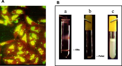
At 48 h postinfection (hpi), fix the shell vial in absolute methanol for 10 min and stain with a fluorescein isothiocyanate (FITC)-labeled chlamydial specific antibody (Pathfinder; Bio-Rad Laboratories, Hercules, Calif.) for 30 min. Wash with distilled water and examine by epifluorescence microscopy to verify an adequate level of infection. In general, >95% of the HEp-2 monolayer should contain detectable C. pneumoniae inclusions (Fig. 1A).
FIG. 1.
(A) Epifluorescence micrograph of HEp-2 cell monolayer infected with C. pneumoniae and stained with FITC-labeled anti-Chlamydia antibody (Pathfinder; Bio-Rad Laboratories) at 48 hpi. Greater than 95% of the monolayer contains C. pneumoniae inclusions, a level that is considered sufficient for continuation of the propagation and purification process (see detailed protocol in text). Magnification, ×340. (B) Renografin gradient containing the C. pneumoniae EB layer at the 20%-50% interphase (a); pellet of C. pneumoniae EB following a washing step (b); final suspension of C. pneumoniae EBs following trituration (c).
Passage of chlamydia.
At 72 hpi, remove the medium from the flask by aspiration, replace it with 5.0 ml of fresh IGM, and gently disrupt the monolayer with a cell scraper. Collect all cells and rinse the flask with 5.0 ml of IGM. Pool both samples into a 50.0-ml sterile conical tube and sonicate three to four times for 15 s each with a sonicator (Fisher Scientific model 100 cell dismembrator) to disrupt the HEp-2 cells.
Inoculate each of two 75-cm2 flasks (with a confluent HEp-2 monolayer prepared as described in “Seeding of HEp2 cells” above) with 5 ml of inoculum obtained as described above and add 10.0 ml of IGM. Inoculate a control shell vial, as described above, with 200 μl of the material obtained from the passage. Rotate and centrifuge the flask as described above and incubate the flask and shell vial at 37°C in 5% CO2.
Harvest of EBs.
At 24 hpi, aspirate the medium and replace it with 15.0 ml of fresh IGM in each flask. At 48 hpi, fix and stain the shell vial as described above to confirm a significant level of infection (>95%). At 72 hpi, begin the EB harvesting process by removing all medium from each flask and replacing it with 2.5 ml of harvest cocktail, which has been chilled on ice and filtered through a 0.22-μm syringe filter, and gently free the monolayer with a cell scraper. The harvest cocktail contains 7.0 mg of DNase (for degrading host cell DNA contaminants), 5.0 mg of heparin (to inhibit attachment of EBs to host cell debris), and one tablet of Complete Mini protease inhibitor (Roche) (to prevent proteolytic degradation of EBs) in 10 ml of Hank's balanced salt solution with Ca++ and Mg++ (Sigma Chemical Co. St. Louis, Mo.). Keep the preparation cold by holding the flasks on ice when not manipulating. Rinse flasks with 2.5 ml of harvest cocktail. Collect the entire cell suspension and store it on ice.
Sonicate the cell suspension at a power and duration necessary to disrupt 99% of the cells (three times for approximately 15 s each in the model 100 cell dismembrator [Fisher Scientific]) and incubate it for 30 min at 37°C. Chill the suspension on ice.
Purification of EBs.
Centrifuge the disrupted cell suspension for 5 min at 3,500 × g (1,800 rpm on a Sorvall RT 6000D) to sediment cellular debris.
Remove the supernatant and transfer it to sterile 38.0-ml Nalgene high-speed centrifuge tubes (no. 3117-0380 polycarbonate). Sediment the EBs by centrifugation at 15,700 rpm (40,000 × g) for 30 min. Discard the supernatant and suspend the pellet in 5.0 ml of Hank's balanced salt solution.
Prepare a step 20%-50% (vol/vol) Renografin gradient in sterile 38.0-ml Nalgene high-speed centrifuge tubes (no. 3117-0380). The gradient should be made with 4.0 ml of 50% Renografin and 15.0 ml of 20% Renografin carefully layered above the 50% Renografin. Add 5.0 ml of the C. pneumoniae suspension onto the top of the 20% phase.
Centrifuge at 18,500 rpm (60,000 × g) for 1 h at 4°C in a swinging bucket rotor. Collect the band at the 20%-50% interphase (Fig. 1B, image a). This EB band should be visibly turbid and appear as a tan gummy disk. Transfer this layer to a 12.0-ml high-speed centrifuge tube (Nalgene 3117-0120) and dilute it in a total of 7.0 ml of cold sucrose-phosphate-glutamic acid (SPG) buffer (10 mM sodium phosphate [8 mM Na2HPO4-2 mM NaH2PO4], 220 mM sucrose, 0.50 mM l-glutamic acid). Mix well and centrifuge at 40,000 × g for 30 min at 4°C in a swinging bucket rotor.
Carefully tip the tube and aspirate the supernatant. The pellet (Fig. 1B, image b) is very sticky and can be sucked away in the aspirator if touched. Add 10.0 ml of SPG buffer and repeat the centrifugation (it is not necessary to completely suspend the pellet at this point).
Aspirate the supernatant as described above and add 0.3 to 0.7 ml (depending on the size of the pellet and the desired final titer) of cold SPG buffer to the pellet. Suspend the pellet by trituration by using a fine-gauged sterile needle until there are no clumps and a uniform turbidity exists (Fig. 1B, image c). Aliquot it in 50.0-μl volumes and freeze it at −80°C.
Titer determination.
Determine the bacterial titer by seeding a 48-well plate with 2 × 10⁵ HEp-2 cells/well in IMM and incubating it for 24 h to allow for confluence. Thaw and dilute a 50.0-μl aliquot of the EB preparation with 450 μl of IGM and serially dilute it at 1:10 through 1:10−10. Inoculate duplicate wells of a 48-well tissue culture plate with 200 μl of dilute EBs and 300 μl of IGM. Centrifuge the plate for 1 h at 3,500 × g at 18°C. Incubate it for 48 h, fix it with methanol for 10 min, and stain it with FITC-labeled antibody as described above. Observe it under epifluorescence microscopy and scan the duplicate wells that have between 30 and 300 inclusions. Determine the dilution of the wells counted and multiply this by the total number of dilutions performed to determine the viability titer (in IFU per milliliter). Table 1 shows the yields of C. pneumoniae EBs from seven representative purifications.
TABLE 1.
Yields of C. pneumoniae EBs from seven representative purifications
| Run no. | Titer of EB purification (IFU/ml) |
|---|---|
| 1 | 2.3 × 10⁸ |
| 2 | 1.5 × 10⁸ |
| 3 | 2.6 × 10⁹ |
| 4 | 3.6 × 10⁹ |
| 5 | 1.9 × 10⁹ |
| 6 | 1.5 × 10⁸ |
| 7 | 1.4 × 10⁹ |
This protocol can accommodate adjustments to yield a higher final titer of EBs. This can be accomplished by adjusting the volume of SPG added to the final washed EB pellet, as described two paragraphs above. The production of a greater total amount of EBs can be achieved by extending the protocol to include a second passage into four additional 75-cm2 flasks (two infected flasks into four new flasks [see “Passage of chlamydia” above]). These four flasks are allowed to incubate for an additional 72 h prior to proceeding to the purification step. This will result in a significant increase in the amount of harvested EBs to, in most cases, 1.0 × 10⁹ to 1.0 × 10¹⁰ IFU/ml.
We describe here the use of a flask-to-flask passage methodology to obtain high titers of purified C. pneumoniae EBs in a short period of time (approximately 8 days). This method is more beneficial for a researcher who has stock suspensions of C. pneumoniae isolates; however, as mentioned above, high-titer stock suspensions can be purchased commercially. This method is probably not applicable for the initial isolation and propagation of clinical isolates, as significant bacterial numbers are required to achieve the initial MOI described in this protocol. In addition, it may be necessary for each laboratory using this protocol to make minor adjustments to accommodate for differences in individual laboratory practices.
REFERENCES
- 1.Caldwell, H. D., J. Kromhout, and J. Schachter. 1981. Purification and partial characterization of the major outer membrane protein of Chlamydia trachomatis. Infect. Immun. 31:1161-1176. [DOI] [PMC free article] [PubMed] [Google Scholar]
- 2.Campbell, L. A., J. Moncada, W. E. Stamm, and C.-C. Kuo. 2001. Chlamydiae, p. 795-822. In N. Cimolai (ed.), Laboratory diagnosis for bacterial infection. Marcel Dekker, Inc., New York, N.Y.
- 3.Kuo, C.-C. 1999. Chlamydia pneumoniae, p. 9-15. In L. Allegra and F. Blasi (ed.), The lung and the heart. Springer Verlag, Milan, Italy.
- 4.Kuo, C.-C., and T. Grayson. 1976. Interaction of Chlamydia trachomatis organisms and HeLa 229 cells. Infect. Immun. 13:1103-1109. [DOI] [PMC free article] [PubMed] [Google Scholar]
- 5.Mahony, J. B., S. Chong, B. K. Coombes, M. Smieja, and A. Petrich. 2000. Analytical sensitivity, reproducibility of results, and clinical performance of five PCR assays for detecting Chlamydia pneumoniae DNA in peripheral blood mononuclear cells. J. Clin. Microbiol. 38:2622-2627. [DOI] [PMC free article] [PubMed] [Google Scholar]
- 6.Tondella, M. L. C., D. F. Talkington, B. P. Holloway, S. F. Dowell, K. Cowley, N. S. Gabarro, M. S. Elkind, and B. S. Fields. 2002. Development and evaluation of real-time PCR-based fluorescence assays for detection of Chlamydia pneumoniae. J. Clin. Microbiol. 40:575-583. [DOI] [PMC free article] [PubMed] [Google Scholar]


