Abstract
PURPOSE
The aim of the present study was to investigate whether coracoacromial arch angle is a predisposing factor for rotator cuff tears.
METHODS
Shoulder magnetic resonance imaging (MRI) examinations of 40 patients having shoulder arthroscopy due to rotator cuff tears and 28 patients with normal MRI findings were evaluated retrospectively. Acromio-humeral distance, coraco-humeral distance, the angle between the longitudinal axis of the coracoacromial ligament and longitudinal axis of the acromion (coracoacromial arch angle), and thickness of the coracoacromial ligament were measured.
RESULTS
In patients with rotator cuff pathology the mean coraco-humeral distance was 7.88±2.37 mm, the mean acromio-humeral distance was 7.89±2.09 mm, and the mean coracoacromial arch angle was 132.38°±6.52° compared to 11.67±1.86 mm, 11.15±1.84 mm, and 116.95°±7.66° in the control group, respectively (P < 0.001, for all). In regression analysis, all three parameters were found to be significant predictors of rotator cuff tears. The mean thickness of the coracoacromial ligament was not significantly different between the patient and control groups (0.95±0.30 mm vs. 1.00±0.33 mm, P > 0.05).
CONCLUSION
Acromio-humeral and coraco-humeral distances are narrower than normal limits in patients with rotator cuff tears. In addition, coracoacromial arch angle may be a predisposing factor for rotator cuff tears.
The most common cause of shoulder pain is rotator cuff pathology, especially in advanced age. Repetitive overhead arm activities, advanced age, morphology of the glenohumeral joint, acromion type, and soft tissue pathologies surrounding the joint have been introduced among its etiologies (1, 2). Neer and et al. (3) defined impingement as a cause of rotator cuff tear in 1972. They also showed that other than shape of the acromion, the coracoacromial ligament and acromioclavicular ligament were associated with tears (3). In later studies it was determined that shoulders with rotator cuff tear had smaller supraspinatus outlet area (4). Burns and Whipple (5) found that the coracoacromial ligament was more effective on impingement than acromion type. Therefore, coracoacromial arch geometry has gained importance and numerous studies, mostly on cadavers, have been performed.
The aim of the present study was to investigate whether the coracoacromial arch angle is a predisposing factor for rotator cuff injury.
Methods
Study population
The Institutional Ethics Committee approved the study protocol and all patients gave informed consent to participate in the study. Shoulder magnetic resonance imaging (MRI) examinations of patients having shoulder arthroscopy due to rotator cuff tears were retrospectively evaluated in our institution between October 2010 and December 2012. Examinations with previously defined predisposing etiologies of rotator cuff pathology including trauma, degeneration, and acromion type 2, 3, and 4 were excluded from the study. In addition, examinations in which coracoacromial ligament was not visible were excluded from the study. Preoperative shoulder MRI examinations of 40 patients with type 1 acromion operated due to rotator cuff tears and, shoulder MRI examinations of 28 unoperated patients with normal imaging findings were included. There were 18 males and 22 females with a mean age of 57±10 years (range, 36–75 years) in the study group and 14 males and 14 females with a mean age of 46±14 years (range, 17–75 years) in the control group.
MRI protocol
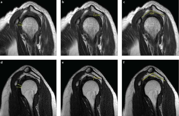
All MRI examinations were performed using surface coils by 1.5 Tesla (T) MRI systems (Achieva and Intera Nova, Philips Healthcare, Best, the Netherlands). T1-weighted images in coronal oblique plane (TR/TE interval, 540–720/14–26 ms), fat-suppressed proton density-weighted images in coronal oblique plane (TR/TE interval, 2600–3000/20–30 ms), fat-suppressed proton density-weighted images in axial plane (TR/TE interval, 2600–3000/20–30 ms), T1-weighted images in sagittal plane (TR/TE interval, 450–640/12–24 ms) and T2-weighted SPAIR images in sagittal plane (TR/TE interval, 2000– 4471/45–50 ms) were obtained. Imaging parameters were as follows: field of view, 18–20 cm; matrix, 256×182 pixels; slice thickness, 4 mm; section gap, 0.3 mm. MRI examinations were reevaluated by using the picture archiving and communication system (Extreme PACS, Ankara, Turkey). Acromio-humeral distance, coraco-humeral distance, the angle between the longitudinal axis of the coracoacromial ligament and longitudinal axis of the acromion (coracoacromial arch angle), and thickness of the coracoacromial ligament were measured in all subjects (Fig. 1). The slice where all three parameters were most clearly visible was selected as the standard. In this slice, the angle (coracoacromial arch angle) between the coracoacromial ligament axis (which extends from the coracoid process to acromion) and the line tangential to the inferior surface of the acromion (acromial axis) was measured. In the same slice acromio-humeral and coraco-humeral distances were measured where they are shortest. The thickness of the coracoacromial ligament was measured from the thickest part of the ligament. An expert musculoskeletal radiologist, having at least three years of competency, repeated the measurements twice, and mean values of the measurements were calculated. Following the initial evaluation, all images were randomly reevaluated two days later by the same radiologist.
Figure 1. a–f.
Sample measurements from a patient with rotator cuff pathology: (a), coraco-humeral distance; (b), acromio-humeral distance; (c), coracoacromial arch angle. Sample measurements from a patient with normal MRI findings: (d), coraco-humeral distance; (e), acromio-humeral distance; (f), coracoacromial arch angle.
Since this is a retrospective study, we did not have special plain roentgenograms in sagittal plane for the evaluation of coracoacromial arch angle.
Statistical analysis
Data were analyzed with the SPSS software version 15.0 for Windows (SPSS Inc., Chicago, Illinois, USA). Categorical variables were presented as frequency and percentage. The chi-square test and Fisher’s exact test were used to compare categorical variables. The Kolmogorov–Smirnov test was used to assess the distribution of continuous variables. Student’s t test was used for variables with normal distribution and the values were presented as mean±standard deviation. Intraobserver reliability was calculated and presented as correlation coefficient for the study parameters. Correlation was investigated among measured parameters using Pearson correlation analysis. Logistic regression analysis was used to evaluate the independent associates of the risk of rotator cuff pathology. Receiver operating characteristics (ROC) analysis was used to determine the cutoff values and the sensitivity and specificity of measured parameters. Odds ratios (OR) and 95% confidence intervals (CI) were calculated. A two-tailed P value of less than 0.05 was considered statistically significant.
Results
The mean age of the study population was significantly higher compared with the mean age of the control group (57±10 years vs. 46±14 years, P < 0.001). Gender distribution was not significantly different between the two groups (P > 0.05).
Among patients with rotator cuff pathology, according to MRI and arthroscopy findings, 32 patients had an isolated tear in the supraspinatus muscle tendon, six patients had tears in both the supraspinatus and subscapularis muscle tendons, one patient had a tear in the supraspinatus muscle tendon and tendinitis of subscapularis muscle tendon, and one patient had tears in both the supraspinatus and infraspinatus muscle tendons. Therefore, all study patients had tears in the supraspinatus muscle tendon.
Patients with rotator cuff pathology had significantly lower mean coraco-humeral distance compared with the control group (7.88±2.37 mm vs. 11.67±1.86 mm, P < 0.001). Similarly, the study patients had narrower acromio-humeral distance than the control group patients (7.89±2.09 mm vs. 11.15±1.84 mm, P < 0.001). There was also statistically significant difference between the two groups in terms of coracoacromial arch angle, which was measured as 132.38°±6.52° for the study group and 116.95°±7.66° for the control group (P < 0.001) (Table 1).
Table 1.
Rotator cuff parameters in the study and control groups
| Patients with rotator cuff pathology (n=40) | Patients with normal MRI findings (n=28) | P | |
|---|---|---|---|
| Acromio-humeral distance (mm) | 7.89±2.09 | 11.15±1.84 | < 0.001 |
| Coraco-humeral distance (mm) | 7.88±2.37 | 11.67±1.86 | < 0.001 |
| Coraco-acromial angle (°) | 132.38±6.52 | 116.95±7.66 | < 0.001 |
| Coracoacromial ligament thickness (mm) | 0.95±0.30 | 1.00±0.33 | > 0.05 |
MRI, magnetic resonance imaging.
Intraobserver reliability was nearly excellent for all measured study parameters (Table 2).
Table 2.
Correlation coefficients for intraobserver reliability of measured parameters
| Correlation coefficient for intraobserver reliability | 95% CI | P | |
|---|---|---|---|
| Acromio-humeral distance | 0.99 | 0.99–1.00 | < 0.001 |
| Coraco-humeral distance | 0.99 | 0.99–1.00 | < 0.001 |
| Coraco-acromial angle | 0.99 | 0.99–0.99 | < 0.001 |
| Coracoacromial ligament thickness | 0.97 | 0.96–0.98 | < 0.001 |
CI, confidence interval.
In regression analysis, with the addition of age factor, all three measured parameters were found to be independent associates of the risk of rotator cuff pathology (Table 3).
Table 3.
Independent associates of the risk of rotator cuff pathology
| OR | 95% CI | P | |
|---|---|---|---|
| Acromio-humeral distance (for each 1 mm) | 0.40 | 0.26–0.63 | < 0.001 |
| Coraco-humeral distance (for each 1 mm) | 0.45 | 0.30–0.66 | < 0.001 |
| Coracoacromial arch angle (for each 1°) | 1.51 | 1.22–1.88 | < 0.001 |
OR, odds ratio; CI, confidence interval.
In ROC analysis, coraco-humeral distance <9.90 mm had 87% sensitivity and 86% specificity, acromio-humeral distance <10.25 mm had 89% sensitivity and 75% specificity, and coracoacromial arch angle >127.08° had 87% sensitivity and 93% specificity in predicting rotator cuff pathology.
Rotator cuff pathology risk increased 24 times with a coraco-humeral distance <9.90 mm, 14 times by an acromio-humeral distance <10.25 mm, and 52 times by a coracoacromial arch angle >127.08° (Table 4).
Table 4.
Relative risks of measured parameters for binary values
| Patients with rotator cuff pathology (n=40) | Patients with normal MRI findings (n=28) | OR (95% CI) | P | |
|---|---|---|---|---|
| Acromio-humeral distance <10.25 mm | 33 (83%) | 7 (25%) | 14.142 (4.337–46.115) | < 0.001 |
| Coraco-humeral distance <9.90 mm | 32 (80%) | 4 (14%) | 24.000 (6.464–89.105) | < 0.001 |
| Coracoacromial arch angle >127.08° | 32 (80%) | 2 (7%) | 52.000 (10.151–266.371) | < 0.001 |
MRI, magnetic resonance imaging; OR, odds ratio; CI, confidence interval.
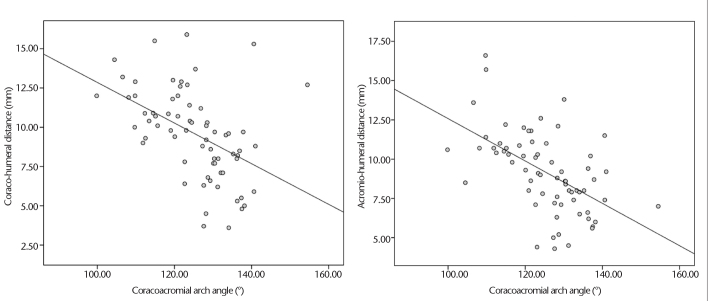
In correlation analysis, there was a statistically significant negative correlation between coraco-humeral distance and coracoacromial arch angle (r=−0.463, P < 0.001) and between acromio-humeral distance and coracoacromial arch angle (r=−0.537, P < 0.001) (Fig. 2).
Figure 2.
Correlations between the coraco-humeral distance and coracoacromial arch angle, and between the acromio-humeral distance and coracoacromial arch angle.
Discussion
Rotator cuff pathology is among the most common causes of shoulder pain, especially in the elderly. High soft tissue resolution and multiplanar imaging of MRI contributes to the identification of pathology, although arthroscopy is still the gold standard in diagnosis (6). We included only patients with arthroscopically confirmed diagnosis to increase the reliability of our study.
Subacromial impingement is a pain syndrome with an unclear natural history and cause. Many studies have asserted that the cause of the pain is due to acromial reasons (3, 7–9). However, there are also studies claiming the opposite (10). In the present study, only those patients with a flat-bottom surface type (type 1) acromion were included; other acromion types that have been subject to controversies in the literature were not included so as to prevent possible influences in the interpretation of the results.
In their study Neer and Poppen described the supraspinatus outlet and speculated that its narrowing is a primary cause of rotator cuff tear (4, 11). Zuckermann et al. (12) found that patients with rotator cuff tear had 22.5% narrower supraspinatus outlet area compared to the normal group. Burns and Whipple (5) performed studies on cadavers to investigate the association between the coracoacromial ligament and the supraspinatus. They concluded that impingement occurred due to coracoacromial ligament compared to the acromion (5). Therefore, in addition to the acromion, coracoacromial arch geometry has gained increasing importance.
Coracoacromial arch is an osteofibrous structure resulting from the continuity of the acromion, coracoacromial ligament, and coracoid process with each other (13). In case of lower localization of coracoacromial arch, the pressure on the rotator cuff may increase (13). In light of this possibility, we investigated the relationship between the structures constituting the coracoacromial arch and the rotator cuff pathology. Our primary aim was to show whether coracoacromial arch angle is a predisposing factor for the rotator cuff pathology.
In our study, measured acromio-humeral distance was 7.89±2.09 mm in the study group compared to 11.15±1.84 mm in the control group (P < 0.001). Similarly, in the literature, an association was shown between narrowing of the acromio-humeral distance and the rotator cuff pathology (14). In addition, we found that the coraco-humeral distance was 7.88±2.37 mm in the study group compared to 11.67±1.86 mm in the control group (P < 0.001). This finding was also compatible with previous studies (15).
We measured coracoacromial ligament thickness as 0.95±0.30 mm in the study group compared to 1.00±0.33 mm in the control group. However, this difference was not statistically significant (P > 0.05). There was no data about the normal or pathologic range of coracoacromial ligament thickness in the literature.
Coracoacromial arch angle was measured as 132.38°±6.52° in the study group compared to 116.95°±7.66° in the control group (P < 0.001). The mean difference between the two groups was 15.43°. In a cadaveric study, coracoacromial arch angle was measured as 131.9° and 134.7° in patients with and without rotator cuff tear, respectively, with no significant difference between them (12). However, in their measurements the lower and upper edges of the acromion and the lower edge of the coracoid process were taken as reference and acromion types were not considered. In contrast, we measured the angle between the axis of the coracoacromial ligament and the axis of the acromion. We think that our measurement technique is more accurate in terms of the pressure on the supraspinatus outlet.
Limited sample size and the retrospective nature of our study constitute its major limitations. A single radiologist performing all measurements was also a limitation although this was needed to ensure standardization of the measurements. To overcome this limitation the measurements were repeated at two different times and the averages were used. Age difference between the two groups also appears to be a limitation. However, when we included age in the multivariate logistic regression analysis, its significance disappeared. In addition, patients having acromion types affecting supraspinatus outlet, trauma or surgery, and acromioclavicular joint pathology such as degenerative changes due to aging were excluded from the study.
In conclusion, we demonstrated that coraco-humeral distance, acromio-humeral distance, and especially, coracoacromial arch angle are significantly and independently associated with the rotator cuff pathology in living cases. Therefore, in patients with shoulder pain the measurement of coraco-humeral distance, acromio-humeral distance, and coracoacromial arch angle can aid to determine the possible etiology of rotator cuff pathology. We measured the coracoacromial arch angle as the angle between the coracoacromial ligament axis (that extends from coracoid process to acromion) and the line tangential to the inferior surface of the acromion (acromial axis). However, further prospective studies with larger sample sizes and different measurement techniques are needed to consolidate our findings.
Footnotes
Conflict of interest disclosure
The authors declared no conflicts of interest.
References
- 1.Michener LA, McClure PW, Karduna AR. Anatomical and biomechanical mechanisms of subacromial impingement syndrome. Clin Biomech (Bristol, Avon) 2003;18:369–379. doi: 10.1016/s0268-0033(03)00047-0. [DOI] [PubMed] [Google Scholar]
- 2.McClure PW, Bialker J, Neff N, Williams G, Karduna A. Shoulder function and 3-Dimensional kinematics in people with shoulder impingement syndrome before and after a 6-week exercise program. Phys Ther. 2004;84:832–848. [PubMed] [Google Scholar]
- 3.Neer CS., II Anterior acromioplasty for the chronic impingement syndrome in the shoulder. J Bone Joint Surg Am. 1972;54A:41–50. [PubMed] [Google Scholar]
- 4.Neer CS, Poppen NK. Supraspinatus outlet. Orthop Trans. 1987;11:234. [Google Scholar]
- 5.Burns WC, Whipple TL. Anatomic relationships in the shoulder impingement syndrome. Clin Orthop Relat Res. 1993;294:96–102. [PubMed] [Google Scholar]
- 6.Craig EV. Shoulder arthroscopy in the throwing athlete. Clin Sports Med. 1996;15:673–700. [PubMed] [Google Scholar]
- 7.Bigliani LU, Morrison DS, April EW. The morphology of the acromion and its relationship to the rotator cuff tears. Orthop Trans. 1986;10:216. [Google Scholar]
- 8.Edelson JG, Taitz C. Anatomy of the coraco-acromial arch: relation to degeneration of the acromion. J Bone Joint Surg. 1992;74B:589–594. doi: 10.1302/0301-620X.74B4.1624522. [DOI] [PubMed] [Google Scholar]
- 9.Kassarjian A, Bencardino JT, Palmer WE. MR imaging of the rotator cuff. Radiol Clin North Am. 2006;44:503–523. doi: 10.1016/j.rcl.2006.04.005. [DOI] [PubMed] [Google Scholar]
- 10.Edelson JG. The ‘hooked’ acromion revisited. J Bone Joint Surg. 1995;77B:284–287. [PubMed] [Google Scholar]
- 11.Neer CS., II . Cuff tears, biceps lesions and impingement. In: Neer CS II, editor. Shoulder reconstruction. Philadelphia: WB Saunders; 1990. pp. 44–48. [Google Scholar]
- 12.Zuckerman JD, Kummer FJ, Cuoma F, Simon J, Rosenblum S, Katz N. The influence of coracoacromial arch anatomy on rotator cuff tears. J Shoulder Elbow Surg. 1992;1:4–14. doi: 10.1016/S1058-2746(09)80010-4. [DOI] [PubMed] [Google Scholar]
- 13.Vaz S, Soyer J, Pries P, Clarac JP. Subacromial impingement: influence of coracoacromial arch geometry on shoulder function. Joint Bone Spine. 2000;67:305–309. [PubMed] [Google Scholar]
- 14.Cay N, Tosun O, Dogan M, Karaoglanoglu M, Bozkurt M. The effect of morphometric relationship between the glenoid fossa and the humeral head on rotator cuff pathology. Acta Orthop Traumatol Turc. 2012;46:325–331. doi: 10.3944/aott.2012.2807. [DOI] [PubMed] [Google Scholar]
- 15.Richards DP, Burkhart SS, Campbell SE. Relation between narrowed coracohumeral distance and subscapularis tears. Arthroscopy. 2005;21:1223–1228. doi: 10.1016/j.arthro.2005.06.015. [DOI] [PubMed] [Google Scholar]


