Summary
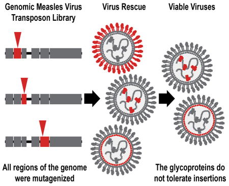
Measles virus undergoes error-prone replication like other RNA viruses, but over time it has remained antigenically monotypic. The constraints on the virus that prevent the emergence of antigenic variants are unclear. As a first step in understanding this question, we subjected the measles virus genome to unbiased insertional mutagenesis and viruses that could tolerate insertions were rescued. Only insertions in the nucleoprotein, phosphoprotein, matrix protein, as well as intergenic regions were easily recoverable. Insertions in the glycoproteins of measles virus were severely under-represented in our screen. Host immunity depends on developing neutralizing antibodies to the hemagglutinin and fusion glycoproteins; our analysis suggests that these proteins occupy very little evolutionary space and therefore have difficulty changing in the face of selective pressures. We propose that the inelasticity of these proteins prevents the sequence variation required to escape antibody neutralization in the host, allowing for long-lived immunity after infection with the virus.

Introduction
Measles (MeV) is an enveloped, single-stranded negative-sense RNA virus of the genus Morbillivirus in the family Paramyxoviradae (Griffin et al., 2012). MeV enters a cell via the actions of two surface glycoproteins, the hemagglutinin (H) and the fusion protein (F) (Yanagi et al., 2006). H acts as the receptor binding protein, while F is the actual fusogenic protein responsible for mediating viral envelope and cell membrane fusion (Griffin, 2013). The cellular receptor for MeV is CD150/SLAM, however some strains (including vaccine strains), can also use the ubiquitously expressed CD46 protein (Yanagi et al., 2009). Neutralizing antibodies against MeV are thought to solely target the F and H proteins, with H being the major neutralizing antigenic target (de Swart et al., 2005). Known to be vaccine preventable, both vaccination and clinical infection confer long-lived immunity (Anders et al., 1996; Moss and Griffin, 2006).
MeV has an error-prone RNA dependent RNA polymerase (RdRP) that in vitro has a mutation rate similar to that of other RNA viruses (Drake, 1993; Parvin et al., 1986; Sanjuan et al., 2010; Schrag et al., 1999). Over a cycle of infection and transmission between humans, MeV is likely to be exposed to similar selection pressures by the immune system as other respiratory transmitted viruses (Braciale et al., 2012; Griffin, 1995; Kohlmeier and Woodland, 2009). Therefore, a priori, one might expect that MeV would acquire sequence diversity in surface exposed protein epitopes to evade the adaptive immune response. Certainly this happens with many viruses, including influenza A virus. However, this antigenic “drift” does not appreciably occur in the MeV F and H glycoproteins. The molecular basis for the lack of emergent antigenically distinct strains of MeV, relative to other related negative sense RNA virus like influenza A virus, is currently unclear (Fayolle et al., 1999; Lech et al., 2013; Xu et al., 2013). Given that MeV does not undergo major antigenic changes, it is possible that the glycoproteins of MeV are under a rigid, but as of yet undefined, constraint that prevents this evolution from occurring.
Previously, we reported a 15-nucleotide insertional mutagenesis on the influenza A virus genome (Heaton et al., 2013). Most regions of the influenza A virus genome were found to be resistant to insertion, but the head domain of the hemagglutinin protein was identified as highly tolerant of transposon insertion. We speculated that the observed mutational tolerance was an experimental readout for evolutionary flexible protein domains; and that this flexibility was the underlying basis for the rapid antigenic drift observed with influenza A virus. However, we had no comparison to a virus that does not undergo rapid antigenic evolution in its surface glycoproteins.
In this study, we sought to determine where in the antigenically stable MeV genome insertions could be tolerated and, in particular, if any insertions could be made in the glycoproteins. By performing these experiments under standard cell culture conditions, we asked where MeV had the potential to change in the absence of immune selection. Mutant measles viruses with insertions in distinct domains in the nucleoprotein (N), phosphoprotein (P), and matrix protein (M), as well as some intergenic regions, were viable. No measles viruses with insertions in either the F or H glycoproteins, or in the large polymerase gene (L), were highly represented in the screen. This data suggests that the MeV hemagglutinin and fusion proteins are very rigid when compared to the influenza A virus hemagglutinin under essentially the same experimental conditions. We hypothesize that this inelasticity may be a contributor to the observed lack of MeV antigenic variation.
Results
Construction and rescue of the MeV transposon library
As a test of viral rescue efficiency, a modified Edmonston strain MeV genomic construct expressing GFP was transfected into BSR-T7 cells with four accessory helper plasmids (Fig. S1A). The transfected cells were then co-cultured with permissible human adenocarcinoma alveolar basal epithelial cells (A549s). Passage of viruses on A549s caused large GFP positive syncytia to form only if all helper plasmids were added to the transfection (Fig. S1B). Recovery of infectious particles was high, with the viral titer of MeV GFP after co-culture at 5.2×03 TC ID50/mL (Fig. S1C).
To generate a high-coverage MeV mutant library, the genome containing plasmid was mutagenized in vitro with Mu transposase and an artificial transposon with a kanamycin selectable marker as previously described (Heaton et al., 2013). The mutagenesis was scaled to generate >105 individual insertional mutants, which would represent >5-fold coverage of the possible insertion positions. The template for the mutagenesis was a “MeV+3” genomic construct, which does not encode GFP and has an extra 3-nucleotide stop codon behind the original stop codon of N (Fig. S1D). After mutagenesis and removal of the transposon body, a 15-nt insert remains in the genome (10 of which serve as a unique molecular tag) making the antigenome once again follow the rule of six required by paramyxoviruses (Kolakofsky et al., 1998) (Fig. S1E).
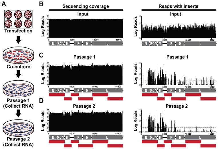
To determine where MeV could tolerate insertions, multiple independent rescues of the mutant libraries were performed (Fig. 1A). Five days post-transfection, the cells were pooled and co-cultured with A549s for three days to propagate rescued viruses. After co-culture, both supernatant and cell-associated virus was collected and passaged on fresh A549 cells to select for fully infectious virus mutants (passage 1). The propagated viruses from passage 1 were passaged 72 hours post-infection onto fresh A549 cells for passage 2. RNA extracted from the cells of both passages was subjected to RT-PCR with MeV specific primers that amplified the genome in six overlapping segments. The subsequent cDNA was prepped and submitted for Illumina HiSeq next-generation sequencing.
Figure 1. Insertional mutagenesis of MeV genome.
(A) Mutant viruses were rescued by transfecting BSR-T7 cells with the transposon library and then co-culturing the cells with A549s. The resulting viruses were passaged on A549 cells and viral RNA was sequenced. (B–D) The input library and both passages were subjected to deep sequencing. The total sequencing coverage of the genome is shown on the left panels, while the number of reads containing transposon insertions are indicated on the right. The numbers along the x-axis of the graphs indicate the genomic nucleotide position. The red bars under the genome diagrams in B–D indicate the individual RT-PCR products amplified for Illumina sequencing. Dashed lines indicate a threshold of 0.01% of the total reads.
Sequencing of our input library demonstrated there was good sequencing coverage and insertions were evenly distributed throughout the MeV genome, with the vast majority of codons containing an insertion (Fig. 1B, Supplementary Dataset). Although the genome of the input library was evenly covered with transposons, only insertions in distinct regions of the MeV genome were recovered after selection (Fig. 1C,D). Insertion sites in intergenic regions except those between the hemagglutinin (H) and large (L) genes were readily recoverable. Sites in the nucleoprotein (N) and the phosphoprotein (P), and matrix protein (M) were also abundant. Despite select regions of the MeV genome tolerating insertions, less than 1% of total reads with insertions were detected in the fusion (F), H, or L genes. Insertions in the 5’ and 3’ distal untranslated regions (UTRs) of the genome were also only rarely recovered. These data are representative of three independent rescue and passaging experiments.
MeV surface glycoproteins are intolerant of insertional mutagenesis
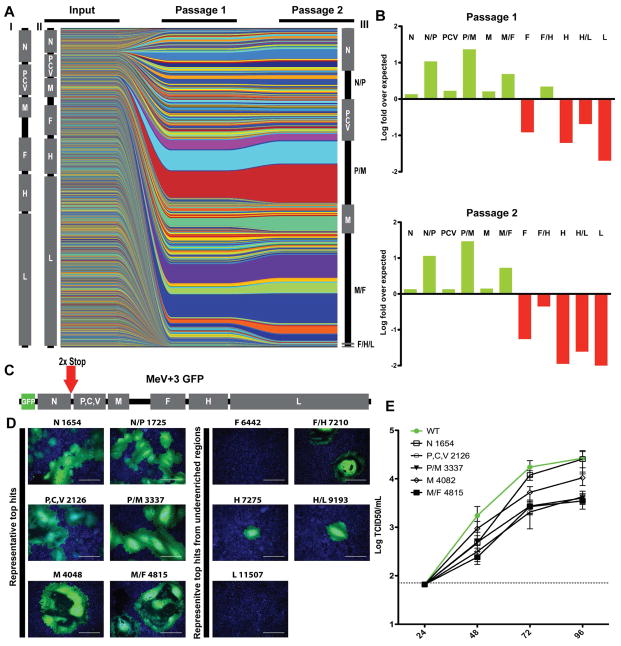
Analyzing the abundance of insertions sites across the passages showed that all sites had relatively the same abundance in the input library, but were recovered with varying efficiencies (Fig. 2A). Viruses rescued in the first round of passaging were generally carried through to the second round in similar proportions, although some were lost or greatly reduced due to additional selection (Fig. 2A). We next normalized the percentage of insertions to the size of each genomic region (Fig. 2B). The N/P, P/M, and M/F intergenic regions were greatly enriched compared to open reading frames. Although enrichment is seen in N, P, and M, few insertions are seen in the H/L intergenic region or the F, H, and L ORFs.
Figure 2. MeV glycoproteins and polymerase genes are intolerant of insertions.
(A) Plots representing individual insertion sites. Individual viruses in the input and the passages are represented by different colors; the thickness of the lines is representative of the proportion in the population. The MeV genomes are drawn either to scale (I), distorted to represent the actual coverage of insertions in the input (II), or distorted to represent the total percentage of reads in each region after the second passage (III). (B) Percent of reads in a region divided by the size of that region to give the fold over predicted values (as if there was no biological selection). Under-represented areas are displayed as negative values (red) while over-represented areas are displayed as positive values (green). (C) Individual insertion sites were cloned into a MeV+3 GFP construct to validate the sequencing results. 2x Stop indicates the presence of an additional stop codon after the normal stop codon of the N gene. (D) Top hits for the screen, plus the top hits for each region were cloned and rescued. For each panel title, the letter represents the genomic region while the number represents the genomic nucleotide position preceding the insertion. Scale bar=400μm. (E) Growth curves of viruses with insertions in the indicated sites. Values and error bars represent the mean and standard error of the mean, respectively. Green filled circles and line=Parental MeV.
To validate the screen data, the top insertion sites were cloned into a MeV+3 GFP construct (Fig. 2C), rescued, and sequence verified. All of these viruses were easily rescued and formed syncytia on A549 cells similar to the parental virus (Fig. 2D, Table 1). The top hits in F, F/H, H, H/L and L in our screen were also cloned despite the fact that our analysis predicted that they would be significantly attenuated. No viruses were rescuable from the top insertion sites in L and in F (Fig. 2D, Table 1). Insertions in sites in the F/H and H/L intergenic regions as well as in H were detectable by fluorescence microscopy after rescue, but viral titer was almost undetectable (Fig. 2D, Table 1). Growth curves were then performed on A549 cells with a panel of rescued viruses that grew to sufficiently high titers in the N, P, P/M, M, and M/F regions. We were unable to obtain high-titer viral stocks of H or F protein mutants, so these viruses were excluded from our growth curve analysis. The insertional mutants predicted by our analysis to be viable had only minor attenuation (less than 1log10 of TCID50) in multicycle growth relative to the parental MeV (Fig. 2E). We therefore conclude that the abundance of an insertional mutant detected in our sequencing data is generally correlated with the fitness of the mutant virus.
Table 1. Verified recoverable transposon insertion sites.
| Genomic location | Genomic nucleotide position | Codon preceding insertion | Translation of insert | Rescue titer (log fold over MeV +3) |
|---|---|---|---|---|
| N | 1654 | 515 | IAAAPI | +++ |
| N | 1660 | 517 | YAAAVY | ++ |
| N/P | 1720 | N/A | N/A | ++ |
| N/P | 1721 | N/A | N/A | ++ |
| N/P | 1725 | N/A | N/A | ++ |
| P/C/V | 2126 | 105 | GAAATG | ++ |
| P/V | 2703 | 298 | CGRTQ | ++ |
| P | 3329 | 506 | NAAAMK | ++ |
| P/M | 3334 | N/A | N/A | + |
| P/M | 3337 | N/A | N/A | ++ |
| M | 4048 | 202 | GAAAPG | ++ |
| M | 4082 | 214 | CGRTE | +++ |
| M/F | 4789 | N/A | N/A | ++ |
| M/F | 4801 | N/A | N/A | ++ |
| M/F | 4815 | N/A | N/A | +++ |
| M/F | 4826 | N/A | N/A | ++ |
| M/F | 4843 | N/A | N/A | + |
| M/F | 4846 | N/A | N/A | ++ |
|
| ||||
| F | 6442 | 330 | MRPHSN | − |
| F | 6953 | 500 | IAAAYI | − |
| F | 7035 | 528 | CGRKG | − |
| F/H | 7120 | N/A | N/A | + |
| F/H | 7210 | N/A | N/A | + |
| F/H | 7212 | N/A | N/A | + |
| H | 7275 | N/A | N/A (Start codon) | + |
| H | 7355 | 27 | VRPHRE | + |
| H | 8697 | 474 | SAAAPR | − |
| H/L | 9153 | N/A | N/A | − |
| H/L | 9163 | N/A | N/A | + |
| H/L | 9193 | N/A | N/A | + |
| L | 11507 | 757 | CGRNL | − |
| L | 11846 | 870 | CGRSN | − |
| L | 14740 | 1834 | DAAAVE | − |
Individual mutant viruses were rescued. The top insertions sites from the screen are shown in the top half of the table. The top three insertions sites in each region from F through L are shown in the bottom half of the table. The titer over the background rescue of the MeV+3 GFP virus was calculated. +++ ≥ 2logs over background, ++ ≥ 1log over background, + ≤1 log over background, − no virus was detected. N/A indicates the insertion was in a non-coding region.
Analysis of insertional mutagenesis of other RNA viruses reveals exceptional intolerance of the MeV F and H glycoproteins
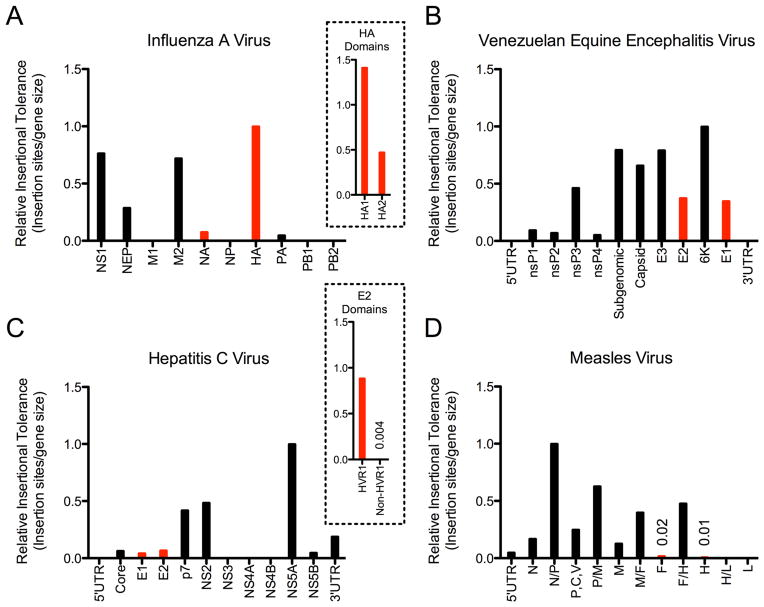
In order to put our MeV insertional mutagenesis into perspective, we took advantage of previously published datasets and analyzed the transposon insertional profiles of influenza A virus (Heaton et al., 2013), hepatitis C virus (HCV) (Remenyi et al., 2014), and Venezuelan equine encephalitis virus (VEEV) (Beitzel et al., 2010) along with our current study on MeV. As each study was performed slightly differently, we plotted each virus individually with each genomic region assigned a value out of a maximum of 1 to show the relative tolerance to insertion. While we observed general consensus on some genomic areas, i.e. polymerase proteins tolerate very few insertions, we observed highly divergent results across the different viral surface glycoproteins responsible for attachment and entry. At one end of the spectrum, the influenza A virus hemagglutinin (HA), and specifically the HA1 domain, which encodes the head region, was the most mutable gene in the entire genome (Fig. 3A and Fig. 3A Inset). VEEV and HCV accepted some insertions in the functionally similar envelope glycoproteins (E2/E1 and E1/E2 respectively), but to a lesser extent than influenza A virus (Fig. 3B,C). For the HCV E2 glycoprotein, insertions were for the most part limited to the N-terminal hyper-variable region 1 (HVR1) (Hijikata et al., 1991); when analyzed alone in fact, the HVR1 becomes the second most tolerant region in the entire genome (Fig 3C, Inset). In contrast to the other viruses, strikingly, the MeV F and H glycoproteins were among the most transposon intolerant genes in the entire MeV genome (Fig. 3D).
Figure 3. Comparative analysis reveals MeV glycoproteins are exceptionally resistant to insertional mutation.
The total number of sites that could tolerate insertions in each region were normalized to region size and graphed. Red columns indicate the major viral surface glycoproteins. (A) Influenza A virus. The HA1 head domain and HA2 stalk domain of the influenza A virus hemagglutinin are separated in the insert. (B) VEEV (C) HCV. The hyper-variable region of E2 is separated from the rest of the E2 protein in the insert. (D) MeV. All values are out of an arbitrary value of 1.
Discussion
MeV is an antigenically monotypic virus. We began this study in an attempt to gain insight into the molecular basis underlying that phenomenon. Mutagenesis revealed that certain domains of the genome can tolerate insertions. In particular, the intergenic regions towards the 5’ end of the genome were highly enriched for insertions. In addition, certain regions of N, P and the M genes were also tolerant of mutations. No domains in the glycoproteins or the polymerase genes tolerated insertions, likely due to the effects of the insertion on the structure and therefore function of the gene products.
It is well known that the C-terminus of the N protein is one of the most variable regions across MeV isolates (Xu et al., 1998), with variability also frequently observed in the P gene (Baczko et al., 1992; Bankamp et al., 2008). Previous reports have also shown that measles viruses not expressing the M protein, characteristic during subacute sclerosing panencephalitis (SSPE) infections, are viable in vitro (Cathomen et al., 1998). Therefore it is not surprising that viral mutants with insertions in these genes were frequently recovered during this screen.
Previous studies have shown that the insertion of GFP into the MeV L (Duprex et al., 2002) as well as insertions at the end of the H protein (Hammond et al., 2001; Nakamura et al., 2005; Plemper et al., 2002; Schneider et al., 2000) are viable. We did not detect high levels of transposon insertions at those particular locations in our study. This is likely a reflection of multiple factors. First of all, the specific sequence of the insertion likely contributes to the tolerance (or lack of tolerance) of the insertion. Secondly, the experimental conditions like specific cell lines used for growth of the virus may also change the acceptance of an insertion. Finally, the competitive nature of our viral rescue “hides” some tolerated insertion sites. When all the mutants are rescued and passaged at the same time, only the most fit viruses are highly represented in the output sequencing. This phenomenon can be observed in with the H gene and H/L intergenic region; these areas were significantly under-enriched for transposon insertions in the screen, however rescue of individual mutants did show some viable (but highly attenuated) viruses (Fig. 2D, Table 1). Viral competition shrinking the representation of viable, but attenuated, mutants likely explains why the H gene was devoid of transposons in our study but variability of H is observed in nature (Bellini and Rota, 1998). We therefore utilize insertional mutagenesis as a readout of generally flexible or inflexible regions of a viral genome, not a comprehensive list of the specific locations that can tolerate insertions. This strategy explores a phenotypic and fitness landscape that is not accessible via normal processes of RdRp-dependent mutation, and reveals the most flexible or inflexible regions of the MeV genome.
While wild-type viruses rarely have large insertions or deletions, our data is consistent with the known features of extant MeV strains. For example, distinct MeV strains isolated from clinical infections contained additions in the 3’ UTR of the M gene and deletions in the F 5’ UTR in the M/F intergenic region (Bankamp et al., 2014). Our study showed that this intergenic region contained the most abundant number of transposons in our screen, totaling ~34% of all reads with insertions. It is generally accepted that the high sequence diversity frequently associated with RNA viruses is the result of rapid, error-prone replication and strong selection pressures applied by the immune system. However these factors alone cannot explain why such a large range of antigenic diversity is observed across different RNA viruses. Despite similar viral polymerase error rates and immune system pressures, virus diversity can vary greatly, from antigenically monotypic viruses like MeV to highly drifted viruses like influenza A virus. It stands to reason that evolutionary constraints on the viral protein scaffolds themselves, on which the variation has to arise, could be the missing molecular explanation.
We have provided evidence that in fact, the glycoproteins of MeV are intolerant of (or highly attenuated by) five amino-acid insertions, which is a fairly significant mutational lesion. When we compare our dataset to other RNA viruses mutagenized with the same technique however, we find that the glycoproteins of VEEV, HCV, and especially influenza A virus tolerate the same 5 amino acid insertions to a much greater extent. It is currently unclear why some viral glycoproteins have evolved to be rigid while others remain flexible. One possibility is the nature of the receptors utilized by the viruses. Influenza A virus requires only a sialic acid moiety to act as a receptor (Skehel and Wiley, 2000) and is by far the most flexible surface glycoprotein. In contrast, HCV, MeV, and VEEV all utilize at least one proteinaceous receptor (Lindenbach and Rice, 2013; Ludwig et al., 1996; Yanagi et al., 2009), which likely put significant constraints on how flexible the proteins can become. It is also worth noting that the intergenic regions of MeV are highly tolerant to insertions, which may appear to minimize the magnitude of the insertions in the genes themselves. If we exclude the intergenic regions from the analysis however, the H and F proteins are the second and third least tolerant open reading frames in the genome, respectively, after the L protein.
From a practical standpoint, this technology may allow the prediction of antigenically invariant epitopes in viral proteins for vaccine purposes. Previously, our research demonstrated the HA2 conserved “stalk” region of the influenza A virus HA protein was refractory to transposon mutagenesis while the head region was very tolerant (Heaton et al., 2013). Work in the influenza A virus vaccine field has found stalk-reactive antibodies to be broadly neutralizing to influenza virus subtypes (Krammer and Palese, 2013), due to a high degree of stalk domain conservation. With respect to MeV, our data predicts that the lack of an evolutionary flexible domain in MeV H or F limits the antigenic variation potential of the virus. This may be part of the reason why the MMR live-attenuated vaccine remains effective against currently circulating strains of MeV.
In summary, our data suggests that MeV is antigenically conserved, at least in part, because the H and F proteins themselves are fundamentally unable to tolerate a large range of mutations. This is in contrast to evolutionary flexible proteins like the HA encoded by influenza A virus. The evolutionary constraints imposed by the viral proteins themselves likely represent a key parameter (along with intrinsic viral polymerase mutation rate) in determining long-term viral diversity and potentially the efficacy of a vaccine over long time scales.
Experimental Procedures
Cells
The human alveolar basal epithelial cell (A549) and the baby hamster kidney cell expressing T7 (BSR-T7) cell lines were maintained in DMEM containing 10% FBS by volume and penicillin/streptomycin (Invitrogen).
Generation of MeV constructs and viral rescue
The MeV GFP rescue system was generated by the lab of Benhur Lee using the GFP-N Edmonston antigenomic clone generously provided by W. Paul Duprex (Boston University)(Duprex et al., 1999), and heterologous N, P and L helper genes (a generous gift from Richard Plemper, Georgia State University). See supplemental experimental procedures for further details.
To test the number of viruses we could rescue from a single transfection, the MeV GFP genomic construct was transfected into BSR-T7 cells with four optimized plasmids expressing codon optimized T7 polymerase and the N, P and L helper genes as previously published (Krumm et al., 2013; Radecke et al., 1995). The transfected cells were co-cultured with permissible A549s. Cell associated and free virus was titered on A549 cells by 50% tissue culture infectious dose (TCID50) as described by Reed and Muench (Reed and Muench, 1938) (Fig. S1C). Individual transposon mutant viruses were rescued in a similar manner. Their cloning, rescue and characterization are fully described in the supplemental material.
Generation of MeV mutant libraries
A MeV+3 genomic construct suitable for mutagenesis was generated as described in supplemental material. The Mutation Generation System (Fisher Scientific) was used to randomly insert transposons in the MeV+3 plasmid according to the manufactures protocol (Fig. S1E). Four independent in vitro transposon insertion reactions were performed on 760 ng MeV+3 plasmid, which were then pooled, and transformed into Stbl2 cells. Following transformation, the cells were plated on 15 cm plates with LB agar containing ampicillin and kanamycin, and allowed to grow for 30 hours at 30 °C. The subsequent colonies were scraped and pooled. DNA was extracted from the pooled colonies using a HiPure maxiprep kit (Invitrogen) and digested with NotI (New England Biolabs) to remove the transposon body. The restricted plasmid was then gel purified using the QIAquick kit (Qiagen) and 1 ug of DNA was relegated at 16 °C for 16 hours using T4 Ligase (Ne w England Biolabs). The entire ligation mixture was transformed into Stbl2 cells and plated on 15 cm plates containing ampicillin and allowed to grow 30 hours at 30 °C. The colonies were again pooled and the DNA was extracted using the HiPure maxiprep kit.
Viral mutant library rescue
The mutant MeV library was rescued using standard protocols with modifications (Krumm et al., 2013; Radecke et al., 1995). 5μg of MeV genome and four helper plasmids were transfected into 70% confluent BSR-T7 cells in 6-well dishes using the Lipofectamine LTX with Plus (Invitrogen) reagent. A total of five 6-well plates were used for rescuing the viral libraries. At 16 hours post-transfection, the media was replaced with fresh DMEM. Five days post-transfection, the transfected cells were scraped into their media, pooled, and then co-cultured with 60% confluent A549s in 15 cm dishes. After 3 days of co-culture, the cells were again scraped into their media freeze thawed at −80°C and pelleted by centrifugation 4000xg for 5 m ins. The clarified supernatant was used to infect 70% confluent A549s in 15 cm dishes. After 3 days (sufficient time to allow high levels of viral replication to occur), the infected cells were scraped into their media. Half of the cells were pelleted, lysed in 3 mL TRIzol, and frozen at −80°C. The remaining cells were freez e thawed and pelleted by centrifugation (4000xg for 5 mins). The clarified supernatant was again used to infect 70% confluent A549s for a second passage of 3 days in 15 cm dishes. After 3 days, the supernatant was removed and the infected cells were lysed in 6 mL TRIzol and frozen at −80°C.
RT-PCR and Illumina sequencing
Samples in TRIzol were thawed and RNA was extracted according to manufacture’s protocols. The measles genomic RNA was then amplified in six segments using overlapping primers sets as described in the supplemental experimental procedures with Invitrogen’s SuperScript III RT-PCR kit with Platinum Taq. The cDNA segments from each sample were pooled in equal molar amounts, sheared with Covaris sonication, and prepped for sequencing using TruSeq DNA LT Sample Prep Kit (Illumina) according to the manufacturer’s instructions. Barcoded and multiplexed samples were sequenced on a HiSeq2000 using 100 nt single-end reads in Rapid Run mode.
Analysis of Illumina sequencing data
Analysis of the transposon insertions was done as previously described (Heaton et al., 2013). Please see supplemental experimental materials for details.
Metaanalysis of HCV, VEEV, and influenza A virus insertional mutation datasets
Previously published datasets were downloaded from the supplemental materials of (Beitzel et al., 2010; Heaton et al., 2013; Remenyi et al., 2014). For VEEV, the 30°C dataset was used (Beitzel et al., 2010). For the MeV, HCV and VEEV data, insertion positions containing >0.01% of total reads were deemed “hits” and divided by the size of the genomic region. For the influenza A virus data, discreet insertion positions in the coding regions (i.e. at least one nucleotide away from a previous “hit” location) ≥9X above background were deemed hits and similarly normalized. For the influenza A virus HA domain analysis, HA1 was defined as the region between nucleotides 81–1121 and HA2 was defined as 1122–1730. For the HCV E2 domain analysis, the hypervariable domain 1 was defined as the first 81 nucleotides of the E2 coding region.
Supplementary Material
Acknowledgments
The authors would like to acknowledge Andrew Varble for his help with the graphical representation of the deep sequencing data. We would also like to acknowledge the genomics and microscopy cores at the Icahn School of Medicine at Mount Sinai. NSH is a Merck fellow of the Life Sciences Research Foundation. SMB was supported by the Viral-Host Pathogenesis Training Grant T32 AI07647. This work was supported in part by the following grants: NIH R21AI115226-01 (BL), R33AI102267-03 (BL), U54AI065359 (BL), U19AI109946-01 (PP), CEIRS HHSN272201400008C (PP), NIH P01AI097092-02 (PP).
Footnotes
Accession Numbers. The raw sequencing data has been deposited and is available via NCBI GEO under the accession numbers: GSE67666, GSM1653192, GSM1653193, and GSM1653194.
Author Contributions.
S.M.B., S.T.W., and B.L. conceptualized, designed and characterized the measles virus rescue system. B.O.F. and N.S.H performed the mutagenesis experiments and analyzed data. D.S. performed the bioinformatics analysis of the deep sequencing data. B.O.F., B.L, P.P., and N.S.H. wrote the manuscript.
Publisher's Disclaimer: This is a PDF file of an unedited manuscript that has been accepted for publication. As a service to our customers we are providing this early version of the manuscript. The manuscript will undergo copyediting, typesetting, and review of the resulting proof before it is published in its final citable form. Please note that during the production process errors may be discovered which could affect the content, and all legal disclaimers that apply to the journal pertain.
References
- Anders JF, Jacobson RM, Poland GA, Jacobsen SJ, Wollan PC. Secondary failure rates of measles vaccines: a metaanalysis of published studies. The Pediatric infectious disease journal. 1996;15:62–66. doi: 10.1097/00006454-199601000-00014. [DOI] [PubMed] [Google Scholar]
- Baczko K, Pardowitz I, Rima BK, ter Meulen V. Constant and variable regions of measles virus proteins encoded by the nucleocapsid and phosphoprotein genes derived from lytic and persistent viruses. Virology. 1992;190:469–474. doi: 10.1016/0042-6822(92)91236-n. [DOI] [PubMed] [Google Scholar]
- Bankamp B, Liu C, Rivailler P, Bera J, Shrivastava S, Kirkness EF, Bellini WJ, Rota PA. Wild-type measles viruses with non-standard genome lengths. PloS one. 2014;9:e95470. doi: 10.1371/journal.pone.0095470. [DOI] [PMC free article] [PubMed] [Google Scholar]
- Bankamp B, Lopareva EN, Kremer JR, Tian Y, Clemens MS, Patel R, Fowlkes AL, Kessler JR, Muller CP, Bellini WJ, et al. Genetic variability and mRNA editing frequencies of the phosphoprotein genes of wild-type measles viruses. Virus research. 2008;135:298–306. doi: 10.1016/j.virusres.2008.04.008. [DOI] [PubMed] [Google Scholar]
- Beitzel BF, Bakken RR, Smith JM, Schmaljohn CS. High-resolution functional mapping of the venezuelan equine encephalitis virus genome by insertional mutagenesis and massively parallel sequencing. PLoS pathogens. 2010;6:e1001146. doi: 10.1371/journal.ppat.1001146. [DOI] [PMC free article] [PubMed] [Google Scholar]
- Bellini WJ, Rota PA. Genetic diversity of wild-type measles viruses: implications for global measles elimination programs. Emerging infectious diseases. 1998;4:29–35. doi: 10.3201/eid0401.980105. [DOI] [PMC free article] [PubMed] [Google Scholar]
- Braciale TJ, Sun J, Kim TS. Regulating the adaptive immune response to respiratory virus infection. Nature reviews Immunology. 2012;12:295–305. doi: 10.1038/nri3166. [DOI] [PMC free article] [PubMed] [Google Scholar]
- Cathomen T, Mrkic B, Spehner D, Drillien R, Naef R, Pavlovic J, Aguzzi A, Billeter MA, Cattaneo R. A matrix-less measles virus is infectious and elicits extensive cell fusion: consequences for propagation in the brain. Embo J. 1998;17:3899–3908. doi: 10.1093/emboj/17.14.3899. [DOI] [PMC free article] [PubMed] [Google Scholar]
- de Swart RL, Yuksel S, Osterhaus AD. Relative contributions of measles virus hemagglutinin- and fusion protein-specific serum antibodies to virus neutralization. Journal of virology. 2005;79:11547–11551. doi: 10.1128/JVI.79.17.11547-11551.2005. [DOI] [PMC free article] [PubMed] [Google Scholar]
- Drake JW. Rates of spontaneous mutation among RNA viruses. Proceedings of the National Academy of Sciences of the United States of America. 1993;90:4171–4175. doi: 10.1073/pnas.90.9.4171. [DOI] [PMC free article] [PubMed] [Google Scholar]
- Duprex WP, Collins FM, Rima BK. Modulating the function of the measles virus RNA-dependent RNA polymerase by insertion of green fluorescent protein into the open reading frame. Journal of virology. 2002;76:7322–7328. doi: 10.1128/JVI.76.14.7322-7328.2002. [DOI] [PMC free article] [PubMed] [Google Scholar]
- Duprex WP, McQuaid S, Hangartner L, Billeter MA, Rima BK. Observation of measles virus cell-to-cell spread in astrocytoma cells by using a green fluorescent protein-expressing recombinant virus. Journal of virology. 1999;73:9568–9575. doi: 10.1128/jvi.73.11.9568-9575.1999. [DOI] [PMC free article] [PubMed] [Google Scholar]
- Fayolle J, Verrier B, Buckland R, Wild TF. Characterization of a natural mutation in an antigenic site on the fusion protein of measles virus that is involved in neutralization. Journal of virology. 1999;73:787–790. doi: 10.1128/jvi.73.1.787-790.1999. [DOI] [PMC free article] [PubMed] [Google Scholar]
- Griffin DE. Immune responses during measles virus infection. Current topics in microbiology and immunology. 1995;191:117–134. doi: 10.1007/978-3-642-78621-1_8. [DOI] [PubMed] [Google Scholar]
- Griffin DE. Measles Virus. In: Knipe DM, Howley PM, editors. Fields Virology. 6. Philadelphia PA: Lippincot Williams & Wilkins; 2013. pp. 1042–1069. [Google Scholar]
- Griffin DE, Lin WH, Pan CH. Measles virus, immune control, and persistence. FEMS microbiology reviews. 2012;36:649–662. doi: 10.1111/j.1574-6976.2012.00330.x. [DOI] [PMC free article] [PubMed] [Google Scholar]
- Hammond AL, Plemper RK, Zhang J, Schneider U, Russell SJ, Cattaneo R. Single-chain antibody displayed on a recombinant measles virus confers entry through the tumor-associated carcinoembryonic antigen. Journal of virology. 2001;75:2087–2096. doi: 10.1128/JVI.75.5.2087-2096.2001. [DOI] [PMC free article] [PubMed] [Google Scholar]
- Heaton NS, Sachs D, Chen CJ, Hai R, Palese P. Genome-wide mutagenesis of influenza virus reveals unique plasticity of the hemagglutinin and NS1 proteins. Proceedings of the National Academy of Sciences of the United States of America. 2013;110:20248–20253. doi: 10.1073/pnas.1320524110. [DOI] [PMC free article] [PubMed] [Google Scholar]
- Hijikata M, Kato N, Ootsuyama Y, Nakagawa M, Ohkoshi S, Shimotohno K. Hypervariable regions in the putative glycoprotein of hepatitis C virus. Biochemical and biophysical research communications. 1991;175:220–228. doi: 10.1016/s0006-291x(05)81223-9. [DOI] [PubMed] [Google Scholar]
- Kohlmeier JE, Woodland DL. Immunity to respiratory viruses. Annual review of immunology. 2009;27:61–82. doi: 10.1146/annurev.immunol.021908.132625. [DOI] [PubMed] [Google Scholar]
- Kolakofsky D, Pelet T, Garcin D, Hausmann S, Curran J, Roux L. Paramyxovirus RNA synthesis and the requirement for hexamer genome length: the rule of six revisited. Journal of virology. 1998;72:891–899. doi: 10.1128/jvi.72.2.891-899.1998. [DOI] [PMC free article] [PubMed] [Google Scholar]
- Krammer F, Palese P. Influenza virus hemagglutinin stalk-based antibodies and vaccines. Current opinion in virology. 2013;3:521–530. doi: 10.1016/j.coviro.2013.07.007. [DOI] [PMC free article] [PubMed] [Google Scholar]
- Krumm SA, Takeda M, Plemper RK. The measles virus nucleocapsid protein tail domain is dispensable for viral polymerase recruitment and activity. The Journal of biological chemistry. 2013;288:29943–29953. doi: 10.1074/jbc.M113.503862. [DOI] [PMC free article] [PubMed] [Google Scholar]
- Lech PJ, Tobin GJ, Bushnell R, Gutschenritter E, Pham LD, Nace R, Verhoeyen E, Cosset FL, Muller CP, Russell SJ, et al. Epitope dampening monotypic measles virus hemagglutinin glycoprotein results in resistance to cocktail of monoclonal antibodies. PloS one. 2013;8:e52306. doi: 10.1371/journal.pone.0052306. [DOI] [PMC free article] [PubMed] [Google Scholar]
- Lindenbach BD, Rice CM. The ins and outs of hepatitis C virus entry and assembly. Nature Reviews Microbiology. 2013;11:688–700. doi: 10.1038/nrmicro3098. [DOI] [PMC free article] [PubMed] [Google Scholar]
- Ludwig GV, Kondig JP, Smith JF. A putative receptor for venezuelan equine encephalitis virus from mosquito cells. Journal of virology. 1996;70:5592–5599. doi: 10.1128/jvi.70.8.5592-5599.1996. [DOI] [PMC free article] [PubMed] [Google Scholar]
- Moss WJ, Griffin DE. Global measles elimination. Nature reviews Microbiology. 2006;4:900–908. doi: 10.1038/nrmicro1550. [DOI] [PMC free article] [PubMed] [Google Scholar]
- Nakamura T, Peng KW, Harvey M, Greiner S, Lorimer IA, James CD, Russell SJ. Rescue and propagation of fully retargeted oncolytic measles viruses. Nature biotechnology. 2005;23:209–214. doi: 10.1038/nbt1060. [DOI] [PubMed] [Google Scholar]
- Parvin JD, Moscona A, Pan WT, Leider JM, Palese P. Measurement of the mutation rates of animal viruses: influenza A virus and poliovirus type 1. Journal of virology. 1986;59:377–383. doi: 10.1128/jvi.59.2.377-383.1986. [DOI] [PMC free article] [PubMed] [Google Scholar]
- Plemper RK, Hammond AL, Gerlier D, Fielding AK, Cattaneo R. Strength of envelope protein interaction modulates cytopathicity of measles virus. Journal of virology. 2002;76:5051–5061. doi: 10.1128/JVI.76.10.5051-5061.2002. [DOI] [PMC free article] [PubMed] [Google Scholar]
- Radecke F, Spielhofer P, Schneider H, Kaelin K, Huber M, Dotsch C, Christiansen G, Billeter MA. Rescue of measles viruses from cloned DNA. Embo J. 1995;14:5773–5784. doi: 10.1002/j.1460-2075.1995.tb00266.x. [DOI] [PMC free article] [PubMed] [Google Scholar]
- Reed LJ, Muench H. A simple method for estimating fifty percent end points. The American Journal of Hygiene. 1938;27:493–497. [Google Scholar]
- Remenyi R, Qi H, Su SY, Chen Z, Wu NC, Arumugaswami V, Truong S, Chu V, Stokelman T, Lo HH, et al. A comprehensive functional map of the hepatitis C virus genome provides a resource for probing viral proteins. mBio. 2014;5:e01469–01414. doi: 10.1128/mBio.01469-14. [DOI] [PMC free article] [PubMed] [Google Scholar]
- Sanjuan R, Nebot MR, Chirico N, Mansky LM, Belshaw R. Viral mutation rates. Journal of virology. 2010;84:9733–9748. doi: 10.1128/JVI.00694-10. [DOI] [PMC free article] [PubMed] [Google Scholar]
- Schneider U, Bullough F, Vongpunsawad S, Russell SJ, Cattaneo R. Recombinant measles viruses efficiently entering cells through targeted receptors. Journal of virology. 2000;74:9928–9936. doi: 10.1128/jvi.74.21.9928-9936.2000. [DOI] [PMC free article] [PubMed] [Google Scholar]
- Schrag SJ, Rota PA, Bellini WJ. Spontaneous mutation rate of measles virus: direct estimation based on mutations conferring monoclonal antibody resistance. Journal of virology. 1999;73:51–54. doi: 10.1128/jvi.73.1.51-54.1999. [DOI] [PMC free article] [PubMed] [Google Scholar]
- Skehel JJ, Wiley DC. Receptor binding and membrane fusion in virus entry: the influenza hemagglutinin. Annual review of biochemistry. 2000;69:531–569. doi: 10.1146/annurev.biochem.69.1.531. [DOI] [PubMed] [Google Scholar]
- Xu S, Zhang Y, Zhu Z, Liu C, Mao N, Ji Y, Wang H, Jiang X, Li C, Tang W, et al. Genetic characterization of the hemagglutinin genes of wild-type measles virus circulating in china, 1993–2009. PloS one. 2013;8:e73374. doi: 10.1371/journal.pone.0073374. [DOI] [PMC free article] [PubMed] [Google Scholar]
- Xu W, Tamin A, Rota JS, Zhang L, Bellini WJ, Rota PA. New genetic group of measles virus isolated in the People's Republic of China. Virus research. 1998;54:147–156. doi: 10.1016/s0168-1702(98)00020-3. [DOI] [PubMed] [Google Scholar]
- Yanagi Y, Takeda M, Ohno S. Measles virus: cellular receptors, tropism and pathogenesis. The Journal of general virology. 2006;87:2767–2779. doi: 10.1099/vir.0.82221-0. [DOI] [PubMed] [Google Scholar]
- Yanagi Y, Takeda M, Ohno S, Hashiguchi T. Measles virus receptors. Current topics in microbiology and immunology. 2009;329:13–30. doi: 10.1007/978-3-540-70523-9_2. [DOI] [PubMed] [Google Scholar]
Associated Data
This section collects any data citations, data availability statements, or supplementary materials included in this article.


