Abstract
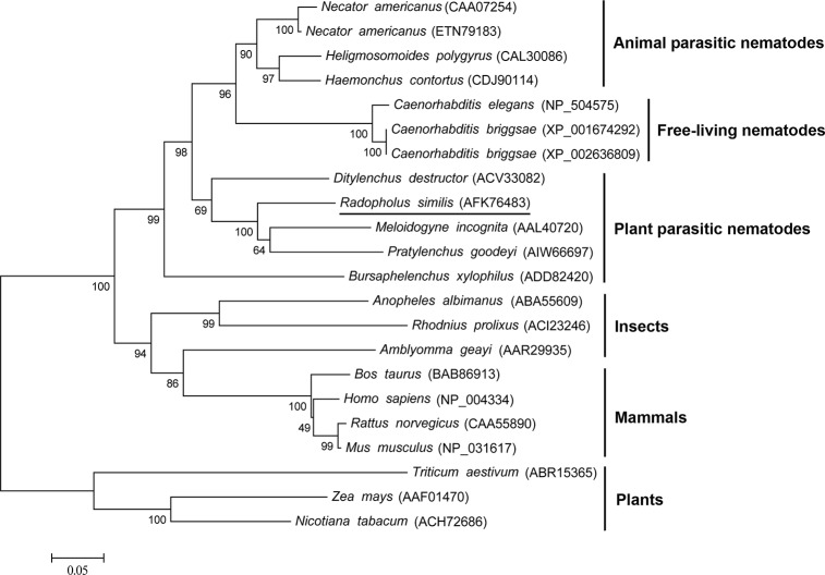
Radopholus similis is a migratory plant-parasitic nematode that causes severe damage to many agricultural and horticultural crops. Calreticulin (CRT) is a Ca2+-binding multifunctional protein that plays key roles in the parasitism, immune evasion, reproduction and pathogenesis of many animal parasites and plant nematodes. Therefore, CRT is a promising target for controlling R. similis. In this study, we obtained the full-length sequence of the CRT gene from R. similis (Rs-crt), which is 1,527-bp long and includes a 1,206-bp ORF that encodes 401 amino acids. Rs-CRT and Mi-CRT from Meloidogyne incognita showed the highest similarity and were grouped on the same branch of the phylogenetic tree. Rs-crt is a multi-copy gene that is expressed in the oesophageal glands and gonads of females, the gonads of males, the intestines of juveniles and the eggs of R. similis. The highest Rs-crt expression was detected in females, followed by juveniles, eggs and males. The reproductive capability and pathogenicity of R. similis were significantly reduced after treatment with Rs-crt dsRNA for 36 h. Using plant-mediated RNAi, we confirmed that Rs-crt expression was significantly inhibited in the nematodes, and resistance to R. similis was significantly improved in transgenic tomato plants. Plant-mediated RNAi-induced silencing of Rs-crt could be effectively transmitted to the F2 generation of R. similis; however, the silencing effect of Rs-crt induced by in vitro RNAi was no longer detectable in F1 and F2 nematodes. Thus, Rs-crt is essential for the reproduction and pathogenicity of R. similis.
Introduction
The burrowing nematode, Radopholus similis [(Cobb, 1893) Thorne, 1949] is a migratory plant parasitic nematode, that has been listed as a quarantine plant pest in many countries and regions [1, 2]. R. similis is known to reproduce on more than 250 plant species [3] and causes severe economic losses [4–6]. At present, the inability to control R. similis is a worldwide problem. Therefore, it is of particular importance to study and establish effective methods for controlling R. similis.
RNA interference (RNAi), which was first discovered in Caenorhabditis elegans, has been shown to be effective in most eukaryotic organisms [7]. RNAi mediated by dsRNA soaking (in vitro RNAi) has been developed as an effective tool for studying gene function in many plant nematodes and other organisms [8–13]. However, the silencing effect of in vitro RNAi on some genes in plant-parasitic nematodes is highly time-limited [11, 14]. As an alternative, plant-mediated RNAi has been successfully applied for gene knockdown with the aim of studying gene function and controlling many pests that impact crop production [15–20]. Plant-mediated RNAi may likewise serve as an effective technique for controlling plant nematodes in agriculture. However, most previous studies have focused on sedentary plant endoparasitic nematodes [21–26], and there is limited information available regarding the use of plant-mediated RNAi to control migratory plant parasitic nematodes.
Calreticulin (CRT) is a Ca2+-binding multifunctional protein that is highly conserved in animals and plants [27]. CRT has been shown to be secreted into host tissues by many animal parasites and to play important roles in skin penetration, infection, parasitism, pathogenesis and immune evasion [28–33]. In C. elegans, CRT is required for the stress response and for fertility [34]. In plant parasitic nematodes, CRT has only been isolated from the oesophageal secretions of Meloidogyne incognita [35–37], Ditylenchus destructor (GenBank accession number ACV33082) and Bursaphelenchus xylophilus [38].
In plant parasitic nematodes, oesophageal secretions (also referred to as stylet secretions) are produced by oesophageal subventral and dorsal glands cells and are secreted through the stylet during parasitism[39]. These stylet secretions are thought to play key roles throughout the process of parasitism [39–41]. At present, in addition to the calreticulin gene (crt), a variety of genes expressed in oesophageal gland cells have been identified and studied in plant nematodes. It has been reported that CRT plays important roles in parasitism and the suppression of plant defences in M. incognita [36, 37, 39] and in the reproduction of B. xylophilus [38]. As a promising target for controlling parasites, CRT has attracted much attention. However, the functions of the calreticulin gene in R. similis (Rs-crt) have not yet been examined. In this study, Rs-crt was first cloned from R. similis, and its structure and features were analyzed. The localization and expression of Rs-crt were determined in R. similis through in situ hybridization and qPCR, and the role of Rs-crt in reproduction and pathogenesis was investigated using in vitro RNAi and plant-mediated RNAi. In addition, we obtained transgenic tomato plants expressing Rs-crt dsRNA that showed obvious resistance to R. similis.
Materials and Methods
Ethics statement
We collected the nematodes in areas where banana-burrowing nematodes occured and no specific permits were required. The land used as the collection area is neither privately owned nor protected in any way, and the field studies did not involve endangered or protected species.
Nematode cultivation and extraction
R. similis was collected from the roots of Anthurium andraeanum, an ornamental plant, and cultured on carrot disks at 25°C as previously described [42]. At 50 d after inoculation, the cultured nematodes were extracted from the carrot disks as described elsewhere [12].
Plant materials
Tomato seeds were purchased from Changhe, Guangzhou, and surface sterilized as previously described [20]. The sterilized seeds were sown in 1.5 L of sterilized soil and grown in a greenhouse for 30 d. The sterilized seeds were also germinated on half-strength MS medium (pH 5.8) with 0.3% Phytagel [43] and cultured in a 25°C chamber (16 h-light/8 h-dark cycle) [20].
Cloning of Rs-crt from R. similis
Total RNA and genomic DNA (gDNA) were extracted from 20,000 mixed-stage nematodes using TRIzol reagent (Invitrogen, Carlsbad, CA, USA) and a tissue DNA kit (Magen, China), respectively. A partial Rs-crt cDNA sequence was amplified using the degenerate primers Cal1F and Cal2R (Table 1), which target crt from M. incognita [35]. Based on the sequence of the obtained fragment, 3′ RACE primers (Rscrt-3F/Outer primer) and 5′ RACE primers (SL/Rscrt-5R) (Table 1) [44] were synthesized to amplify the cDNA sequence of Rs-crt using the SMART RACE cDNA amplification kit (Clontech, Japan). The purified PCR products (5′ end, middle fragment and 3′ end) were sequenced and spliced into the complete sequence. Based on the spliced complete sequence, two pairs of primers, Rscrt-FullF/Rscrt-FullR and Rscrt-cdsF/Rscrt-cdsR (Table 1), were designed to amplify the full-length cDNA and gDNA sequences of Rs-crt.
Table 1. Primers used in this study.
| Primer name | Sequence | Primer use |
|---|---|---|
| Cal1F | 5′- GAAGTCTTYTTCAARGAGGAG -3′ | PCR amplification |
| Cal2R | 5′- GTTSTCAATYTCTGGRTGKATCCA -3′ | PCR amplification |
| Rscrt-3F | 5′- ACAAAGCAAAGAACCACCTGAT -3′ | 3′- RACE |
| Outer primer | 5′- TACCGTCGTTCCACTAGTGATTT -3′ | 3′- RACE |
| SL | 5′- GGTTTAATTACCCAAGTTTGAG -3′ | 5′- RACE |
| Rscrt-5R | 5′- TAACCTTCACATAGCCTCCTCC -3′ | 5′- RACE |
| Rscrt-FullF | 5′- GGTTTAATTACCCAAGTTTG -3′ | cDNA amplification |
| Rscrt-FullR | 5′- CTTGATGTCTGTGTCCATTCC -3′ | cDNA amplification |
| Rscrt-cdsF | 5′- GGAAATGATTAAATCAGTTGCACTC -3′ | DNA amplification |
| Rscrt-cdsR | 5′- TCTGCTTGTCGGTCTCAGAGCT -3′ | DNA amplification |
| Southern-F | 5′- ATCTGTGGCCCTGGTACTAAA -3′ | Southern blot |
| Southern-R | 5′- CCATTCTCCGTCCATCTCGT -3′ | Southern blot |
| ISH-T7S a | 5′- GGATCCTAATACGACTCACTATAGGGggaggctatgtgaaggtta -3′ | ISH template |
| ISH-A | 5′- CGGCAGTAGTTCCCAAT -3′ | ISH template |
| ISH-S1 | 5′- ggaggctatgtgaaggtta-3′ | ISH template |
| ISH-T7A1 a | 5′- GGATCCTAATACGACTCACTATAGGGCGGCAGTAGTTCCCAAT-3′ | ISH template |
| qPCR-F | 5′- aagcaaagaaccacctga -3′ | qPCR |
| qPCR-R | 5′- CGGCAGTAGTTCCCAAT -3′ | qPCR |
| Actin-F | 5′- GAAAGAGGGCCGGAAGAG -3′ | qPCR |
| Actin-R | 5′- AGATCGTCCGCGACATAAAG -3′ | qPCR |
| Rscrt-T7S a | 5′- GGATCCTAATACGACTCACTATAGGGatcgtaagggctgaggtatt-3′ | dsRNA template |
| Rscrt-A | 5′- CAGGTGGTTCTTTGCTTTGTA-3′ | dsRNA template |
| Rscrt-S | 5′- atcgtaagggctgaggtatt-3′ | dsRNA template |
| Rscrt-T7A a | 5′-GGATCCTAATACGACTCACTATAGGGACCGTTGCATCCCTGGCT -3′ | dsRNA template |
| eGFP-T7S a | 5′-GGATCCTAATACGACTCACTATAGGGCAGTGCTTCAGCCGCTACC-3′ | dsRNA template |
| eGFP-A | 5′-AGTTCACCTTGATGCCGTTCTT-3′ | dsRNA template |
| eGFP-S | 5′-CAGTGCTTCAGCCGCTACC-3′ | dsRNA template |
| eGFP-T7 A a | 5′- GGATCCTAATACGACTCACTATAGGGAGTTCACCTTGATGCCGTTCTT-3′ | dsRNA template |
| RNAi-F | 5′- CCGCTCGAGTCTAGAATCTGTGGCCCTGGTACTAAA -3′ | Vector construction |
| RNAi-R | 5′- CTAGCCATGGATCCCCATTCTCCGTCCATCTCGT-3′ | Vector construction |
| eGFP-F | 5′-CCGCTCGAGTCTAGATGCTTCAGCCGCTACCC-3′ | Vector construction |
| eGFP-R | 5′-CATGCCATGGATCCAGTTCACCTTGATGCCGTTC-3′ | Vector construction |
| CHSA-F | 5′- ACTTGCCTTGGAGTTTATGTT -3′ | PCR detection |
| OCS-R | 5′- TTGTTATTGTGGCGCTCTATC -3′ | PCR detection |
a The T7 promoter sequence is underlined.
Sequence analysis, alignment and phylogenetic analyses
Sequence homology comparisons were conducted using the BLASTX and BLASTN programs from NCBI (http://blast.ncbi.nlm.nih.gov/Blast.cgi). The ORF finder program was employed to predict the open reading frame (http://www.ncbi.nlm.nih.gov/gorf/gorf.html). The protein transmembrane regions, molecular weight, theoretical isoelectric point, glycosylation sites and hydrophobicity sites were predicted using the Protein Machine software available at ExPASy (http://www.expasy.ch/tools/). The signal peptide for secretion and the cleavage site were predicted using the SignalP 3.0 server [45]. The amino acid (aa) sequences of Rs-CRT (AFK76483) and six other CRT proteins from M. incognita (AAL40720), C. elegans (NP_504575), Necator americanus (CAA07254), D. destructor (ACV33082), B. xylophilus (ADD82420) and Pratylenchus goodeyi (AIW66697) were aligned using DNAMAN. Based on the aa sequences of 22 CRT proteins from 20 species, a neighbour-joining phylogenetic tree was constructed using MEGA5.0 [46].
Southern blot hybridization
The Southern-F and Southern-R primers (Table 1) were designed to amplify a 378-bp digoxigenin (DIG)-labelled probe using the PCR DIG Probe Synthesis Kit (Roche, Germany). Approximately 10 μg of R. similis gDNA was digested with EcoR I and Xba I. The digested DNA was separated via electrophoresis and then transferred to a Hybond N+ membrane (Amersham) [38]. Hybridization and detection were carried out using the Dig High Primer DNA Labeling and Detection Starter Kit I (Roche). An equal amount of gDNA from carrot callus was used as a control.
Tissue localization and expression of Rs-crt in R. similis
The specific primers ISH-T7S/ISH-A and ISH-S1/ISH-T7A1 (Table 1) were designed to synthesize DIG-labelled sense and antisense RNA probes (294-bp) using DIG RNA labelling mix (Roche). In situ hybridization was performed as described elsewhere [13, 47]. After hybridization, the nematodes were examined by microscopy (Nikon 90i).
RNA samples were extracted from 100 eggs, juveniles, females and males using the RNeasy Micro kit (Qiagen, Germany), respectively. Total RNA was then treated and quantified as previously described [13]. The RNA extracted from each sample served as a template for synthesizing cDNA using the iScript cDNA synthesis kit (Bio-Rad, USA). The primers qPCR-F/qPCR-R (Table 1) were designed to assay Rs-crt expression. The primers Actin-F/Actin-R (Table 1) were employed to amplify β-actin as a reference gene [48]. qPCR was performed using a CFX-96 qPCR instrument (Bio-Rad) with iTaq Universal SYBR Green Supermix (Bio-Rad). The initial data analysis was carried out using Bio-Rad CFX-96 manager software, which created Ct values and extrapolated the relative levels of PCR products from standard curves. Melt curves were obtained routinely, which allowed the possibility of both contamination and primer dimers to be discounted [12, 13]. All expression experiments were performed in triplicate with three biological replicates.
Knockdown of Rs-crt by soaking with target-specific dsRNA
The primers Rscrt-T7S/Rscrt-A and Rscrt-S/Rscrt-T7A, which contained a T7 promoter (Table 1), were designed to transcribe Rs-crt sense and antisense single-stranded RNA (ssRNA) using a T7 Transcription Kit (TOYOBO, Japan). The corresponding dsRNA was synthesized and purified as previously described [13, 49]. Non-endogenous control dsRNA (enhanced green fluorescent protein gene, egfp) was synthesized using the primers eGFP-T7S/eGFP-A and eGFP-S/eGFP-T7A (Table 1).
Approximately 1,000 mixed-stage nematodes were soaked in Rs-crt dsRNA solution (2.0 mg/mL) at 25°C for 12 h, 24 h, 36 h and 48 h, respectively. egfp dsRNA solution (2.0 mg/mL) was used as a non-endogenous control. The soaking times for the control were the same as those for the Rs-crt dsRNAs. Additionally, untreated nematodes were used as a blank control. Tests were subsequently performed as follows: (1) Total RNA was extracted from the nematodes in each treatment group, and qPCR was performed to analyze the suppression of Rs-crt mRNA expression in R. similis, as described above; (2) the total number of nematodes was calculated after culturing 30 different treated females on carrot disks for 50 d at 25°C.
After treatment with Rs-crt dsRNA for 36 h, 1,000 mixed-stage nematodes were inoculated onto each of the selected tomato plantlets, which had been grown under the same conditions. Nematodes treated with egfp dsRNA for 36 h were used as a control. Additionally, untreated nematodes were used as a blank control. These plants were cultivated ina greenhouse as described elsewhere [18]. At 60 d after inoculation, three growth parameters (plant height, fresh shoot weight and fresh root weight) were measured. The nematodes in the rhizosphere were extracted and quantified as previously described [12, 50]. Five biological replicates were performed.
Vector construction and plant transformation
The RNAi-F/RNAi-R primers (Table 1) were designed to amplify a 407-bp target sequence of Rs-crt. The digested PCR products were inserted into the XhoI/NcoI and XbaI/BamHI sites of pFGC5941 at inverted repeat sequences to obtain the plant RNAi vector pFGC-RS-crt2, which can generate a hairpin RNAi construct. The non-endogenous control vector pFGC-egfp2 was constructed using the primers eGFP-F/eGFP-R (Table 1). The pFGC-RS-crt2 and pFGC-egfp2 vectors and an empty pFGC5941 vector were transformed into Agrobacterium tumefaciens (strain EHA105) via the freeze-thaw method [51]. Tomato transformation was performed according to the methods described by Arshad et al. [20], and the transformants were selected on MS medium containing kanamycin. The regenerated transgenic plants were transferred to soil and grown in the greenhouse to obtain seeds.
Molecular analysis of putative transgenic tomato plants
gDNA was extracted from kanamycin-tolerant T0 Rs-crt transgenic tomato leaves using the HiPure Plant DNA Maxi Kit (Magen, China) and was assessed via PCR and sequencing using the CHSA-F/OCS-R and RNAi-F/RNAi-R primers (Table 1). The egfp transgenic plants were similarly checked using the CHSA-F/OCS-R and eGFP-F/eGFP-R primers (Table 1). For Southern blot analysis, 15 μg of gDNA from PCR-positive Rs-crt transgenic plants was digested with EcoR I and transferred to a Hybond-N+ membrane. Hybridization and detection were performed as described above. Equal amounts of gDNA from T0 egfp transgenic plants and empty transformation vector plants were used as controls.
Total RNA was extracted from the leaves of PCR- and Southern-positive Rs-crt transgenic plants and assessed via RT-PCR using the RNAi-F/RNAi-R primers (Table 1). Positive Rs-crt transgenic plants were cultured to obtain seeds. After approximately 60 d, 20 seeds (T1) collected from a single plant were sown in pots. After 30 d of culture, gDNA was extracted from these T1 transgenic plants and checked via PCR using the RNAi-F/RNAi-R primers (Table 1). Positive T1 plants were cultured to obtain T2 homozygous transgenic plants for further analyses.
Analysis of transgenic tomato plant resistance
A total of 1,000 mixed-stage nematodes were inoculated onto each of the selected T2 Rs-crt transgenic plantlets. All plantlets were grown under the same conditions. At 60 d after inoculation, the resistance of the Rs-crt transgenic plants was examined according to the method described in a previous section of this report (“Knockdown of Rs-crt by soaking with target-specific dsRNA”). T2 egfp transgenic plants, empty transformation vector plants and wild-type tomato plants inoculated with nematodes were used as controls. Five biological replicates were performed.
Analysis of Rs-crt expression in R. similis feeding on transgenic plants
Total RNA was extracted from 100 mixed-stage nematodes isolated from T2 Rs-crt transgenic tomato roots using the RNeasy Micro Kit (Qiagen). qPCR was performed to analyze Rs-crt suppression in R. similis as described above. Nematodes isolated from T2 egfp transgenic plants, empty transformation vector plants and wild-type tomato plants were used as controls.
Persistence and inheritance of Rs-crt silencing induced by in vitro RNAi and plant-mediated RNAi
To determine the transcript recovery of Rs-crt, mixed-stage nematodes collected from different sources (treated with Rs-crt dsRNA for 36 h and isolated from T2 Rs-crt transgenic tomato roots) were washed and maintained in sterile water for 1, 3, 5, 7, 9, 11, 13, and 15 d, respectively. Then, 100 nematodes in each group were collected for qPCR analysis as described above. The remaining nematodes were maintained in water at 25°C, and the water was changed daily [14]. Untreated nematodes were used as a control.
Approximately 100 females (P0 generation) collected from different sources were inoculated onto carrot disks. After being cultured at 25°C for 20 d, the first-generation (F1) juveniles and immature females were extracted from the carrot disks and used for the following experiments: (1) A total of 100 mixed-stage F1 nematodes were used for RNA extraction, and qPCR was performed to analyze Rs-crt expression in R. similis as described above; (2) 30 F1 females were inoculated onto carrot disks and incubated at 25°C for 30 d, and the total number of nematodes was subsequently calculated; and (3) wild-type tomato plantlets were selected as described above, and each plantlet was inoculated with 200 mixed-stage nematodes (F1). At 45 d after inoculation, three growth parameters were measured in these plants. The rhizosphere nematodes were extracted and quantified. Plantlets inoculated with untreated nematodes were used as controls. Five biological replicates were performed.
Approximately 100 F1 females collected from different sources were cultured as described above. The obtained second-generation (F2) juveniles and immature females were used for the following experiments: (1) qPCR was performed to analyze Rs-crt expression in F2 nematodes as described above; (2) 30 F2 females from different sources were inoculated onto carrot disks and incubated at 25°C for 30 d, and the number of nematodes was calculated; and (3) these F2 female nematodes respectively were cultured to obtain F3 nematodes. qPCR was used to analyze Rs-crt expression in F3 nematodes as described above.
Statistical analysis
The data collected from the experiments were analyzed using SAS 9.2 (SAS Institute, Cary, NC, USA). All of the data obtained in this study were subjected to one-way analysis of variance (ANOVA) and tested for differences between treatments at the 5% level using Duncan’s Multiple Range Test (DMRT).
Results
Cloning and characterization of the Rs-crt gene in R. similis
An 832-bp partial cDNA sequence of Rs-crt was amplified via qPCR (S1 Fig). Based on the sequence of this fragment, the full-length cDNA sequence of Rs-crt was obtained through RACE-PCR (1,527-bp; JX067552). The Rs-crt cDNA was composed of a 52-bp 5′-untranslated region (5′-UTR), a 1,206-bp ORF, and a 269-bp 3′-UTR containing a typical polyadenylation signal (ATTAAA) (S2 Fig). Amplification of Rs-crt using gDNA as a template yielded a 1,515-bp fragment (from theATG to the stop codon). Introns were identified by aligning the genomic sequence to the corresponding cDNA sequence. Rs-crt gDNA contained six introns and seven exons (S3 Fig).
The Rs-CRT protein encodes 401 aa with a theoretical molecular mass of 46.99 kDa and an isoelectric point of 4.73. The molecular formula of Rs-CRT is C2089H3175N545O672S10. An 18 aa signal peptide with a cleavage site between Ala18 and Glu19 was predicted by SignalP 3.0 at the N-terminus of the deduced Rs-CRT sequence. BLASTP searches revealed that the Rs-CRT aa sequence showed high similarity to CRT sequences from five other nematode species. Rs-CRT (AFK76483) had the highest similarity with Mi-CRT from M. incognita (AAL40720, 84% identity and 92% similarity, E-value = 0) and Pg-CRT from P. goodeyi (AIW66697, 84% identity and 92% similarity, E-value = 0). Rs-CRT also showed very higher similarity with Dd-CRT-1 from D. destructor (ACV33082, 82% identity and 92% similarity, E-value = 0) and Bx-CRT from B. xylophilus (ADD82420, 73% identity and 84% similarity, E-value = 0). Additionally, Rs-CRT exhibited high similarity with Na-CRT from N. americanus (CAA07254, 77% identity and 88% similarity, E-value = 0) and Ce-CRT-1 from C. elegans (NP_504575, 73% identity and 86% similarity, E-value = 0) (Fig 1).
Fig 1. Multiple sequence alignment of the predicted Radopholus similis Rs-CRT protein with other nematode CRT proteins.
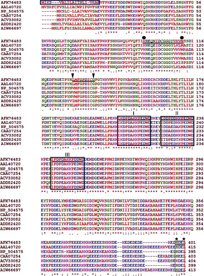
GenBank accession numbers: R. similis (AFK76483), Meloidogyne incognita (AAL40720), Caenorhabditis elegans (NP_504575), Necator americanus (CAA07254), Ditylenchus destructor (ACV33082), Bursaphelenchus xylophilus (ADD82420), Pratylenchus goodeyi (AIW66697). Box, the putative signal peptide sequences of Rs-CRT; I-III, the conserved regions characteristic of the calreticulin class; black circle, possible Ca2+-binding signal motif one; black triangle, possible Ca2+-binding signal motif two; black pentagram, C-terminal endoplasmic reticulum (ER) retention signal sequences; (*), highly conserved amino acid residues; (:), conserved amino acid residues; (.) conserved cysteine residues.
A phylogenetic tree was constructed based on the aa sequences of 22 CRT proteins from 20 different species (Fig 2). Rs-CRT from R. similis, Mi-CRT from M. incognita and Pg-CRT from P. goodeyi were grouped on the same branch, suggesting that they have a closer phylogenetic relationship. In addition, Rs-CRT and the other 21 CRT proteins were divided into 6 groups: animal parasitic nematodes, free-living nematodes, plant parasitic nematodes, insects, mammals, and plants.
Fig 2. Neighbor-joining phylogenetic tree of CRT proteins.
The phylogram was constructed according to the amino acid sequences of 22 CRT proteins from 20 different species using MEGA 5.0. R. similis CRT is underlined. The accession numbers of the sequences are shown in brackets.
Southern blotting was carried out to investigate the gene copy number of Rs-crt in R. similis. The 378-bp-long DIG-labelled probe hybridized to multiple fragments in both the EcoRI- and XbaI-digested gDNA of R. similis. No hybridization signal was detected when using carrot callus gDNA. Neither the genomic coding region nor the cDNA contained an EcoRI or XbaI site. These results indicate that Rs-crt exist as a multi-copy gene in the R. similis genome (Fig 3J).
Fig 3. Localization, expression and Southern blot analysis of Rs-crt in Radopholus similis.
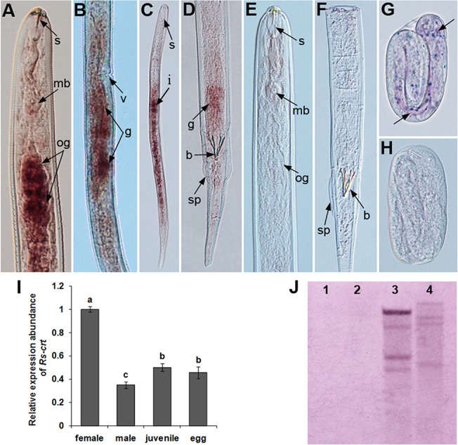
(A-H) Tissue localization of Rs-crt mRNA via in situ hybridization. Rs-crt was localized in the oesophageal glands (A) and gonads (B) of females, the gonads of males (D), the intestines of juveniles (C) and the eggs (G). (E, F and H) No hybridization signal was observed in nematodes hybridized with the DIG-labelled sense RNA probe. b, bursa; g, gonads; mb, medium bulb; og, oesophageal glands; s, stylet; sp, spicules; v, vulva. (I) Rs-crt expression in females, males, juveniles and eggs of R. similis. Bars indicate the standard errors of the mean data (n = 3), and different letters indicate significant differences (p<0.05) between treatments. (J) Southern blot analysis of Rs-crt. Lanes 1–2, gDNA from carrot callus digested with EcoR I and Xba I; lanes 3–4, gDNA from R. similis digested with EcoR I and Xba I.
Tissue localization and expression of Rs-crt in R. similis
In situ hybridization was performed to localize Rs-crt expression. A DIG-labelled antisense probe specifically hybridized with Rs-crt transcripts in the oesophageal glands and gonads of females (Fig 3A and 3B), the gonads of males (Fig 3D), the intestines of juveniles and the eggs of R. similis (Fig 3C and 3G). No hybridization signal was observed in the nematodes when the control sense probe was used (Fig 3E, 3F and 3H). The qPCR results showed that the Rs-crt mRNA transcript was present in all developmental stages of R. similis, and the expression was significantly highest in females (p < 0.05). Compared to its level in females, percentage reduction of Rs-crt expression were 65.1%, 50.0% and 54.5% in males, juveniles and eggs, respectively. Rs-crt expression in males was significantly lower (p < 0.05) than that in juveniles and eggs. No significant difference in expression (p > 0.05) was observed between juveniles and eggs (Fig 3I).
Induction of RNAi in R. similis by soaking with target-specific Rs-crt dsRNA
qPCR was employed to measure the Rs-crt silencing efficiency in R. similis after treatment with Rs-crt dsRNA for 12 h, 24 h, 36 h and 48 h, and it was shown that Rs-crt expression was decreased significantly (p < 0.05) by 53.0%, 58.9%, 79.0% and 58.5%, respectively, compared to that observed in the corresponding egfp dsRNA treatments. The silencing efficiency was enhanced with increasing incubation durations within a certain range and was highest at 36 h. The egfp dsRNA-treated and untreated nematodes showed no significant difference (p > 0.05) in Rs-crt expression (Fig 4A). After being cultured on carrot disks for 50 d, R. similis treated with Rs-crt dsRNA for 12 h, 24 h, 36 h and 48 h had significantly lower reproduction (p < 0.05) than untreated and egfp dsRNA-treated nematodes. Nematodes treated with Rs-crt dsRNA for 36 h had the lowest reproduction, showing significant differences (p < 0.05) compared with the 12 h and 24 h treatments. There was no significant difference (p > 0.05) in reproduction among the untreated and egfp dsRNA-treated nematodes (Fig 4B).
Fig 4. Inducing RNAi in Radopholus similis by soaking with Rs-crt dsRNA.
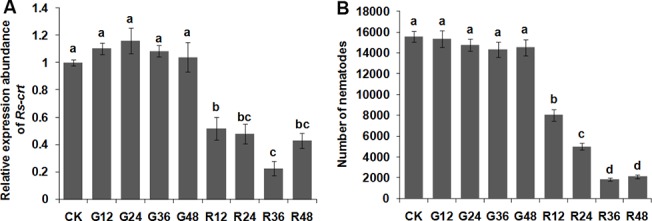
(A) Expression of Rs-crt mRNA in R. similis treated with Rs-crt dsRNA. Bars indicate the standard errors of the mean (n = 3). (B) Number of nematodes on carrot disks 50 d after the inoculation of 30 females. Bars indicate the standard errors of the mean data (n = 5). CK: untreated nematodes; G12-G48: nematodes treated with egfp dsRNA for 12 h, 24 h, 36 h and 48 h, respectively; R12-R48: nematodes treated with Rs-crt dsRNA for 12 h, 24 h, 36 h and 48 h, respectively. Different letters indicate significant differences (p<0.05) between treatments.
At 60 d after inoculation, the tomato plants inoculated with nematodes treated with Rs-crt dsRNA for 36 h showed significant increases in growth parameters compared with plants inoculated with egfp dsRNA-treated nematodes for 36 h and plants inoculated with untreated nematodes (i.e.,the two control treatments); however, the values of these growth parameters were still significantly lower than those in uninoculated healthy plants (p < 0.05) (Fig 5A–5C). The number of nematodes in the rhizosphere of plants inoculated with nematodes treated with Rs-crt dsRNA for 36 h was significantly lower (p < 0.05) than that in the two control treatments (Fig 5D). There was no significant difference between the two control groups (p > 0.05) in terms of the three growth parameters or nematode numbers (Fig 5). These results confirmed that the pathogenicity of R. similis was significantly reduced after treatment with Rs-crt dsRNA for 36 h.
Fig 5. The pathogenicity of Radopholus similis to tomato plants is decreased significantly after soaking with Rs-crt dsRNA.
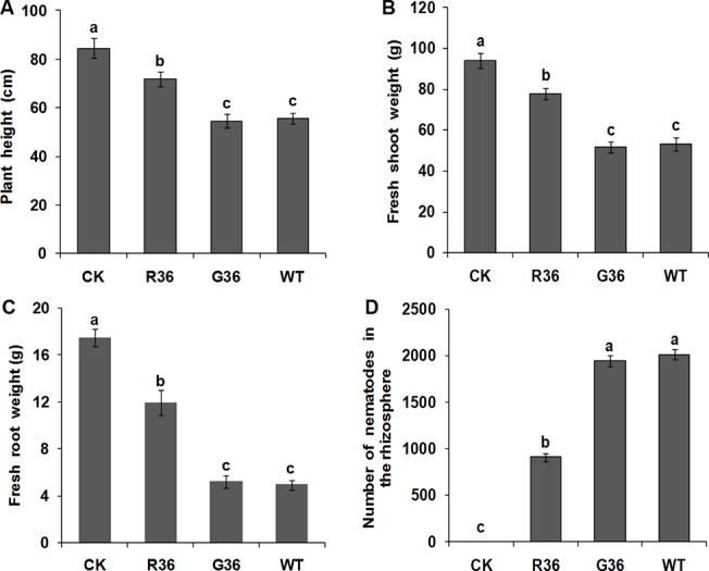
Plant height (A), fresh shoot weight (B), fresh root weight (C) and number of nematodes in the rhizosphere (D) of plants at 60 d after nematodes inoculation. CK: uninoculated healthy plants; R36 and G36: plants inoculated with nematodes treated with Rs-crt dsRNA and egfp dsRNA for 36 h, respectively; WT: plants inoculated with untreated nematodes. Bars indicate the standard errors of the mean data (n = 5), and different letters indicate significant differences (p <0.05) between treatments.
Production and molecular analysis of transgenic RNAi plants
The constructed RNAi expression vector pFGC-RS-crt2 contained a CaMV35S promoter, 407-bp sense and antisense fragments of Rs-crt cDNA, a CHSA intron and an OCS terminator. The inverted repeats present in Rs-crt were separated by the CHSA intron (S4 Fig). Transgenic tomato plants were regenerated from transformed calli, which were selected based on resistance to kanamycin (S5 Fig). Using this approach, 27 Rs-crt transgenic plants, 18 egfp transgenic plants and 13 empty vector transgenic plants were obtained. These transgenic lines did not show obvious morphological differences compared with wild-type tomato plants (S5 Fig).
Two fragments with lengths of 407-bp and 777-bp were amplified from the DNA of T0 Rs-crt transgenic tomato plants. A 407-bp fragment was also amplified from the empty transformation vector plants, but no bands were amplified from the wild-type plants (Fig 6A). Two fragments with lengths of 315-bp and 685-bp were amplified from the egfp transgenic plants (results not shown). These results revealed that the hairpin dsRNAs were successfully inserted into the tomato gDNA. Southern blot analysis showed that PCR-positive Rs-crt transgenic plants carried 1–3 copies of the target coding sequence. Conversely, no hybridization band was observed when using gDNA from the egfp transgenic plants and empty transformation vector plants (Fig 6C). RT-PCR analysis indicated that the integrated Rs-crt dsRNA was successfully expressed in transgenic plants (Fig 6B). The ratio of positive and negative T1 Rs-crt transgenic plants (Nos. 1, 2 and 4) was 3:1, and a 407-bp fragment corresponding to the Rs-crt sequence was amplified from positive plants. These results revealed that the integrated Rs-crt could be stably inherited in transgenic tomato gDNA (S6 Fig). Previous reports have shown that the effectiveness of RNAi is higher in single-copy lines than in other lines [52, 53]. Therefore, we selected the single-copy Rs-crt transgenic plant (No. 2) for further analyses.
Fig 6. Molecular analysis of transgenic tomato plants.

(A) Independently derived transgenic lines were detected via PCR using the primers RNAi-F/RNAi-R and CHSA-F/OCS-R (lanes 1–9: independent Rs-crt transgenic lines). (B) Independent transgenic lines were detected via RT-PCR using the primers RNAi-F/RNAi-R (lanes 1–5: RNA from Rs-crt transgenic lines 1, 2, 3, 4 and 5). M, DNA marker (DL2000); B, blank control without template; V, empty transformation vector plant; W, wild-type plant (negative control); P, positive plasmid control. (C) Southern blot analysis of NdeI-digested gDNA from T0 transgenic plant leaves (lanes v and g: gDNA from empty transformation vector plants and egfp transgenic plants (controls); lanes 1–5, gDNA from Rs-crt transgenic lines 1, 2, 3, 4 and 5).
T2 generation transgenic tomato plants expressing Rs-crt dsRNA exhibit greater resistance to R. similis
At 60 d after inoculation, the growth parameters of Rs-crt transgenic plants (No. 2) were significantly (p < 0.05) increased compared to the growth parameters of three types of inoculation control plants (egfp transgenic plants, empty transformation vector plants and wild-type tomato plants); however, no significant difference (p > 0.05) was observed between the Rs-crt transgenic plants and the uninoculated wild-type plants. The percentage reduction of plant height, fresh shoot weight and fresh root weight of Rs-crt transgenic plants were only 1.64%, 3.63% and 10.92%, respectively, compared to the uninoculated wild-type tomato plants (CK), (Fig 7A–7C). The number of nematodes in the rhizosphere of the Rs-crt transgenic plants was significantly lower (p < 0.05) than that observed in the rhizosphere of the three inoculation control plants (Fig 7D). Additionally, the degree of root damage was much lower and there was no obvious root rot in the Rs-crt transgenic plants compared with the three inoculation control plants (Fig 7E). There was no significant difference (p > 0.05) in these pathogenicity measures among the three types of inoculation control plants, and their roots were severely damaged, showing obvious root rot (Fig 7). The inoculation tests demonstrated that resistance to R. similis was significantly increased in Rs-crt transgenic plants.
Fig 7. T2 transgenic tomato plants expressing Rs-crt dsRNA showed improved resistance to Radopholus similis.
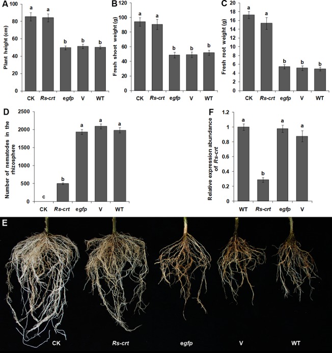
Plant height (A), fresh shoot weight (B), fresh root weight (C), number of nematodes in the rhizosphere (D) and root infection symptoms (E) in different plants at 60 d after inoculation with 1,000 nematodes. Bars indicate the standard errors of the mean (n = 5). (F) qPCR assay to detect Rs-crt expression in R. similis individuals collected from T2 transgenic plants. Bars indicate the standard errors of the mean (n = 3). CK, uninoculated wild-type plants; Rs-crt, Rs-crt transgenic plants; egfp, egfp transgenic plants; V, empty transformation vector plants; WT, wild-type plants. Different letters indicate significant differences (p<0.05) between treatments.
Rs-crt expression is significantly suppressed in R. similis feeding on T2 Rs-crt transgenic tomato plants
The percentage reduction of Rs-crt expression in R. similis fed on T2 Rs-crt transgenic plants were 70.6%, 67.3% and 71.3% compared with that in nematodes fed on the egfp transgenic plants, empty transformation vector plants and wild-type tomato plants, respectively. There were no significant differences (p > 0.05) among the three inoculation control treatments (Fig 7F). Therefore, it can be concluded that the suppression of Rs-crt expression in R. similis, which was caused by feeding on the roots of Rs-crt dsRNA-expressing transgenic tomato plants, resulted in a reduction of pathogenicity.
Persistence and inheritance of Rs-crt silencing induced by in vitro RNAi and plant-mediated RNAi
Following recovery in sterile water for 1–7 d, Rs-crt expression in R. similis soaked with Rs-crt dsRNA for 36 h was significantly (p < 0.05) reduced by 50.6–73.9% compared with Rs-crt expression in untreated nematodes (CK group); however, no significant difference (p > 0.05) was found between the different recovery times. After recovery for 9 d and 11 d, Rs-crt expression was significantly (p < 0.05) increased to 396% and 174% of that in the CK group, respectively. After recovery for 13 d and 15 d, Rs-crt expression returned to normal levels (Fig 8A). However, Rs-crt expression in nematodes isolated from T2 Rs-crt transgenic tomato plants and recovered in water for 1–15 d was significantly (p < 0.05) reduced by 55.2%-66.8% compared with the CK group, but no significant difference (p > 0.05) was found between the different recovery times (Fig 8B).
Fig 8. Persistence and inheritance of Rs-crt silencing induced by RNAi.
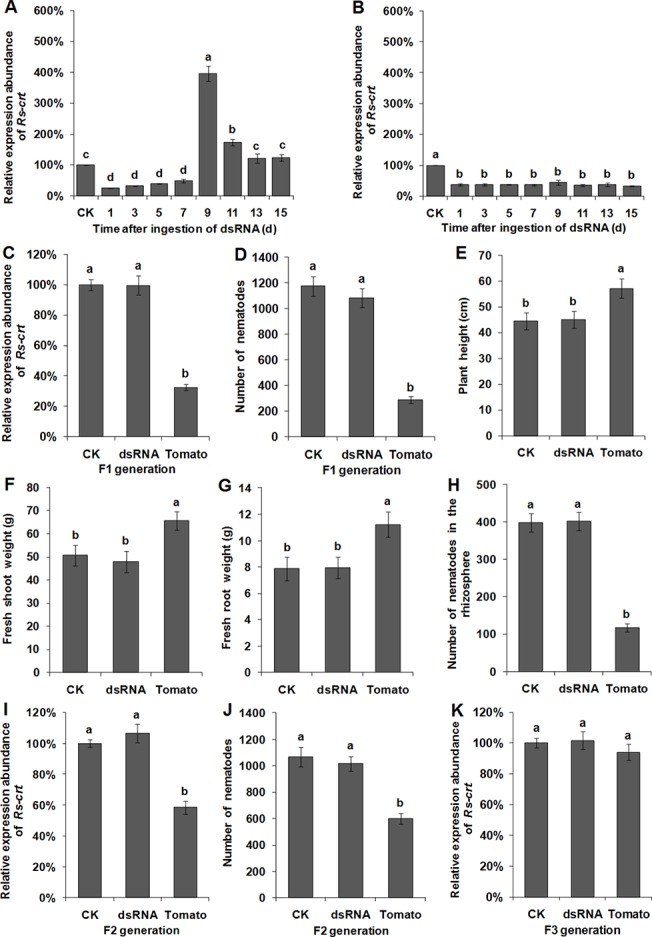
The recovery of Rs-crt expression in Radopholus similis at different times after soaking in Rs-crt dsRNA for 36 h (A) and in R. similis collected from T2 Rs-crt transgenic tomato roots (B). 1–15, Rs-crt expression levels in nematodes maintained in water and sampled at 1, 3, 5, 7, 9, 11, 13 and 15 d post-treatment, respectively. qPCR assay to detect Rs-crt expression in F1 (C), F2 (I) and F3 (K) nematodes. Bars indicate the standard errors of the mean (n = 3). Number of F1 (D) and F2 (J) nematodes on carrot disks at 30 d after the inoculation of 30 females. Plant height (E), fresh shoot weight (F), fresh root weight (G) and the number of nematodes in the rhizosphere (H) at 45 d after the plants were inoculated with 200 F1 nematodes. CK, untreated nematodes; dsRNA, nematodes treated with Rs-crt dsRNA for 36 h; Tomato, nematodes collected from T2 Rs-crt transgenic tomato roots. Bars indicate the standard errors of the mean (n = 5), and different letters indicate significant differences (p<0.05) between different treatments.
Rs-crt expression in F1 nematodes derived from T2 Rs-crt transgenic plants was reduced by 67.4% compared with the CK group. This reduction was significantly greater (p < 0.05) than that observed in the Rs-crt dsRNA soaking treatment and CK groups; however no significant difference (p > 0.05) was observed between the latter two groups (Fig 8C). After being cultured for 30 d, the F1 nematodes derived from Rs-crt transgenic plants exhibited significantly lower reproduction than the F1 nematodes derived from the Rs-crt dsRNA soaking treatment and CK groups (p < 0.05), but there was no significant difference between the latter two groups (p > 0.05) (Fig 8D). At 45 d after inoculation, the growth parameters of wild-type tomato plants inoculated with F1 nematodes derived from Rs-crt transgenic plants were significantly increased compared to the Rs-crt dsRNA soaking treatment and CK groups, and the number of nematodes in the rhizosphere was 118, which was significantly lower than that in the Rs-crt dsRNA soaking treatment (402) and CK groups (398) (p < 0.05). However, there was no significant difference between the last two groups (p > 0.05) (Fig 8E–8H).
Rs-crt expression in F2 nematodes derived from T2 Rs-crt transgenic plants was significantly (p < 0.05) reduced by 41.6% compared with the CK group (Fig 8I). After being cultured for 30 d, the F2 nematodes derived from Rs-crt transgenic plants exhibited significantly lower (p < 0.05) reproduction compared with the F2 nematodes derived from the Rs-crt dsRNA soaking treatment and CK groups (Fig 8J). Rs-crt expression in the F3 nematodes derived from T2 Rs-crt transgenic plants showed no significant difference (p > 0.05) compared with the Rs-crt expression in F3 nematodes derived from the Rs-crt dsRNA soaking treatment and CK groups (Fig 8K).
These results suggested that Rs-crt expression in R. similis isolated from T2 Rs-crt transgenic plants was significantly inhibited after recovery in water and that this inhibition was still observable in the F1 and F2 nematodes. The reproductive capability and pathogenicity of the F1 and F2 nematodes were significantly reduced (p < 0.05). However, Rs-crt expression in F3 nematodes returned to normal levels. Conversely, in vitro RNAi-induced Rs-crt silencing only lasted for a limited time and could be recovered; the F1 and F2 nematodes exhibited normal expression of Rs-crt and showed normal reproductive capacity and pathogenicity. Overall, these results indicate that plant-mediated RNAi-induced Rs-crt silencing could be effectively transmitted to F2 nematodes.
Discussion
This study is the first to describe the cloning of the full-length Rs-crt sequence from R. similis and the identification of its structure, features and function. We confirmed that Rs-crt plays key roles in the reproduction and pathogenicity of R. similis using in vitro and plant-mediated RNAi. Plant-mediated RNAi-induced Rs-crt silencing could be effectively transmitted to F2 nematodes, whereas the silencing effect of Rs-crt induced by in vitro RNAi was no longer detectable in F1 and F2 nematodes.
Park et al. [34] reported that crt-1 was expressed in the pharynx, intestine, body-wall muscles, coelomocytes and sperm of C. elegans. Khalife et al. [28] confirmed that crt was mainly expressed in the genital organs and digestive duct epithelia of Schistosoma mansoni. Gao et al. [33] demonstrated that crt was ubiquitously expressed in different tissues and at different developmental stages of Haemaphysalis qinghaiensis. In plant parasitic nematodes, Mi-crt is expressed in the female gonads and in the subventral oesophageal glands of M. incognita [39]. The present study revealed that the expression and localization of Rs-crt are associated with the biological functions of CRT. CRT and other related proteins are necessary for R. similis to break the host defence response and complete the infection process as well as to obtain nutrients for metabolism and reproduction. R. similis females are responsible for both infection and reproduction; consequently, Rs-crt expression was found to be highest in females. CRT plays important roles in infection, the establishment of the parasitic relationship, reproduction and cell differentiation in nematodes [36–39]. The successful invasion of host plants and establishment of the parasitic relationship by R. similis is a precondition for other functions to be implemented; therefore, Rs-crt expression was observed to be slightly higher in juveniles than in eggs, but this difference was not significant. R. similis males, which present a degraded stylet and oesophagus, are non-parasitical and are not necessary for reproduction, as this species can reproduce via parthenogenesis. Therefore, Rs-crt expression was found to be lowest in males. Cheng et al. [13] reported that the Ab-far-1 expression level was lowest in males and was only 4.7% of the Ab-far-1 expression level in females. In the current study, the Rs-crt expression level was also lowest in males and was 34.9% of the Rs-crt expression level in females. This result reveals that CRT is a multifunctional protein that also plays important roles in R. similis males. Previous studies have shown that CRT plays key roles in protein export, cell adhesion, mRNA degradation and cellular calcium homeostasis [39]. Mi-crt from M. incognita and Rs-eng-1b from R. similis are genes that are expressed in the oesophageal gland and help nematodes to break the host defence, establish the parasitic relationship, and digest host tissue to obtain necessary nutrients [12, 39]. In this study, Rs-crt was found to be localized in the oesophageal glands of R. similis; therefore, it is likely to have functions related to Mi-crt and Rs-eng-1b. Additionally, Rs-crt was found to be localized in the reproductive system, possibly because CRT plays roles in reproduction, development and cell differentiation in R. similis. A previous report showed that Bx-crt-1 has functions related to B. xylophilus development and reproduction [38]. Similarly, the present study revealed that silencing of Rs-crt through in vitro RNAi significantly reduced the reproductive capability and pathogenicity of R. similis. These results were consistent with the expression and localization patterns of Rs-crt described above.
In this study, we confirmed that Rs-crt expression was significantly inhibited in R. similis that feed on Rs-crt transgenic tomato plants, and the pathogenicity of the nematodes was significantly reduced on transgenic plants expressing Rs-crt dsRNA. These results were consistent with the roles of Rs-crt in R. similis, which were validated by in vitro RNAi. Plant-mediated RNAi has been used to validate gene functions in cyst nematodes, root-knot nematodes and other sedentary endoparasites [23–26, 54]. As there is limited information available regarding in planta RNAi against migratory endoparasites [55] compared to sedentary endoparasites [21–26], the current investigation will enrich the database of RNAi against migratory endoparasites.Rosso et al. [11] reported that the in vitro RNAi-induced silencing effect was temporary, and transcript depletion of mi-crt and Mi-pg-1 was undetectable 68 h after soaking. The expression of hg-eng-1 in Heterodera glycines (J2) was severely inhibited 16 h after forced ingestion of dsRNA; however, this expression level was significantly increased after recovery in water for 10 d and returned to normal levels after recovery for 15 d [14]. The present study revealed that Rs-crt expression in R. similis soaked with Rs-crt dsRNA for 36 h was significantly inhibited after recovery in water for 1–7 d; however, Rs-crt expression was significantly increased to 3.96-fold of that in the control group after 9 d of recovery and returned to normal levels after 13 d of recovery. These results indicate that in vitro RNAi-induced gene silencing is time-limited, possibly because the amount of ingested dsRNA is limited and its effect decrease as dsRNA degrades in nematodes. Following treatment with dsRNA, the expression levels of the target geneswere highest in R. similis and H. glycines after recovery for 9 d and 10 d, respectively. This result may have occurred because the regulation system of nematodes triggers an increase in mRNA synthesis when the expression of the corresponding target gene is low for an extended period of time after the loss of RNAi efficiency. Subsequently, the expression levels return to normal levels under the control of standard gene regulation mechanisms.
Steeves et al. [22] reported that the number of H. glycines eggs was significantly reduced after the nematodes were fed on transgenic soybean roots expressing specific MSP dsRNA, and ability of the progeny (F1) to successfully reproduce was also impaired. These results provided the first confirmation of the inheritability of gene silencing induced by plant-mediated RNAi in sedentary plant nematodes; however, the question of whether the progeny of migratory plant nematodes can inherit this RNAi effect has not yet been addressed. The present study revealed that Rs-crt expression in F1 and F2 R. similis derived from T2 Rs-crt transgenic tomato plants was severely inhibited, and the reproductive capability and pathogenicity of the nematodes was significantly reduced. The effect of RNAi treatment disappeared in F3 nematodes. Conversely, in vitro RNAi-induced Rs-crt silencing lasted only for a limited time, and Rs-crt expression could be recovered. Thus, we confirmed that plant-mediated RNAi-induced Rs-crt silencing could be effectively inherited by F2 R. similis, whereas the silencing effects induced by in vitro RNAi were not heritable. These results suggest that plant-mediated RNAi could overcome the limitations of in vitro RNAi and might be successfully applied in both sedentary and migratory plant nematodes. These findings provide a scientific basis for further studies of the relationships between nematodes and their hosts and for the development of new methods for controlling plant parasitic nematodes and other pests.
Supporting Information
An 832-bp PCR fragment was amplified using the degenerate primers Cal1Fand Cal1R.
(TIF)
The 5′- and 3′- untranslated regions (UTR) are shown in lowercase letters, and the open reading frame is shown in uppercase letters. The putative polyadenylation signal (attaaa) is boxed. ATG, initiation codon; TGA, stop codon.
(TIF)
The Rs-crt genomic coding region contains six introns and seven exons. Introns are marked in dark grey. ATG, initiation codon; TGA, stop codon.
(TIF)
The constructed pFGC-RS-crt2 vector contains a CaMV 35S promoter, 378-bp sense and antisense fragment of Rs-crt cDNA, a CHSA intron and an octopine synthase (ocs) terminator.
(TIF)
(A-E) Development of transgenic plants expressing Rs-crt dsRNA. (A) Preculture of explants. (B) Putative transformed calli growing on selection medium. (C) Transgenic plantlets germinated from transformed calli. (D, E) Transgenic plants growing on rooting medium. (F) Growth morphology of the transgenic tomato leaves. No obvious differences were observed between the transgenic and wild-type tomato plants. 1, 2, 3 and 4: expression of wild-type, Rs-crt transgenic, egfp transgenic and empty transformation vector tomato leaves, respectively.
(TIF)
(A-C) Expression of genomic DNA from Rs-crt transgenic lines 1, 2 and 4, respectively; M: DNA marker (DL2000); 1–15: independent T1 Rs-crt transgenic lines from the same T0 transgenic tomato seeds.
(TIF)
Acknowledgments
We thank Dr. Guozhang Kang (National Engineering Research Centre for Wheat, Henan Agricultural University, China) for providing the binary vector pFGC5941 and Prof. Huaping Li (South China Agricultural University) for contributing Agrobacterium tumefaciens strain EHA105.
Data Availability
All relevant data are within the paper and its Supporting Information files.
Funding Statement
This work was funded by the National Natural Science Foundation of China (Nos. 31071665 and 31371920). The funders had no role in study design, data collection and analysis, decision to publish, or preparation of the manuscript.
References
- 1. Cotton J, Van RH. Quarantine: Problems and Solutions In: Evans K, Trudgill DL, Webster JM, editors. Plant Parasitic Nematodes in Temperate Agriculture. Oxford: CAB International; 1993. pp. 593–607. [Google Scholar]
- 2. Smith IM, Charles LMF. Distribution Maps of Quarantine Pests for Europe: CABI and EPPO: CABI Publishing; 1998. [Google Scholar]
- 3. Haegeman A, Elsen A, De Waele D, Gheysen G. Emerging molecular knowledge on Radopholus similis, an important nematode pest of banana. Mol Plant Pathol. 2010; 11: 315–323. 10.1111/j.1364-3703.2010.00614.x [DOI] [PMC free article] [PubMed] [Google Scholar]
- 4. Richardson PN, Grewal PS. Nematode pests of glasshouse crops and mushrooms In: Evans K, Trudgill DL, Webster JM, editors. Plant Parasitic Nematodes in Temperate Agriculture. Oxford: CAB International, 1993. pp. 515–516. [Google Scholar]
- 5. Ssango F, Speijer PR, Coyne DL, De Waele D. Path analysis: a novel approach to determine the contribution of nematode damage to East African Highlan banana (Musa spp., AAA) yield loss under two crop management practices in Uganda. Field Crop Res. 2004; 90: 177–187. [Google Scholar]
- 6. Luc M, Sikora RA, Bridge J. Plant parasitic nematodes in subtropical and tropical agriculture Oxford: CAB International; 2005. pp. 365–366, 448–451, 467–476, 504–516. [Google Scholar]
- 7. Wianny F, Zernicka-Goetz M. Specific interference with gene function by double-stranded RNA in early mouse development. Nat Cell Biol. 2000; 2: 70–75. [DOI] [PubMed] [Google Scholar]
- 8. Urwin PE, Lilley CJ, Atkinson HJ. Ingestion of double-stranded RNA by preparasitic juvenile cyst nematodes leads to RNA interference. Mol Plant Microbe Interact. 2002; 15: 747–752. [DOI] [PubMed] [Google Scholar]
- 9. Cottrell TR, Doering TL. Silence of the strands: RNA interference in eukaryotic pathogens. Trends Microbiol. 2003; 11: 37–43. [DOI] [PubMed] [Google Scholar]
- 10. Chen Q, Rehman S, Smant G, Jones JT. Functional analysis of pathogenicity proteins of the potato cyst nematode Globodera rostochiensis using RNAi. Mol Plant Microbe Interact. 2005; 18: 621–625. [DOI] [PubMed] [Google Scholar]
- 11. Rosso MN, Dubrana MP, Cimbolini N, Jaubert S, Abad P. Application of RNA interference to root-knot nematode genes encoding esophageal gland proteins. Mol Plant Microbe Interact. 2005; 18: 615–620. [DOI] [PubMed] [Google Scholar]
- 12. Zhang C, Xie H, Xu CL, Cheng X, Li KE, Li Y. Differential expression of Rs-eng-1b in two populations of Radopholus similis (Tylenchida: Pratylecnchidae) and its relationship to pathogenicity. Eur J Plant Pathol. 2012; 133: 899–910. [Google Scholar]
- 13. Cheng X, Xiang Y, Xie H, Xu CL, Xie TF, Zhang C, et al. Molecular characterization and functions of fatty acid and retinoid binding protein gene (Ab-far-1) in Aphelenchoides besseyi . PLoS One. 2013; 8: e66011 10.1371/journal.pone.0066011 [DOI] [PMC free article] [PubMed] [Google Scholar]
- 14. Bakhetia M, Urwin PE, Atkinson HJ. qPCR analysis and RNAi define pharyngeal gland cell-expressed genes of Heterodera glycines required for initial interactions with the host. Mol Plant Microbe Interact. 2007; 20: 306–312. [DOI] [PubMed] [Google Scholar]
- 15. Tyagi H, Rajasubramaniam S, Rajam MV, Dasgupta I. RNA-interference in rice against Rice tungro bacilliform virus results in its decreased accumulation in inoculated rice plants. Transgenic Res. 2008; 17: 897–904. 10.1007/s11248-008-9174-7 [DOI] [PMC free article] [PubMed] [Google Scholar]
- 16. Ma J, Song YZ, Wu B, Jiang MS, Li KD, Zhu CX, et al. Production of transgenic rice new germplasm with strong resistance against two isolations of Rice stripe virus by RNA interference. Transgenic Res. 2011; 20: 1367–1377. 10.1007/s11248-011-9502-1 [DOI] [PubMed] [Google Scholar]
- 17. Zha WJ, Peng XX, Chen RZ, Du B, Zhu LL, He GC. Knockdown of midgut genes by dsRNA-transgenic plant-mediated RNA interference in the hemipteran insect Nilaparvata lugens . PLoS One. 2011; 6: e20504 10.1371/journal.pone.0020504 [DOI] [PMC free article] [PubMed] [Google Scholar]
- 18. Zhu JQ, Liu SM, Ma Y, Zhang JQ, Qi HS, Wei ZJ, et al. Improvement of pest resistance in transgenic tobacco plants expressing dsRNA of an insect-associated gene EcR . PloS One. 2012; 7: e38572 10.1371/journal.pone.0038572 [DOI] [PMC free article] [PubMed] [Google Scholar]
- 19. Mao JJ, Zeng FR. Plant-mediated RNAi of a gap gene-enhanced tobacco tolerance against the Myzus persicae . Transgenic Res. 2013; 23: 145–152. 10.1007/s11248-013-9739-y [DOI] [PubMed] [Google Scholar]
- 20. Arshad W, Haq IU, Waheed MT, Mysore KS, Mirza B. Agrobacterium-Mediated transformation of tomato with rolB Gene results in enhancement of fruit quality and foliar resistance against fungal pathogens. PLoS One. 2014; 9: e96979 10.1371/journal.pone.0096979 [DOI] [PMC free article] [PubMed] [Google Scholar]
- 21. Yadav BC, Veluthambi K, Subramaniam K. Host-generated double stranded RNA induces RNAi in plant-parasitic nematodes and protects the host from infection. Mol Biochem Parasitol. 2006; 148: 219–222. [DOI] [PubMed] [Google Scholar]
- 22. Steeves RM, Todd TC, Essig JS, Trick HN. Transgenic soybeans expressing siRNAs specific to a major sperm protein gene suppress Heterodera glycines reproduction. Funct Plant Biol. 2006; 33: 991–999. [DOI] [PubMed] [Google Scholar]
- 23. Huang GZ, Allen R, Davis EL, Baum TJ, Hussey RS. Engineering broad root-knot resistance in transgenic plants by RNAi silencing of a conserved and essential root-knot nematode parasitism gene. Proc Natl Acad Sci USA. 2006; 103: 14302–14306. [DOI] [PMC free article] [PubMed] [Google Scholar]
- 24. Klink VP, Kim KH, Martins V, MacDonald MH, Beard HS, Alkharouf NW, et al. A correlation between host-mediated expression of parasite genes as tandem inverted repeats and abrogation of development of female Heterodera glycines cyst formation during infection of Glycine max . Planta. 2009; 230: 53–71. 10.1007/s00425-009-0926-2 [DOI] [PubMed] [Google Scholar]
- 25. Ibrahim HMM, Alkharouf NW, Meyer SL, Aly MA, Gamal El-Din AelK, Hussein EH, et al. Post-transcriptional gene silencing of root-knot nematode in transformed soybean roots. Exp Parasitol. 2011; 127: 90–99. 10.1016/j.exppara.2010.06.037 [DOI] [PubMed] [Google Scholar]
- 26. Hu LL, Cui RQ, Sun LH, Lin BR, Zhuo K, Liao JL. Molecular and biochemical characterization of the β-1, 4-endoglucanase gene Mj-eng-3 in the root-knot nematode Meloidogyne javanica . Exp Parasitol. 2013; 135: 15–23. 10.1016/j.exppara.2013.05.012 [DOI] [PubMed] [Google Scholar]
- 27. Cabezón C, Cabrera G, Paredesa R, Ferreira A, Galanti N. Echinococcus granulosus calreticulin: molecular characterization and hydatid cyst localization. Mol Immunol. 2008; 45: 1431–1438. [DOI] [PubMed] [Google Scholar]
- 28. Khalife J, Liu JL, Pierce R, Porchet E, Godin C, Capron A. Characterization and localization of Schistosoma mansoni calreticulin Sm58. Parasitology. 1994; 108: 527–532. [DOI] [PubMed] [Google Scholar]
- 29. Kovacs H, Campbell ID, Strong P, Johnson S, Ward FJ, Reid KB, et al. Evidence that C1q binds specifically to CH2-like immunoglobulin gamma motifs present in the autoantigen calreticulin and interferes with complement activation. Biochemistry. 1998; 37: 17865–17874. [DOI] [PubMed] [Google Scholar]
- 30. Kasper G, Brown A, Eberl M, Vallar L, Kieffer N, Berry C, et al. A calreticulin-like molecule from the human hookworm Necator americanus interacts with C1q and the cytoplasmic signaling domains of some integrins. Parasite Immunol. 1998; 23: 141–152. [DOI] [PubMed] [Google Scholar]
- 31. Ferreira V, Molina MC, Valck C, Rojas A, Aguilar L, Ramírez G, et al. Role of calreticulin from parasitesin its interaction with vertebrate hosts. Mol Immunol. 2004; 4: 1279–1291. [DOI] [PubMed] [Google Scholar]
- 32. Suchitra S, Joshi P. Characterization of Haemonchus contortus calreticulin suggests its role in feeding and immune evasion by the parasite. BBA Gen Subjects. 2005; 1722: 293–303. [DOI] [PubMed] [Google Scholar]
- 33. Gao J, Luo J, Fan R, Fingerle V, Guan G, Liu Z, et al. Cloning and characterization of a cDNA clone encoding calreticulin from Haemaphysalis qinghaiensis (Acari: Ixodidae). Parasitol Res. 2008; 102: 737–746. [DOI] [PubMed] [Google Scholar]
- 34. Park BJ, Lee DG, Yu JR, Jung SK, Choi K, Lee J, et al. Calreticulin, a calcium-binding molecular chaperone, is required for stress response and fertility in Caenorhabditis elegans . Mol Biol Cell. 2001; 12: 2835–2845. [DOI] [PMC free article] [PubMed] [Google Scholar]
- 35. Jaubert S, Laffaire JB, Piotte C, Abad P, Rosso MN, Ledger TN. Direct identification of stylet secreted proteins from root-knot nematodes by a proteomic approach. Mol Biochem Parasitol, 2002; 121: 205–211. [DOI] [PubMed] [Google Scholar]
- 36. Dubreuil G, Magliano M, Dubrana MP, Lozano J, Lecomte P, Favery B, et al. Tobacco rattle virus mediates gene silencing in a plant parasitic root-knot nematode. J Exp Bot. 2009; 60: 4041–4050. 10.1093/jxb/erp237 [DOI] [PubMed] [Google Scholar]
- 37. Jaouannet M, Magliano M, Arguel MJ, Gourgues M, Evangelisti E, Abad P, et al. The root-knot nematode calreticulin Mi-CRT is a key effector in plant defense suppression. Mol Plant Microbe Interact. 2013; 26: 97–105. 10.1094/MPMI-05-12-0130-R [DOI] [PubMed] [Google Scholar]
- 38. Li XD, Zhuo K, Luo M, Sun LH, Liao JL. Molecular cloning and characterization of a calreticulin cDNA from the pinewood nematode Bursaphelenchus xylophilus . Exp Parasitol. 2011; 128:121–126. 10.1016/j.exppara.2011.02.017 [DOI] [PubMed] [Google Scholar]
- 39. Jaubert S, Milac AL, Petrescu AJ, de Almeida-Engler J, Abad P, Rosso MN. In planta secretion of a calreticulin by migratory and sedentary stages of root-knot nematode. Mol Plant Microbe Interact. 2005; 18: 1277–1284. [DOI] [PubMed] [Google Scholar]
- 40. Smant G, Goversem A, Stokkermans JP, De Boer JM, Pomp H, Zilverentant JF, et al. Potato root diffusate-induced secretion of soluble, basic protein originating from the subventral esophageal glands of potato cyst nematode. Phytopatholgy. 1997; 87: 839–845. [DOI] [PubMed] [Google Scholar]
- 41. Davis EL, Hussey RS, Baum TJ, Bakker J, Schots A, Rosso MN, et al. Nematode parasitism genes. Annu Rev Phytopathol. 2000; 38: 365–396. [DOI] [PubMed] [Google Scholar]
- 42. Fallas GA, Sarah JL. Effect of storage temperature on the in vitro reproduction of Rahodpholus similis . Nematropica.1994; 24: 175–177. [Google Scholar]
- 43. Chaudhury A, Qu R. Somatic embryogenesis and plant regeneration of turf-type bermudagrass: Effect of 6-benzyladenine in callus induction medium. Plant Cell Tiss Org. 2000; 60: 113–120. [Google Scholar]
- 44. Blaxter M, Liu L. Nematode spliced leaders-ubiquity, evolution and utility. Int J Parasitol. 1996; 26: 1025–1033. [PubMed] [Google Scholar]
- 45. Bendtsen JD, Nielsen H, von Heijne G, Brunak S. Improved prediction of signal peptides: SignalP 3.0. J Mol Biol. 2004; 340: 783–795. [DOI] [PubMed] [Google Scholar]
- 46. Saitou N, Nei M. The neighbor-joining method: a new method for reconstructing phylogenetic trees. Mol Biol Evol. 1987; 4: 406–425. [DOI] [PubMed] [Google Scholar]
- 47. De Boer JM, Yan Y, Smant G, Davis EL, Baum TJ. In-situ hybridization to messenger RNA in Heterodera glycines . J Nematol. 1998; 30: 309–312. [PMC free article] [PubMed] [Google Scholar]
- 48. Jacob J, Vanholme B, Haegeman A, Gheysen G. Four transthyretin-like genes of the migratory plant-parasitic nematode Radopholus similis: Members of an extensive nematode-specific family. Gene. 2007; 402: 9–19. [DOI] [PubMed] [Google Scholar]
- 49. Hannon GJ. RNAi: A Guide to Gene Silencing Beijing: Chemical Industry Publishing; 2004. [Google Scholar]
- 50. Kaplan DT, Vanderspool MC, Opperman CH. Sequence tag site and host range assays demonstrate that Radopholus similis and R. citrophilus are not reproductively isolated. J Nematol 1997; 29: 421–429. [PMC free article] [PubMed] [Google Scholar]
- 51. Chen H, Nelson RS, Sherwood JL. Enhanced recovery of transformants of Agrobacterium tumefaciens after freeze-thaw transformation and drug selection. Biotechniques. 1994; 16: 664–668, 670. [PubMed] [Google Scholar]
- 52. Kerschen A, Napoli CA, Jorgensen RA, Müller AE. Effectiveness of RNA interference in transgenic plants. FEBS Lett. 2004; 566: 223–228. [DOI] [PubMed] [Google Scholar]
- 53. Peng H, Zhai Y, Zhang Q, Zhang Z, Wan S, Guo Y, et al. Establishment and functional analysis of high efficiency RNA interference system in rice. Sci Agr Sin. 2006; 39: 1729–1735. [Google Scholar]
- 54. Fairbairn DJ, Cavallaro AS, Bernard M, Mahalinga-Iyer J, Graham MW, Botella JR. Host-delivered RNAi: an effective strategy to silence genes in plant parasitic nematodes. Planta. 2007; 226: 1525–1533. [DOI] [PubMed] [Google Scholar]
- 55. Walawage SL, Britton MT, Leslie CA, Uratsu SL, Li Y, Dandekar, AM. Stacking resistance to crown gall and nematodes in walnut rootstocks. BMC Genomics. 2013; 14: 668 10.1186/1471-2164-14-668 [DOI] [PMC free article] [PubMed] [Google Scholar]
Associated Data
This section collects any data citations, data availability statements, or supplementary materials included in this article.
Supplementary Materials
An 832-bp PCR fragment was amplified using the degenerate primers Cal1Fand Cal1R.
(TIF)
The 5′- and 3′- untranslated regions (UTR) are shown in lowercase letters, and the open reading frame is shown in uppercase letters. The putative polyadenylation signal (attaaa) is boxed. ATG, initiation codon; TGA, stop codon.
(TIF)
The Rs-crt genomic coding region contains six introns and seven exons. Introns are marked in dark grey. ATG, initiation codon; TGA, stop codon.
(TIF)
The constructed pFGC-RS-crt2 vector contains a CaMV 35S promoter, 378-bp sense and antisense fragment of Rs-crt cDNA, a CHSA intron and an octopine synthase (ocs) terminator.
(TIF)
(A-E) Development of transgenic plants expressing Rs-crt dsRNA. (A) Preculture of explants. (B) Putative transformed calli growing on selection medium. (C) Transgenic plantlets germinated from transformed calli. (D, E) Transgenic plants growing on rooting medium. (F) Growth morphology of the transgenic tomato leaves. No obvious differences were observed between the transgenic and wild-type tomato plants. 1, 2, 3 and 4: expression of wild-type, Rs-crt transgenic, egfp transgenic and empty transformation vector tomato leaves, respectively.
(TIF)
(A-C) Expression of genomic DNA from Rs-crt transgenic lines 1, 2 and 4, respectively; M: DNA marker (DL2000); 1–15: independent T1 Rs-crt transgenic lines from the same T0 transgenic tomato seeds.
(TIF)
Data Availability Statement
All relevant data are within the paper and its Supporting Information files.


