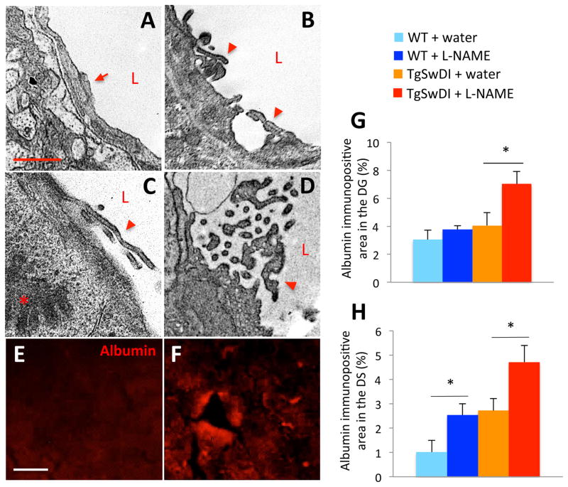
Figure 4. Hypertensive TgSwDI mice have increased BBB disruption.
Compared to capillary structure in water-treated mouse vessels (A), capillaries in brains of hypertensive TgSwDI mice exhibited tight junction alterations (arrowheads in B–D). Compared to the structure of normal tight junctions (arrow in A), those of hypertensive TgSwDI mice were often lifting or fragmented (arrowheads in B–D). The plasma protein albumin, which was present at low levels in the DS of normotensive TgSwDI brains (E), was significantly enriched in the DS (F–H; *p<0.05) and DG (not shown) of hypertensive TgSwDI mice after 3 months of L-NAME treatment. Compared to normotensive WT mice, hypertensive WT mice also exhibited significant leakage of albumin from the vasculature into the DS after 3 months of L-NAME treatment (H, *p<0.05; scale bar in A=1 μm; E=20 μm; n=4–6/group).

