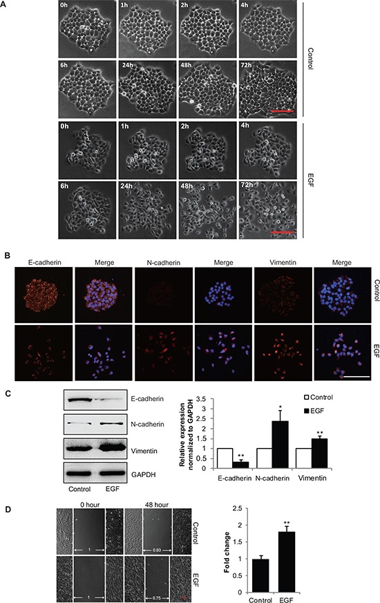
Figure 1. EGF induces EMT in gastric cancer SGC-7901 cells.

(A) SGC-7901 cells were incubated in the absence or presence of EGF (20 ng/mL), cell images were captured by phase-contrast microscopy for indicated times. Scale bar, 100 μm. (B–D) The extracts of SGC-7901 cells incubated with EGF (20 ng/mL) for 48 h, (B) representative microscopy images of SGC-7901 cells stained immunofluorescence for E-cadherin, N-cadherin and Vimentin, scale bar, 100 μm, and (C) the total cellular proteins were extracted and analyzed for expressions of E-cadherin, N-cadherin and Vimentin by immunoblotting assays. *P < 0.05, **P < 0.01 in the cultures with EGF relative to the cultures without EGF. (D) The SGC-7901 cells were scraped by a pipette tip and incubated with or without EGF for additional 48 h, a representative of wound healing assay was presented, and the quantification of cell migration rate was performed. **P < 0.01 in the cultures with EGF relative to the cultures without EGF.
