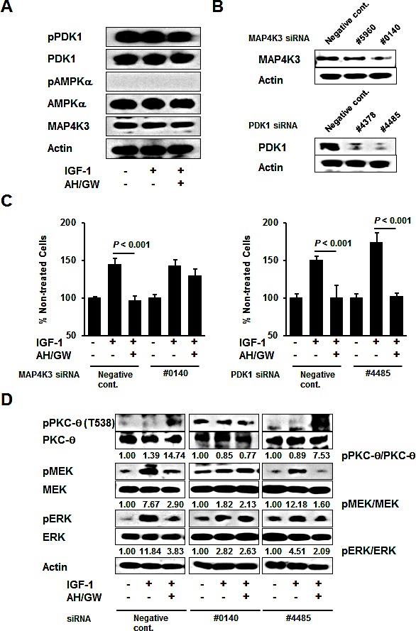
Figure 5. MAP4K3 plays a key role in the activation of PKC-θ by EP2/EP4 antagonism.

A, BxPC-3 cells were stimulated with IGF-1 (20 ng/mL) for 20 min in the absence or presence of AH6809/GW627368X (5 μM each) pretreatment for 3 h. The levels of phosphorylated PDK1, total PDK1, phosphorylated AMPKα, total AMPKα, and MAP4K3 were determined by immunoblotting. B, Knockdown was performed with specific siRNAs for MAP4K3 and PDK1 in BxPC-3 cells and confirmed by immunoblotting. C, Cells transfected with negative control siRNA, MAP4K3 siRNA (#0140), and PDK1 siRNA (#4485) were stimulated with IGF-1 (20 ng/mL) for 48 h in the absence or presence of AH6809/GW627368X (5 μM each) pretreatment for 3 h. Cell growth was measured by the MTT assay. The A550 values for untreated cells were assigned as 100% and the relative percentages for treated cells are shown. Columns, mean percentages (n = 6); bars, SD. D, Cells transfected with negative control siRNA, MAP4K3 siRNA (#0140), and PDK1 siRNA (#4485) were stimulated with IGF-1 (20 ng/mL) for 20 min in the absence or presence of AH6809/GW627368X (5 μM each) pretreatment for 3 h. The levels of phosphorylated PKC-θ, total PKC-θ, phosphorylated MEK, total MEK, phosphorylated ERK, and total ERK were determined by immunoblotting. The relative levels of phospho-PKC-θ, -MEK and -ERK were calculated using ImageJ software.
