Abstract
A rare case of an isolated traumatic palmar dislocation of the distal radioulnar joint is presented. Clinically, there is a loss of pronation and supination. The dislocation was treated using an open reduction, reinsertion of the capsule-ligamentous complex and temporary stabilization using K-wires.
Keywords: Dislocation, distal radioulnar joint, wrist
Introduction
Distal radioulnar joint (DRUJ) dislocation does not occur frequently. Clinical signs are sometimes misleading and radiography does not always provide a clear answer; the diagnosis can therefore be missed. The cause of this radial–ulnar dislocation is often traumatic; it is caused by a hypersupination of the forearm for a palmar dislocation, and hyperpronation for a dorsal dislocation [1, 2]. In 1777, Desault (cited by Albisson et al., and Caranfil [3, 4]) was the first of several authors to describe a case of distal radioulnar dislocation, but neither the physiopathology nor a standard treatment for this type of lesion has yet been described.
Case report
The patient, a 23 years-old, right handed metal worker, was admitted to our accident and emergency services for pain in the left wrist following a fall with forced flexion of the wrist. The pain was located at the level of the distal radioulnar joint. There was no swelling or instability on testing of the distal radioulnar joint (DRUJ). The diagnosis was missed on the initial X-rays, as the lateral view was not well aligned (Figure 1a and b ). One month later, the patient attended our department, as he continued to have a painful wrist. Clinical examination showed a slight depression at the level of the ulnar styloid. The anterior surface of the wrist showed a hard, round and smooth abnormal protuberance, made evident on deep palpation. There was no movement possible of this protuberance. There was a limited supination, and no pronation due to pain. Flexion and extension were normal. Examination of motor and sensory function of the digits did not reveal any abnormality. Because of this atypical clinical presentation, a computed tomography (CT) arthrography of the wrist was requested in order to establish a diagnosis of ligamentous injuries. MRI imaging was performed to assess both the distal radioulnar ligament and the extensor carpi ulnaris tendon.
Figure 1.
(a and b) X-ray image of the left wrist, AP and lateral view.
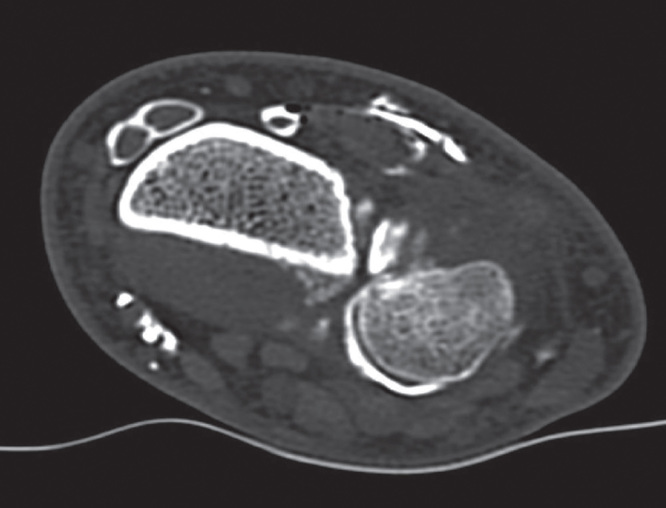
The CT arthrogram showed a palmar luxation of the ulnar head located at the distal radioulnar joint (Figure 2). The Triangular FibroCartilage Complex (TFCC) showed a perforation and irregularities on the palmar side. Elbow x-rays did not reveal any abnormalities. MRI imaging confirmed the palmar luxation and showed an intact extensor carpi ulnaris tendon that was not displaced. There was an interpositon of the TFCC in the distal radioulnar joint. Closed reduction by external manipulation was not possible. We therefore performed an open reduction in combination with ligament reconstruction and pinning. This was performed 2 months after the initial injury. During the operation, we found the TFCC to be completely detached from its medial insertion and interposed in the distal radioulnar space. The ulnar notch of the radius was of good quality but the ulna head, however, showed a grade III chondropathy (Figure 3). Due to the young age of our patient, our goal was an anatomic reinsertion of the TFCC, with keeping salvage procedures open for the future if necessary. First, the TFCC was reinserted at its insertion of the ulnar styloid using a non-absorbable transosseous suture. Reduction of the DRUJ was then secured by two extra-articular K-wires. Finally, the capsuloligamentous complex was re-inserted. Intra- and post-operative x-rays were satisfactory (Figure 4a and b ). K-wires were removed at 6 weeks post-operatively. Unfortunately, 1 K-wire was broken, and was left inside the distal radius. At 2-months follow-up, the patient presented a non-painful wrist with a pronation of 180°, supination of 155° and symmetrical flexion and extension. Final x-rays are shown in Figure 5.
Figure 2.
CT arthrogram.
Figure 3.
Grade III chondropathy of the ulnar head with entrapment of the TFCC.
Figure 4.
(a and b) Post-operative x-rays, AP and lateral view.
Figure 5.
(a and b) Final post-operative x-rays after hardware removal.
Discussion
Isolated palmar dislocation of the ulnar head is an uncommon injury, which explains the scarcity of publications. This type of dislocation is generally associated with fractures of the forearm and distal radius, and seldom presents without an accompanying fracture [5]. Being difficult to diagnose, this injury goes generally unnoticed in 50% of cases at first presentation at the emergency department, notwithstanding a routine radiographic examination [3, 6]. A judicious clinical examination will note an absence of the ulnar styloid at the dorsal surface of the wrist if present [3]. However, radiographic imaging of the wrist is not always easy to interpret, especially if correct positioning of the wrist and forearm is not possible due to pain.
CT arthrography of the wrist [3] and MRI imaging are the preferred diagnostic tools, being able to diagnose both the dislocation and permitting a complete assessment of any adjacent ligamentous injury. Rapid reduction of reducible dislocations offers a good prognosis, as is shown in a case series of Mulford et al. [7]. Eleven closed reductions in a total of nine patients were performed the same day as presentation at the emergency department. Early diagnosis seems to influence the treatment. Singletary [6] reports on a case of early diagnosis of palmar dislocation of the DRUJ in a 39 year-old patient. A closed reduction was performed followed by immobilization in pronation. The follow-up was uneventful with a good outcome. Kumar [8] also reports on a case seen after a 3-day delay in diagnosis, which was successfully treated by closed reduction. In these cases, there was no lesion of the TFCC. Caranafil describes a 27 year old patient with a 10-week delayed diagnosis and an unsuccessful open reduction that was the reason for an ulnar head resection according to Darrach [4]. In this case, the interposition of the TFCC ligament in the distal radioulnar joint was the reason for the failure of closed reduction, as also described by Ellanti and Grieve [9]. P Ellanti and PP Grieve, consider the repair of the TFCC as the most important factor for obtaining a stable reduction of a chronic dislocation. Takahashi et al. [10], confirmed the presence of an adjacent TFCC lesion by wrist arthroscopy. On the other hand, only McMurray and Muralikkutan [11] mention an absence of damage to the TFCC on MRI imaging in his case report of an irreducible DRUJ dislocation.
The recurrent character of these dislocations is reported by Sakota et al. [12], who describe a 73 years-old female patient whose closed reduction ended in a failure and underwent a Sauvé–Kapandji arthrodesis 10 months after her initial injury. Detailed information on the TFCC ligament was not given in this case report. All of the above permits us to emphasize, as does Caranafil [4], the concept of either a simple or complex dislocation: A simple dislocation is easily reduced by external manipulation and remains stable. A complex dislocation is characterized by instability, recurrence or irreducibility, caused by the interposition of soft tissues, especially the TFCC. It is therefore important to always look for lesions of the TFCC.
Conclusion
Isolated palmar dislocation of the distal radioulnar joint is a rare lesion of which the diagnosis is difficult to establish. The x-rays are sometimes non-conclusive; therefore, only a CT arthrogram or MRI can confirm the diagnosis. We would like to stress that even after a late diagnosis; a good result can be obtained by open reduction and repair of the TFCC ligament.
Declaration of interest
The authors report no conflicts of interest. The authors alone are responsible for the content and writing of the paper.
References
- Kihara H, Fortino MD, Palmer AK. The stabilizing mechanism of the distal radioulnar joint during pronation and supination. J Hand Surg [Am] 1995;20:930–6. doi: 10.1016/S0363-5023(05)80139-X. [DOI] [PubMed] [Google Scholar]
- Schernberg F. Le poignet-anatomie radiologique et chirurgie. Paris: Masson; 1992. [Google Scholar]
- Albisson F, Kerdiles N, Taton E, Tovagliaro F, et al. Luxation palmaire isolée de l’articulation radio-ulnaire inférieure. À propos d’un cas et revue de la littérature. J Traumatol Sport. 2003;20:110–13. [Google Scholar]
- Caranfil R. [Luxation traumatique isolée de l’articulation radio-ulnaire distale] Acta Orthop Belg. 2000;66:102–4. [PubMed] [Google Scholar]
- Jenkins NH, Mintowt-Czyz WJ, Fairclough JA. Irreducible dislocation of the distal radioulnar joint. Injury. 1987;18:40–3. doi: 10.1016/0020-1383(87)90384-6. [DOI] [PubMed] [Google Scholar]
- Singletary EM. Volar dislocation of the distal radioulnar joint. Ann Emerg Med. 1994;23:881–3. doi: 10.1016/s0196-0644(94)70328-0. [DOI] [PubMed] [Google Scholar]
- Mulford JS, Jansen S, Axelrod TS. Isolated volar distal radioulnar joint dislocation. J Trauma. 2010;68:E23–5. doi: 10.1097/TA.0b013e3181568db2. [DOI] [PubMed] [Google Scholar]
- Kumar A, Iqbal MJ. Missed isolated volar dislocation of distal radio-ulnar joint: a case report. J Emerg Med. 1999;17:873–5. doi: 10.1016/s0736-4679(99)00098-0. [DOI] [PubMed] [Google Scholar]
- Ellanti P, Grieve PP. Acute irreducible isolated anterior distal radioulnar joint dislocation. J Hand Surg [Eur] 2012;37:72–4. doi: 10.1177/1753193411422024. [DOI] [PubMed] [Google Scholar]
- Takahashi Y, Nakamura T, Sato K, Toyama Y. Subluxation of the palmar radioulnar ligament as a cause of blocked forearm supination: a case of DRUJ Locking. J Wrist Surg. 2013;2:83–6. doi: 10.1055/s-0032-1333065. [DOI] [PMC free article] [PubMed] [Google Scholar]
- McMurray D, Muralikuttan K. Volar dislocation of the distal radio-ulnar joint without fracture: A case report and literature review. Inj Extra. 2008;39:352–5. [Google Scholar]
- Sakota J, Kaneko K, Miyahara S, Mogami A, et al. Recurrent palmar dislocation of the distal radioulnar joint. A case report. Chir Main. 2002;21:301–4. doi: 10.1016/s1297-3203(02)00132-4. [DOI] [PubMed] [Google Scholar]


