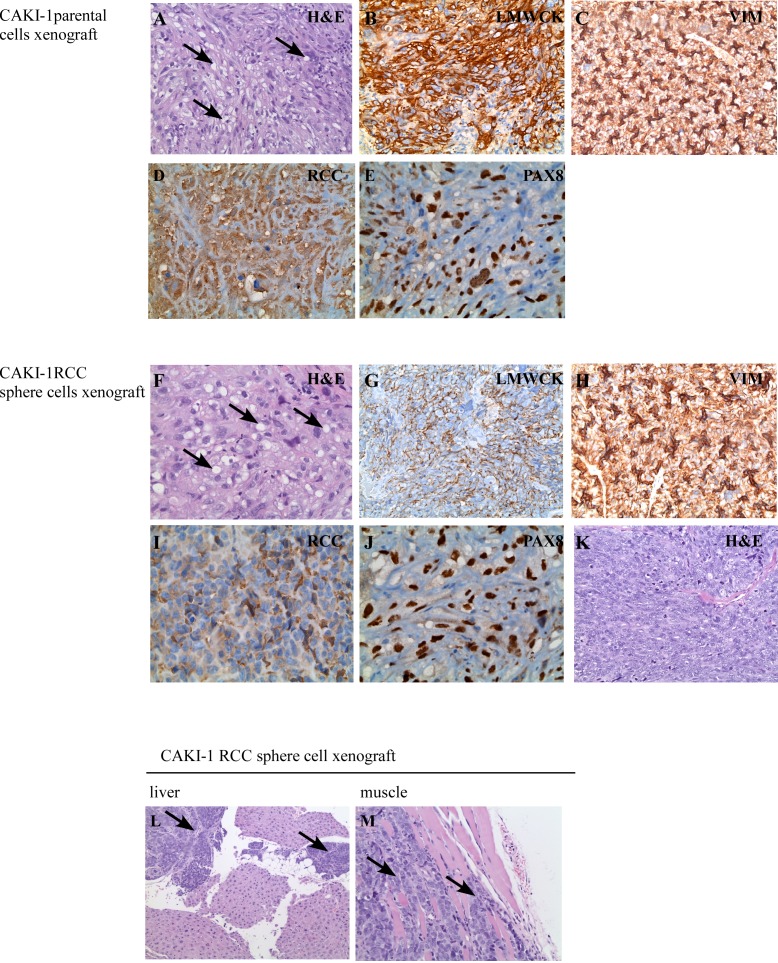
Figure 4. Histological features of xenograft tumors.
CAKI-1 parental cell xenografts developed tumors with typical RCC morphology (H & E staining) (A). Xenografts were positive for the RCC markers low molecular weight cytokeratines (LMWCK) (B), vimentin (VIM) (C), RCC (D) and PAX8 (E). CAKI-1 sphere xenografts developed tumors with typical RCC morphology (H & E staining) (F). Xenografts were positive for the RCC markers low molecular weight cytokeratines (LMWCK) (G), vimentin (VIM) (H), RCC (I) and PAX8 (J). CAKI-1 sphere xenografts also contained areas of undifferentiated, sarcomatoid RCC (H&E staining) (K). CAKI-1 sphere xenografts also developed metastatic lesions in the liver and muscle (L-M).

