
Figure 1. Immunohistochemistry of CaM and AR in human prostate tumor and non-tumor tissues.
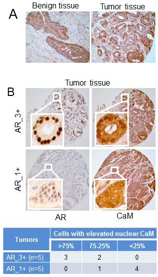
A) Evaluation of CaM expression in non-tumor (benign) and tumor prostate tissues. B) Evaluation of CaM expression in serial sections of prostate tumors with high (AR_3+) and low (AR_1+) AR levels. Immunostaining is as described in Materials and Methods. Images are representative of five each of benign and, AR_3+ and AR_1+ tumor tissue specimens. Table shows percentage of luminal epithelial cells in AR_3+ and AR_1+ tumors with elevated nuclear CaM. Percentage of cells with elevated nuclear CaM was determined by counting more than 300 cells in each tumor tissue.