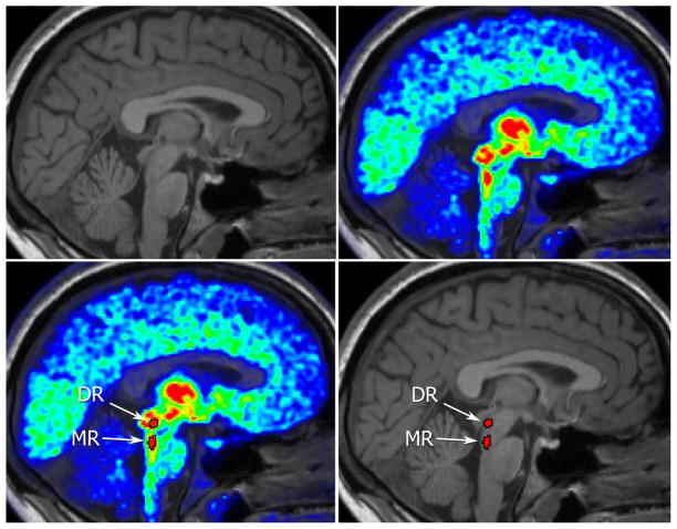
Figure 1.
(A) Structural MRI image. The raphe nuclei are not identifiable. (B) [11C]DASB PET image superimposed on the corresponding structural image, highlighting the 5-HTT system. The raphe nuclei are visible within brainstem as regions of higher binding. (C)Delineation of the DR and MR nuclei based on [11C]DASB PET. (D) DR and MR identified from the [11C]DASB PET image transferred as seeds onto the structural image.

