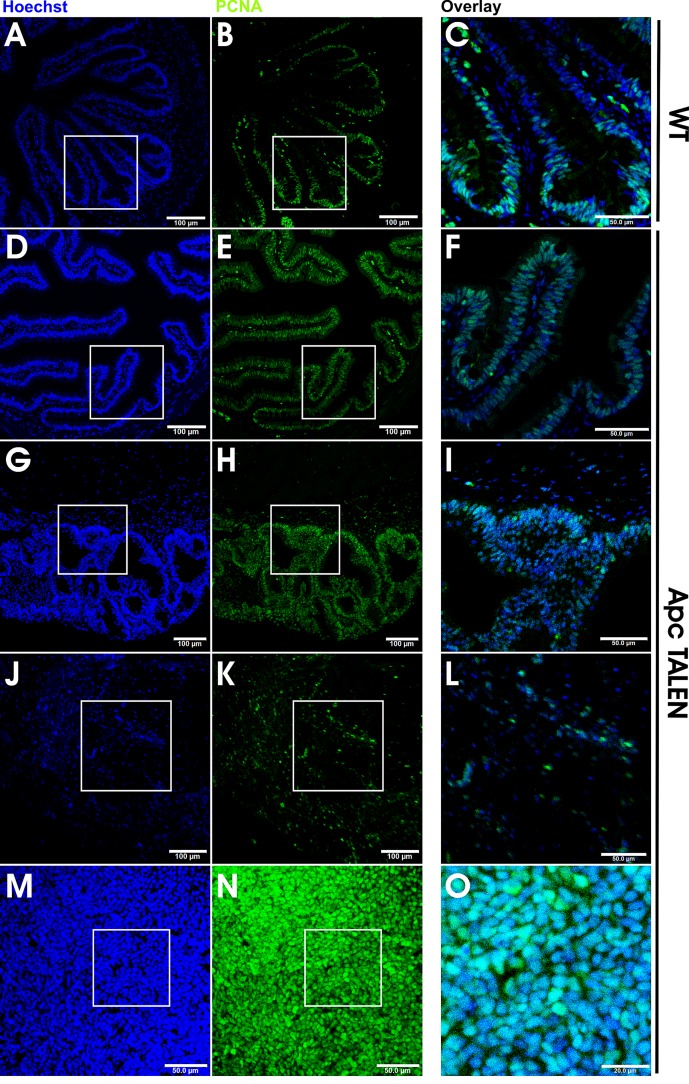
Figure 6. Increased and mislocalized proliferation in apc TALEN induced tumors.
Hoechst (left panels) and PCNA (middle panels) double staining; magnified overlays in right panels. (A, B, C) In WT small intestine PCNA staining is primarily localized at the base of the folds and largely absent in the crests. (D, E, F) Small intestine of apc TALEN injected froglet showing the presence of PCNA staining along the entire trough-crest axis. (G, H, I) Epidermoid cysts showing a high number of proliferating PCNA positive cells. (J, K, L) Desmoid tumor displaying a large fraction of PCNA positive nuclei. (M, N, O) Hind limb-associated tumor with almost all nuclei staining positive for PCNA indicating a very high proliferation rate.

