Abstract
The presence of lower molecular weight species comprising the C-terminal region of TAR DNA-binding protein 43 (TDP-43) is a characteristic of TDP-43 proteinopathy in amyotrophic lateral sclerosis (ALS) and frontotemporal lobar degeneration (FTLD). Here, we have identified a novel splice variant of TDP-43 that is upregulated in ALS and generates a 35-kDa N-terminally truncated species through use of an alternate translation initiation codon (ATGMet85), denoted here as Met85-TDP-35. Met85-TDP-35 expressed ectopically in human neuroblastoma cells exhibited reduced solubility, cytoplasmic distribution, and aggregation. Furthermore, Met85-TDP-35 sequestered full-length TDP-43 from the nucleus to form cytoplasmic aggregates. Expression of Met85-TDP-35 in primary motor neurons resulted in the formation of Met85-TDP-35-positive cytoplasmic aggregates and motor neuron death. A neo-epitope antibody specific for Met85-TDP-35 labeled the 35-kDa lower molecular weight species on immunoblots of urea-soluble extracts from ALS-FTLD disease-affected tissues and co-labeled TDP-43-positive inclusions in ALS spinal cord sections, confirming the physiological relevance of this species. These results show that the 35-kDa low molecular weight species in ALS-FTLD can be generated from an abnormal splicing event and use of a downstream initiation codon and may represent a mechanism by which TDP-43 elicits its pathogenicity.
Electronic supplementary material
The online version of this article (doi:10.1007/s00401-015-1412-5) contains supplementary material, which is available to authorized users.
Keywords: Amyotrophic lateral sclerosis, Frontotemporal lobar degeneration, TDP-43, TDP-35, C-terminal species, Alternative splicing, Alternative translation
Introduction
TAR DNA-binding protein of 43 kDa (TDP-43) is a nuclear DNA/RNA binding protein that has numerous functions in regulating RNA metabolism, including transcription, alternative splicing, miRNA processing and mRNA stabilization [3]. TDP-43 proteinopathy is characterized by the formation of neuronal cytoplasmic inclusions containing TDP-43, and this pathology is common to the majority of amyotrophic lateral sclerosis (ALS) as well as frontotemporal lobar degeneration (FTLD) cases [1, 16]. Biochemically, pathological TDP-43 comprises the normal full-length protein; an abnormally phosphorylated form that runs at 45 kDa; and lower molecular weight species of 25–35 kDa that correspond to N-terminally truncated forms of TDP-43. Expression of lower molecular weight species in cell culture recapitulates features of disease, including formation of cytoplasmic aggregates and recruitment of full-length TDP-43 from the nucleus [9, 24, 26, 29]. For C-terminal species of ~35 kDa, these effects led to decreased cell viability, impaired neurite outgrowth in neuronal cell lines, and a loss of TDP-43 splicing activity [9, 24, 26, 29]. These results suggest that C-terminal species of TDP-43 can drive pathogenesis and as such understanding their etiology is important for therapy development. In this regard, the 35-kDa species described above were generated from different starting amino acids. Prior studies have shown that species of 35 and 25 kDa can be generated by caspase-3 cleavage at DETD89 and DVMD219, respectively, a phenomenon that could be triggered upon suppression of progranulin expression in cell culture [31]. Similarly, another study found that induction of apoptosis in cultured cells with staurosporine also led to the activation of caspase-3 and the subsequent generation of 25-kDa and 35-kDa TDP-43 fragments [11]. However, species of 35 and 25 kDa are also found in cells lacking caspase-3 [17]. As such, although the TDP-43 caspase-3 cleavage product runs at the same molecular weight as the 35-kDa C-terminal species observed in disease tissues on SDS–polyacrylamide gel electrophoresis, one cannot infer with absolute certainty that the 35-kDa species in disease tissues is indeed derived from caspase-3 cleavage. Therefore, given the role of TDP-43 in splicing of other genes [4, 6, 7, 14] and the fact that it is alternatively spliced itself [25], we explored alternative splicing and/or translation as a potential mechanism for generating pathological TDP-43 C-terminal species in ALS. To this end, we have identified an alternatively spliced variant of TDP-43 that is upregulated in ALS and has 91 bp spliced out in exon 2, generating a protein species of 35 kDa through use of a downstream start codon, ATGMet85, denoted here as Met85-TDP-35. Use of ATGMet85 as a TDP-43 start codon has previously been shown in cell culture studies, however, the physiological relevance of this was unknown [17]. Here, using a neo-epitope antibody specifically recognizing Met85-TDP-35, we have shown that the pathologically associated TDP-43 species of 35 kDa in ALS-FTLD can be generated through use of ATGMet85. The Met85-TDP-35 neo-epitope antibody not only labeled the 35-kDa species on immunoblots of disease tissues, but also co-labeled TDP-43-positive inclusions in ALS spinal cord tissue, showing that Met85-TDP-35 is fully integrated within the pathology associated with the disease. These findings indicate that pathological variants of TDP-43 can be generated through a combination of alternative splicing and translation, mechanisms that could be targeted therapeutically.
Materials and methods
ALS and control cases
Spinal cord tissues were collected from eleven sALS cases, four with C9orf72 repeat expansions, and one fALS case also carrying a C9orf72 repeat expansion [27]; and one case each of Alzheimer’s disease, corticobasal degeneration, multisystem atrophy and Huntington’s disease (Table 1). Prefrontal cortex tissues were collected from six ALS cases with pathologically confirmed FTLD, and six neurologically normal control cases (Table 1). ALS cases were diagnosed using the Revised El Escorial Criteria (Brooks 2000), and patient consents were obtained from the legal representatives in accordance with the Ethical Review Boards of Sunnybrook Health Sciences Centre and University of Toronto (Toronto, Canada).
Table 1.
ALS and control cases used in this study
| Cases | Sex | Age (years) | Diagnosis | Gene mutation |
|---|---|---|---|---|
| ALS cases | ||||
| A1 | F | 49 | sALS-FTLD | C9orf72 |
| A2 | F | 46 | sALS | N/A |
| A3 | F | 73 | sALS | N/A |
| A4 | M | 70 | sALS | C9orf72 |
| A5 | F | 58 | sALS-FTLD | C9orf72 |
| A6 | F | 59 | sALS | N/A |
| A7 | M | 52 | sALS | N/A |
| A8 | F | 65 | sALS | N/A |
| A9 | M | 58 | fALS-FTLD | C9orf72 |
| A10 | F | 60 | sALS | N/A |
| A11 | M | 64 | sALS-FTLD | C9orf72 |
| A12 | F | 57 | sALS | N/A |
| A13 | F | 83 | sALS-FTLD | N/A |
| A14 | F | 71 | sALS-FTLD | N/A |
| Control cases | ||||
| C1 | F | 91 | AD | N/A |
| C2 | M | 77 | CBD | N/A |
| C3 | M | 69 | MSA | N/A |
| C4 | F | 78 | HD | N/A |
| C5 | M | 76 | N.N. | N/A |
| C6 | M | 74 | N.N. | N/A |
| C7 | M | 89 | N.N. | N/A |
| C8 | F | 48 | N.N. | N/A |
| C9 | M | 54 | N.N. | N/A |
| C10 | F | 82 | N.N. | N/A |
sALS sporadic ALS, fALS familial ALS, FTLD frontotemporal lobar degeneration, C9orf72 chromosome 9 open reading frame 72, N/A not applicable, AD Alzheimer’s disease, CBD corticobasal degeneration, MSA multiple system atrophy, HD Huntington’s disease, N.N. neurologically normal
RT-PCR and cloning of TDP-43 and its splice variant
Fifty milligrams of gray matter was dissected from the anterior horn of postmortem lumbar spinal cord from twelve ALS and four control cases, from which total RNA was extracted using TRIzol Reagent (Life Technologies) according to the manufacturer’s protocol and treated with DNase to minimize DNA contamination. The RNA transcripts were first normalized with β-actin primers (Life Technologies) after synthesizing complementary DNA (cDNA) from 1 μg of total RNA using random hexamers and the SuperScript III First-strand Synthesis System for RT-PCR (Life Technologies) following manufacturer’s protocol. PCR was performed with 2 μl of template cDNA mixed with 18 μl of reaction mix containing 10 μl of 2× Master Mix (Life Technologies), 6 μl water, 1 μl of 10 μM forward primer (5′-ATG TCT GAA TAT ATT CGG GTA AC-3′) and 1 μl of 10 μM reverse primer (5′-GTC CAT CTA TCA TAT GTC GCT GTG-3′). Amplification of TDP-43 and its splice variant was performed in a 9800 Fast Thermal Cycler (Life Technologies) with an initial denaturation at 95 °C for 10 s, followed by 35 cycles of denaturation at 95 °C for 0 s and annealing at 66 °C for 15 s. The amplified products were visualized on a 1.5 % (w/v) agarose/ethidium bromide gels and subjected to sequence verification.
For cloning of full-length TDP-43 and its splice variant, 1 μg total RNA isolated from ALS spinal cords was reverse transcribed with an adapter-tagged oligo dT primer (oligo(dT)17-GGC CAC GCG TCG ACT AGT AC) into cDNA, which was purified with a Rapid Amplification of cDNA Ends (RACE) Purification System (Life Technologies). Poly-(dC) sequences were added to the N-terminus of 10 μl cDNA using a TdT-tailing reaction (6.5 μl DEPC-treated water, 5 μl of 5× tailing buffer, 2.5 μl of 2 mM dCTP, and 1 μl TdT) according to manufacturer’s protocol (Life Technologies). PCR was performed with 5 μl of tailed cDNA with 45 μl PCR reaction mix containing 1× PfuUltra buffer, 200 μM dNTP, 0.2 μM 5′RACE abridged anchor primer (5′-GGC CAC GCG TCG ACT AGT ACG GGI IGG GII GGG IIG-3′), 0.2 μM adapter primer (5′-GGC CAC GCG TCG ACT AGT AC-3′), and 1.25 U PfuUltra DNA polymerase (Agilent Technologies). Amplification of PCR products was performed with an initial denaturation at 95 °C for 45 s, followed by 25 cycles of denaturation at 95 °C for 45 s, 55 °C for 45 s, and 68 °C for 90 s in GeneAmp PCR System 9700 (Life Technologies). The amplified products were purified with Qiagen PCR Purification Kit (Qiagen) and were subjected to a second round of amplification with a 5′UTR forward primer with a NotI restriction site (5′-ataagaatgcggccgcGTC CGT CGC TGC TTC GGT G-3′) and a 3′UTR reverse primer with a BamHI restriction site (5′-atacgcggatccGCC TGT GAT GCG TGA TGA CG-3′). The PCR conditions were the same as the first round except with 30 cycles. The amplified products were visualized on a 1.0 % (w/v) agarose/ethidium bromide gel, purified with the MiniElute Gel Extraction Kit (Qiagen), and then subcloned into the NotI/BamHI restriction sites of the mammalian expression vector pcDNA3.1(−). The Met85-TDP-35 construct, starting at the 3rd ATG (Met85) of full-length TDP-43, was generated by PCR and cloned into pcDNA3.1(−) expression vector using a forward primer containing a BamHI restriction site (5′-gcggatccaccATG GAT GAG ACA GAT GCT TC-3′) and a reverse primer containing a KpnI restriction site (5′-cacggtacCTA CAT TCC CCA GCC AGA AGA CTT AG-3′). For visualization of the subcellular localization of full-length TDP-43 and Met85-TDP-35, these two sequences were cloned into pEGFP-C2 and pmRFP-C2 vectors, for N-terminal tagging with EGFP and RFP, respectively. Sequence analysis was performed on all constructs to ensure correct sequences.
Silver staining
PCR amplified products were resolved on 10 % native PAGE (polyacrylamide gel electrophoresis) gels followed by fixation in 10 % ethanol, 0.05 % acetic acid for 30 min. Gels were then stained with a 10 mM silver nitrate solution for 30 min, developed in a 350 mM sodium hydroxide, 0.3 % formaldehyde solution, and the reaction stopped using 5 % acetic acid. Gel images were captured and band intensities quantified using Image J software with values expressed as mean ± standard error of the mean (SEM).
Cell culture and microinjection of primary motor neurons
The human neuroblastoma cell line SH-SY5Y was maintained in DMEM/F12 medium (Life Technologies) supplemented with 10 % fetal bovine serum (FBS). Transfection was performed with the corresponding plasmid DNAs using Lipofectamine 2000 (Life Technologies) following manufacturer’s protocol. Generation of primary motor neuron cultures was as described previously [21, 28], and plasmids containing EGFP-tagged full-length TDP-43 and EGFP-tagged Met85-TDP-35 in pEGFP-C2 vector were microinjected at 200 ng/μl into motor neuron nuclei along with 20 μg/μl of the fluorescent marker Dextran FITC (Life Technologies). Cell viability was determined daily by counting the number of FITC-positive motor neurons, which was normalized to the number of FITC-positive motor neurons on day 1 after microinjection. All experiments were performed in triplicate and values were expressed as mean ± SEM.
Immunocytochemistry of cultured cells
To visualize EGFP-tagged construct expression, cultured cells grown on glass coverslips were fixed with 3.7 % (w/v) paraformaldehyde in phosphate-buffered saline (PBS) for 20 min at room temperature (RT) followed by 3 × 5 min PBS washes then counterstained with 4′,6-diamidino-2-phenylindole (DAPI) nucleic acid stain for 1 min (Life Technologies). Immunofluorescence was visualized using a Leica DMI6000 microscope and images captured using Volocity software (PerkinElmer).
Protein extraction and Western blotting
SH-SY5Y cells grown on 35 mm2 dishes (Thermo Scientific) were harvested in 1 ml PBS containing protease inhibitors at 4 °C, and the cell suspensions were centrifuged at 500×g for 5 min at 4 °C. The resulting pellets were homogenized in 1 ml low salt buffer [50 mM Tris, pH 7.5, 150 mM NaCl, 5 mM ethylenediaminetetraacetic acid (EDTA)] containing protease inhibitors (Sigma-Aldrich) and then centrifuged at 16,100×g for 20 min at 4 °C. Supernatants were saved as the low salt fraction, and pellets were homogenized in a high salt Triton X-100 buffer (50 mM Tris, pH 7.5, 750 mM NaCl, 5 mM EDTA, 1 % Triton X-100, and protease inhibitors), followed by centrifugation at 16,100×g for 20 min at 4 °C. Supernatants were saved as the high salt Triton X-100 fraction, and the remaining pellets were solubilized in urea buffer (7 M urea, 2 M thiourea, 4 % 3-[(3-cholamidopropyl)dimethylammonio]-1-propanesulfonate (CHAPS), 30 mM Tris–HCl, pH 8.5) and saved as the urea fraction.
For human tissues, gray matter from ALS and control lumbar spinal cord was dissected and weighed, then homogenized at 200 mg/ml using the same biochemical fractionation procedure as for cultured cells but with one exception: after extraction in high salt Triton X-100 buffer, the resulting pellets were homogenized in a high salt sucrose buffer (50 mM Tris, pH 7.5, 750 mM NaCl, 5 mM EDTA, 30 % sucrose, and protease inhibitors), followed by centrifugation at 16,100×g for 30 min at 4 °C to float and remove myelin. For immunoblotting, sodium dodecyl sulfate (SDS) sample buffer (10 mM Tris–HCl, pH 6.8, 1 mM EDTA, 40 mM DTT, 1 % SDS, and 10 % sucrose) was added to 50 μg of each sample fraction, which was heated at 95 °C for 5 min with the exception of the urea fraction to avoid carbamoylation of proteins. Equivalent amounts of protein samples were loaded onto 10 % SDS-PAGE gels and transferred to polyvinyldiflouride (PVDF) membranes, which were blocked in 5 % skim milk dissolved in Tris-buffered saline (TBS) containing 0.2 % Tween-20 for 1 h at RT, followed by overnight incubation at 4 °C with a polyclonal TDP-43 antibody (1:1000, Proteintech), a polyclonal N-terminal TDP-43 antibody (1:1000, Aviva Systems Biology), or a polyclonal C-terminal TDP-43 antibody (1:1000, generated in-house using a synthetic peptide, FGSSMDSKSSGWG, linked to keyhole limpet hemocyanin as the antigen). Bound antibody was visualized with an ECL Detection System (PerkinElmer) using HRP-conjugated secondary antibodies.
To extract insoluble proteins from the prefrontal cortex, six ALS cases with pathologically confirmed FTLD-TDP-43, and six neurological normal controls of a similar age group were included. Tissue was dissected from Brodmann’s area 10, weighed, and homogenized at 250 mg/ml in low salt buffer (10 mM Tris–HCl, pH 7.5, 5 mM EDTA, 1 mM DTT, 10 % sucrose, and protease inhibitors) with a glass Dounce homogenizer (Sigma-Aldrich). Homogenate was centrifuged at 25,000×g for 30 min at 4 °C, and the resulting supernatant was saved as the low salt fraction. Pellet was homogenized at 250 mg/ml (starting tissue weight) in high salt buffer (low salt buffer + 500 mM NaCl, 1 % Triton X-100, and protease inhibitors) and centrifuged at 180,000×g for 30 min at 4 °C, and the resulting supernatant was saved as the high salt fraction. Pellet was homogenized in 250 mg/ml (starting tissue weight) myelin flotation buffer (10 mM Tris–HCl, pH 7.5, 500 mM NaCl, 5 mM EDTA, 1 mM DTT, 40 % sucrose, 1 % Triton X-100, and protease inhibitors) and centrifuged at 180,000×g for 30 min at 4 °C, and the floating myelin removed. The resulting supernatant was saved as the myelin flotation fraction. Pellet was homogenized in 50 mg/ml (starting tissue weight) sarkosyl buffer (low salt buffer + 500 mM NaCl, 1 % N-lauroyl-sarcosine, and protease inhibitors) and incubated with agitation at 22 °C for 30 min, followed by centrifugation at 180,000×g for 30 min at 22 °C. The resulting supernatant was saved as the sarkosyl fraction. Pellet was sonicated with the Ultrasonic Cell Disrupter (Misonix) in 1 g/ml (starting tissue weight) urea buffer (30 mM Tris–HCl, pH 8.5, 7 M urea, 2 M thiourea, 4 % CHAPS) and saved as the urea fraction. All fractions were resuspended with SDS sample buffer and all samples except for the urea fraction were heated at 95 °C for 5 min prior to SDS-PAGE and immunoblot analyses.
Generation of neo-epitope antibodies
Rabbit polyclonal antibodies specifically recognizing Met85-TDP-35 or caspase-3-cleaved TDP-35 (C3-TDP-35) were generated using synthetic peptides corresponding to the neo-epitopes generated by alternative translation initiation at ATGMet85 (M85DETDASSA) or caspase-3 cleavage at DETD89 (A90SSAVKVKR), respectively, linked to keyhole limpet hemocyanin (ProSci Inc.). Both antibodies were diluted 1:1000 for immunoblotting and 1:500 for immunohistochemistry.
Generation of recombinant TDP-43 protein
Full-length TDP-43 was expressed as a fusion protein with His-tagged Venus YFP at the N-terminus joined by a TEV-cleavable linker. The construct was cloned into the pET-30 vector (EMD Millipore) and expressed in E. coli BL21-AI cells (Life Technologies). Bacterial culture was lysed by sonication on ice with lysis buffer (40 mM HEPES–KOH, pH 7.4, 500 mM KCl, 20 mM MgCl2, 10 % glycerol, 20 mM imidazole, 2 mM β-mercaptoethanol) containing protease inhibitors (Roche Applied Science). Lysate was centrifuged at 15,000 RPM in a Sorvall SS-34 rotor for 30 min at 4 °C. Prior to Ni–NTA agarose bead column (Qiagen) purification of the lysate supernatant, the column was washed with 5 column volumes of 40 mM HEPES–KOH, pH 7.4, 500 mM KCl, 20 mM MgCl2, 10 % glycerol, 20 mM imidazole, 2 mM β-mercaptoethanol, and protease inhibitors. The YFP-TDP-43 fusion protein was eluted from the column with 40 mM HEPES, pH 7.4, 500 mM KCl, 20 mM MgCl2, 250 mM imidazole, 10 % glycerol, 2 mM β-mercaptoethanol, and protease inhibitors. SDS-PAGE analysis of eluent showed that the fusion protein fraction was 95 % pure.
In vitro caspase-3 cleavage of recombinant TDP-43
To generate caspase-3-cleaved TDP-43, 2 μg of recombinant human YFP-tagged TDP-43 was incubated with 2 U of active human recombinant caspase-3 (EMD Millipore) in a reaction mix containing 100 mM NaCl, 50 mM HEPES, 10 mM DTT, 1 mM EDTA, 10 % glycerol, and 0.1 % CHAPS, pH 7.4, at 37 °C for 4 h. The reaction was halted by adding 2× SDS sample buffer.
Immunohistochemistry of human spinal cord tissues
Formalin-fixed paraffin-embedded lumbar spinal cord tissues from ALS and control cases were cut into 4 μm serial sections and then mounted on glass slides. Tissue sections were rehydrated in sequential washes of xylene, xylene:ethanol (1:1), and decreasing concentrations of ethanol, followed by a final wash in PBS. Blocking of endogenous peroxidase activity was achieved by incubation with 3 % (v/v) hydrogen peroxide, and antigens retrieved by incubation in either 10 mM citrate buffer at pH 6.0 for polyclonal TDP-43 antibody (ProteinTech) or Tris–EDTA buffer (20 mM Tris–HCl, 0.65 mM EDTA, 0.0005 % Tween-20, pH 9.0, for Met85-TDP-35 antibody). Sections were incubated overnight at RT with polyclonal TDP-43 antibody (1:5000, Proteintech) or Met85-TDP-35 antibody (1:500) diluted in antibody diluent (0.05 M Tris–HCl, pH 7.4, 0.25 % Tween-20, 0.2 % Triton X-100, 1 % casein, and 0.05 % thimerosol). Bound antibody was visualized using the ImmPRESS Anti-rabbit Ig Peroxidase Kit (Vector Labs) for polyclonal TDP-43 antibody or with the Super Sensitive Polymer HRP Kit (Biogenex) for Met85-TDP-35 antibody labeling. Color development was achieved using NovaRed (Vector Labs) and sections were counterstained with hematoxylin. Images were captured using a QImaging Micropublisher 3.3 RTV digital camera attached to a Leica DM6000 upright microscope and Openlab Imaging software (PerkinElmer).
For double immunofluorescence labeling, formalin-fixed paraffin-embedded spinal cords were cut into 6 μm sections and subjected to the same processing as above, except that the primary antibodies were rabbit polyclonal Met85-TDP-35 antibody (1:500) and mouse monoclonal TDP-43 antibody (2E2-D3, 1:500, Novus Biologicals) and the secondary antibodies were goat anti-rabbit Alexa Fluor 488 and goat anti-mouse Alexa Fluor 594, respectively. Images were captured with an ORCA ER Hamamatsu digital camera mounted on a Leica DMI600 inverted microscope using Volocity Imaging software (PerkinElmer).
Results
Identification of a novel splice variant of TDP-43
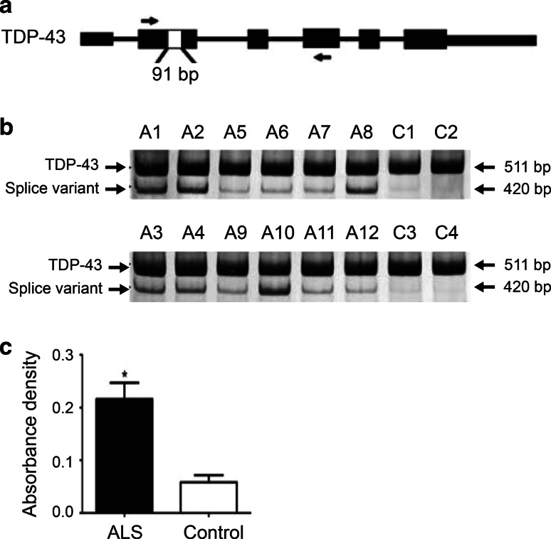
A search of EST sequences in Genbank (http://www.ncbi.nlm.nih.gov/genbank/) yielded two human (accession numbers: BI464234 and BG435185) and fourteen mouse (accession numbers: AV506877, AW 106495, BU610138, BY740492, BY788083, CA319629, CA543520, CA879844, CD553499, CD766458, CN691221, CN692254, CN696163, and DV060848) EST sequences, which revealed the same 91 bp splicing deletion in exon 2 of TARDBP, encoding TDP-43. This splice variant uses a non-canonical splicing pair (UU:AG), thus skipping base pairs 106–196 within exon 2 (denoted here as TDP-43 r.[106_196del]). To investigate the relevance of this splice variant in ALS, we selected twelve cases (11 sALS and one fALS with C9orf72 expansions), all of which exhibited TDP-43- and ubiquitin-positive inclusions upon postmortem examination, and 4 control cases for transcript analysis. RT-PCR was performed using total RNA isolated from lumbar spinal cords, with a forward primer located in exon 2 and a reverse primer in exon 4 of TARDBP (Fig. 1a). A major band of 511 bp, corresponding to the full-length TDP-43 transcript, was present across all ALS and control cases. A lower band of 420 bp that corresponds to the alternatively spliced variant TDP-43 r.[106_196del] was found at much higher levels in ALS cases compared to controls (Fig. 1b). After normalization of total RNA to β-actin, quantification analysis showed that the TDP-43 r.[106_196del] transcript was upregulated by ~4.0 fold (p < 0.0126) in ALS (n = 12; 0.216 ± 0.031) compared to controls (n = 4; 0.059 ± 0.013) (Fig. 1c).
Fig. 1.
TDP-43 r.[106_196del] splice variant is upregulated in ALS. a TARDBP gene structure showing splicing deletion. Black boxes are exons. White box in exon 2 denotes the 91 bp skipped by alternative splicing. Arrows indicate positions of the forward and reverse primers for RT-PCR. b RT-PCR amplification of the TDP-43 r.[106_196del] splice variant using RNA isolated from ALS and control spinal cord. Region amplified has 511 bp in the constitutively spliced transcript and 420 bp in the alternatively spliced transcript. Cases A1, A4, A5, A9, and A11 also carry C9orf72 expansions. c Quantification using ImageJ showed a ~4 fold upregulation of the splice variant in ALS (n = 12; 0.216 ± 0.031) compared to controls (n = 4; 0.059 ± 0.013), p < 0.0126
TDP-35 is an N-terminally truncated protein with reduced solubility
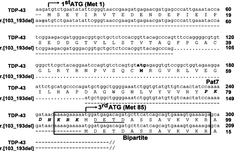
Translation tool analysis (http://www.expasy.org/tools/) of the TDP-43 splice variant transcript showed that if it was transcribed from the first ATGMet1, the 91 bp deletion in exon 2 would cause a frame shift and generate a premature stop codon (TAG) 190–192 bp downstream from ATGMet1, thereby producing a short protein of 63 aa with a pI/MW of 4.32/7.2 kDa. Theoretically, such mRNA transcripts containing premature stop codons would be eliminated through nonsense-mediated mRNA decay [8], which was corroborated in our findings as we did not detect a protein product of this size across our studies. However, translation analysis also revealed the presence of a complete open reading frame (ORF) situated 161 bp downstream from ATGMet1. The ATG initiation codon of this ORF corresponds to the third ATGMet85 of full-length TDP-43, with the loss of the second ATGMet51 through the splicing deletion (Fig. 2).
Fig. 2.
Sequence alignment of TDP-43 and the TDP-43 r.[106_196del] splice variant. The 91 bp splicing deletion is signified by dotted lines. Pat7 (aa 78–84, shown in bold italics) and bipartite (aa 82–98, boxed) nuclear localization sequences are indicated. Note that ATGMet85 immediately precedes the caspase-3 cleavage site DETD89 (underlined)
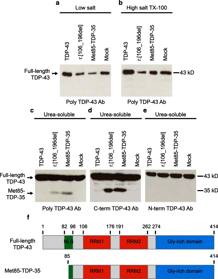
Expression of the TDP-43 r.[106_196del] cDNA in pcDNA3.1(−) in human neuroblastoma (SH-SY5Y) cells resulted in the generation of a 35-kDa protein product on immunoblots of cell lysates, thereby confirming the prediction described above (Fig. 3). To biochemically characterize this 35-kDa species, SH-SY5Y cells expressing full-length TDP-43, TDP-43 r.[106_196del], or Met85-TDP-35 (generated by PCR) were harvested and proteins sequentially extracted with buffers of increasing stringency, followed by immunoblot analysis of all fractions (Fig. 3). Full-length TDP-43 distributed to all fractions, as has been reported previously, whereas the 35-kDa species expressed from TDP-43 r.[106_196] and Met85-TDP-35 was exclusively partitioned to the urea-soluble fraction (Fig. 3), indicating a decreased solubility compared to the full-length protein. Both TDP-43 r.[106_196del] and Met85-TDP-35 were detected with an antibody to the C-terminus of TDP-43, but not to the N-terminus, confirming the loss of the N-terminal domain in these proteins (Fig. 3).
Fig. 3.
TDP-43 r.[106_196del] splice variant generates an N-terminally truncated protein of 35 kDa. SH-SY5Y cells expressing full-length TDP-43, TDP-43 r.[106_196del], Met85-TDP-35, or mock vector were fractionated in buffers of increasing stringency: a low salt; b high salt-TX-100; c–e urea buffer. Full-length TDP-43 was found in all fractions on immunoblots probed with polyclonal TDP-43. A lower molecular weight species of 35 kDa was observed only in urea-soluble fractions of lysates from cells expressing TDP-43 r.[106_196del] or Met85-TDP-35 (c) and was detected using antibody to the C-terminus of TDP-43 (d) but not to the N-terminus (e). f Comparative structures of TDP-43 and Met85-TDP-35
TDP-35 has disrupted nuclear localization signals and forms cytoplasmic aggregates in human neuroblastoma cells
Given that Met85-TDP-35 is translated from the third ATGMet85 of full-length TDP-43, there is disruption of two NLS sequences located within the N-terminal region of the full-length protein, namely, the Pat7 NLS from aa 78–84 (PKDNKRK) and the bipartite NLS from aa 82–98 (KRKMDETDASSAVKVKR) (Fig. 2). To examine the subcellular localization of full-length TDP-43 and Met85-TDP-35, EGFP tags were added to either the N-terminus or the C-terminus of these two constructs to visualize ectopic expression in SH-SY5Y cells. EGFP-tagged full-length TDP-43 exhibited a nuclear distribution that colocalized with DAPI labeling, whereas EGFP-tagged Met85-TDP-35 localized to the cytoplasm exhibiting three distinct patterns: Type I showed that 60 % of transfected cells had a diffuse Met85-TDP-35 localization in both the nucleus and the cytoplasm; Type II showed that 25 % of transfected cells had diffuse Met85-TDP-35 localization accompanied with small cytoplasmic aggregates, and Type III showed that 15 % of transfected cells had large Met85-TDP-35 aggregates in the cytoplasm but no labeling in the nucleus (Online Resource 1). These results recapitulate previous studies [9, 29]. The Met85-TDP-35-positive cytoplasmic aggregates did not label with antibodies to ubiquitin or phosphorylated TDP-43 (409/410) (not shown). Moreover, as observed previously for other TDP-43 species with disrupted NLS [9, 29], Met85-TDP-35 recruited full-length TDP-43 from the nucleus into the cytoplasm, where it formed aggregates (Online Resource 1).
Met85-TDP-35 forms cytoplasmic aggregates and is toxic to primary motor neurons
To examine the effect of expressing Met85-TDP-35 in an ALS-relevant cell type, plasmids encoding N-terminal EGFP-tagged full-length TDP-43, N-terminal EGFP-tagged Met85-TDP-35, or empty vector were expressed in primary motor neurons using intranuclear microinjection. Full-length TDP-43 localized exclusively to the nucleus, whereas Met85-TDP-35 was found in the cytoplasm of primary motor neurons, where it appeared as punctate aggregates similar to those observed in transfected SH-SY5Y cells (Fig. 4a). Cell viability assays conducted over a period of 1 week showed that expression of Met85-TDP-35 induced motor neuron death with a 50 % loss at day 5 post-microinjection, compared to full-length TDP-43, which exhibited no toxicity with a similar cell viability curve as neurons expressing the empty vector (Fig. 4b).
Fig. 4.
Expression of Met85-TDP-35 causes death of primary motor neurons. a Expression of EGFP-tagged TDP-43 and EGFP-tagged Met85-TDP-35 in primary motor neurons resulted in localization of TDP-43 to the nucleus and Met85-TDP-35 to the cytoplasm, where it formed aggregates (arrow). b A viability curve showed that Met85-TDP-35 induced motor neuron death compared to TDP-43 or empty vector alone. Scale bar 20 μm
Met85-TDP-35 expression in ALS
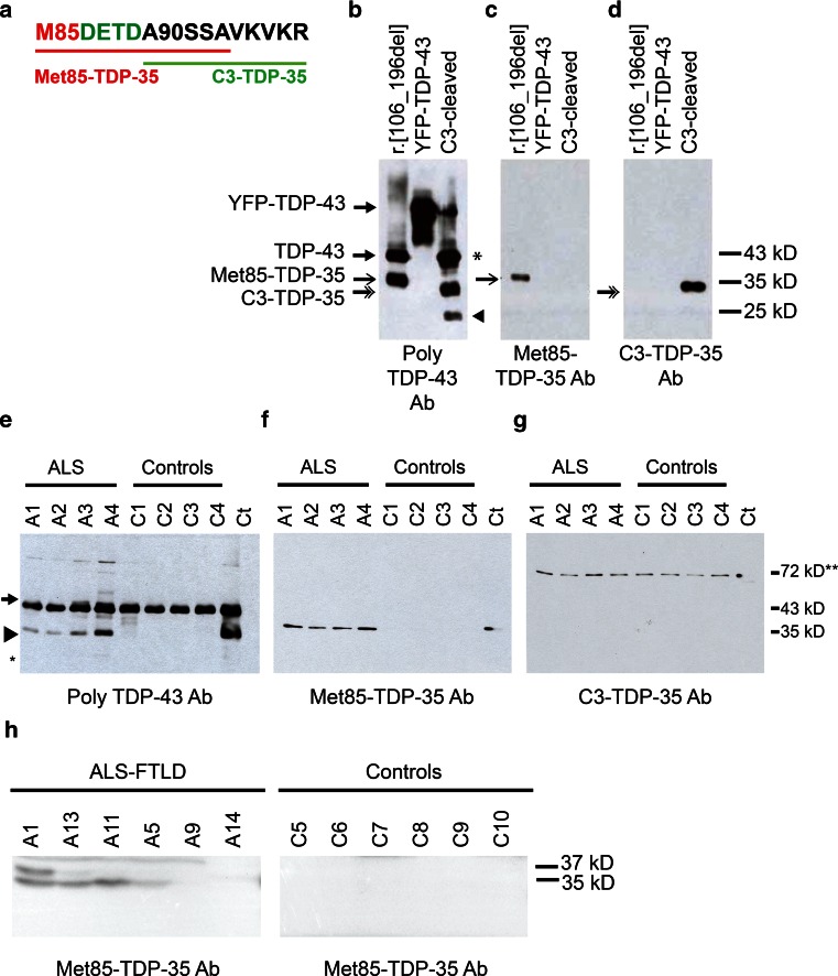
As our studies in cultured cell lines and primary motor neurons indicated that Met85-TDP-35 formed cytoplasmic aggregates, recapitulating a characteristic feature of TDP-43 proteinopathy, we proceeded to assess the pathological relevance of Met85-TDP-35 in ALS disease tissues. To study the presence of Met85-TDP-35, gray matter from the anterior horn of four sALS and four control lumbar spinal cords were sequentially extracted with buffers of increasing stringency to isolate insoluble proteins (Online Resource 2). Equivalent amounts of low salt, high salt Triton X-100 and urea fractions were immunoblotted with polyclonal TDP-43 antibody (Proteintech), which immunolabeled full-length TDP-43 in all fractions but also a distinct 35-kDa species that was only present in the urea fraction of ALS samples (Online Resource 2). This 35-kDa species was immunolabeled with C-terminal TDP-43 antibody but not with N-terminal TDP-43 antibody (Online Resource 2), confirming that this is a lower molecular weight C-terminal species frequently observed in disease tissues. The 25-kDa species could be observed at much longer exposures of the immunoblots (Online Resource 2). To determine whether this 35-kDa C-terminal species was generated through translation from Met85 or from caspase-3 cleavage at DETD89, we generated neo-epitope antibodies specific for the extreme N-terminal sequences of these two proteins, termed Met85-TDP-35 antibody or C3-TDP-35 antibody, respectively (Fig. 5a). Specificity of the antibodies for their respective protein isoforms was confirmed by immunoblotting using cell lysates expressing TDP-43 r.[106_196del] or full-length recombinant TDP-43 cleaved with caspase-3 in vitro (Fig. 5b–d). Immunoblotting of the urea fractions of ALS and control spinal cords tissues with polyclonal TDP-43 antibody revealed a distinct 35-kDa species present only in ALS samples in addition to the full-length TDP-43 (Fig. 5e). This 35-kDa species was immunolabeled by the neo-epitope Met85-TDP-35 antibody (Fig. 5f) but not by C3-TDP-35 antibody (Fig. 5g). Given that the discovery of TDP-43 proteinopathy linked the two previously separate diseases, ALS and FTLD, into one disease spectrum, we examined the presence of Met85-TDP-35 in ALS-FTLD brain tissue. Tissues were extracted from the prefrontal cortex, more specifically the most anterior region of Brodmann’s area 10, since this represents a region that usually exhibits extensive TDP-43 proteinopathy in FTLD [5]. A pathological species of 35 kDa was clearly detected in four of the six ALS-FTLD cases, not in controls, and was detected with Met85-TDP-35 antibody (Fig. 5h).
Fig. 5.
Antibody to Met85-TDP-35 detects Met85-TDP-35 in ALS and ALS-FTLD tissues. a Synthetic peptide sequences used for generation of neo-epitope rabbit polyclonal antibodies specific to Met85-TDP-35 (underlined in red) and C3-TDP-35 (underlined in green). b–d Neo-epitope antibodies were characterized using lysates of cells expressing TDP-43 r.[106_196del], recombinant YFP-tagged full-length TDP-43 (YFP-TDP-43), and recombinant YFP-tagged full-length TDP-43 cleaved with caspase 3 (C3-cleaved). Note that there is a caspase-3 cleavage site at DEND13 of full-length TDP-43, which cleaves the YFP tag (asterisk); and at DVMD19, generating a cleavage product of 25 kDa (arrowhead). e–g Urea-soluble fractions of spinal cord extracts from four ALS cases (A1–A4) and four controls (C1–C4) were immunoblotted with polyclonal TDP-43 antibody (e), Met85-TDP-35 antibody (g), and C3-TDP-35 antibody (g). The 35-kDa TDP-43 species (arrowhead) in ALS tissue was detected with polyclonal TDP-43 antibody and Met85-TDP-35 antibody but not with C3-TDP-35 antibody. Full-length TDP-43 (arrow) was not detected by the Met85-TDP-35 antibody. The location of the 25-kDa species is shown with an asterisk. The 72-kDa band detected by the C3-TDP-35 antibody is non-specific (double asterisk). h Urea-soluble fractions of prefrontal cortex extracts from six patients with ALS-FTLD and six controls were immunoblotted with Met85-TDP-35 antibody. In four out of six patient samples, a 35-kDa species corresponding to Met85-TDP-35 was apparent and was absent from all control samples. An additional band of 37 kDa was also apparent in one patient sample, the identity of which is unknown. Cases A1, A4, A5, A9, and A11 also carry C9orf72 expansions
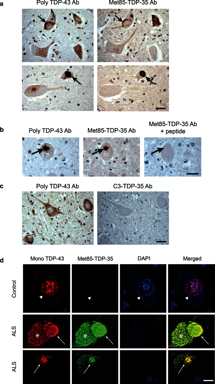
To determine whether Met85-TDP-35 is incorporated within pathological inclusions, immunohistochemical labeling with polyclonal TDP-43 antibody and Met85-TDP-35 antibody was performed on serial sections of ALS lumbar spinal cord. Met85-TDP-35 antibody co-labeled the same pathological inclusions as polyclonal TDP-43 antibody (Fig. 6a), and this labeling could be abrogated by competition with the peptide used to generate the Met85-TDP-35 antibody (Fig. 6b). In contrast, there was no labeling of TDP-43-positive inclusions with C3-TDP-35 antibody (Fig. 6c). Double fluorescence labeling showed labeling of the nucleus in normal motor neurons of control cases with mouse monoclonal TDP-43 antibody, and absence of labeling with Met85-TDP-35 antibody, confirming that Met85-TDP-35 neo-epitope antibody did not cross-react with full-length TDP-43 (Fig. 6d). In contrast, double fluorescence labeling of motor neurons in ALS lumbar spinal cord showed co-labeling of both round and skein like inclusions with the polyclonal TDP-43 antibody and Met85-TDP-35 antibody, confirming that Met85-TDP-35 is incorporated together with full-length TDP-43 within the pathological inclusions characteristic of ALS (Fig. 6d).
Fig. 6.
Immunohistochemical characterization of Met85-TDP-35 in ALS spinal cord. a Immunohistochemical analysis of serial sections from ALS and labeled with polyclonal TDP-43 antibody (poly-TDP-43 Ab) and Met85-TDP-35 antibody (Met85-TDP-35 Ab). Note that Met85-TDP-35 immunoreactivity is localized to TDP-43-positive pathology (arrows). b Met85-TDP-35 immunoreactivity was ablated by competition with the immunizing peptide (arrows). c There was no coincident labeling of TDP-43 pathology with C3-TDP-35 antibody (C3-TDP-35 Ab). d Double immunofluorescence labeling of control and ALS spinal motor neurons with mouse monoclonal TDP-43 antibody (Mono TDP-43; red) and Met85-TDP-35 Ab (green), nuclei stained with DAPI (blue). Note labeling of motor neuron nucleus in control spinal cord with mono TDP-43 antibody but not with Met85-TDP-35 antibody (arrowhead), indicating lack of cross reactivity of Met85-TDP-35 Ab with full-length TDP-43. Conversely, Met85-TDP-35 immunoreactivity co-localized with TDP-43 pathology in motor neurons of ALS cases (arrows). Asterisk indicates lipofuscin. Scale bars 20 μm (a–c); 5 μm (d)
Discussion
TDP-43 proteinopathy is the hallmark of the majority of ALS-FTLD cases, and biochemical characterization of pathological TDP-43 from disease tissues indicates that abnormal lower molecular weight TDP-43 species comprising the C-terminal region may play a role in disease pathogenesis [16]. Understanding the etiology of such species is important to advancing our understanding of TDP-43 pathogenicity. Here, we have provided evidence that the 35-kDa lower molecular weight species observed in diseases tissues can be derived from expression of an alternative splice variant of TDP-43. This splice variant, TDP-43 r.[106_196del], is upregulated approximately four-fold in ALS spinal cord compared to controls and is generated using non-canonical splice sites that skip the base pairs 106–196 in exon 2 of TARDBP, causing a frame shift and generating a premature termination codon 190 bp downstream from the first ATG. However, we have shown that a translation reinitiation event from the third ATGMet85 leads to the expression of an N-terminally truncated TDP-43 protein, denoted here as Met85-TDP-35, that has a disrupted NLS. Use of ATGMet85 as an alternative TDP-43 start site has been reported previously in cell culture studies showing that TDP-43 species of 35 kDa can be generated independent of caspase-3 cleavage [9, 17, 29]. Here, we have established the physiological relevance of this isoform in ALS and FTLD-TDP and shown that it can be generated via an abnormal splicing event.
As has been reported for other N-terminally truncated 35-kDa species of TDP-43 [9, 29], Met85-TDP-35 had reduced solubility, a cytoplasmic localization and formed aggregates when expressed in human neuroblastoma cells, all of which are predominant features of pathological TDP-43 in ALS and FTLD [1, 15]. Moreover, co-expression of Met85-TDP-35 with full-length TDP-43 resulted in a redistribution of full-length TDP-43 from the nucleus to the cytoplasm, where it was sequestered into Met85-TDP-35-positive inclusions. These inclusions were not labeled with ubiquitin or TDP-43 phosphorylation-dependent antibodies, supporting earlier studies showing that aggregation can occur in the absence of these modifications [12, 13, 22]. Given that TDP-43 has been shown to form homodimers [23], Met85-TDP-35 could potentially interact with TDP-43 and sequester it into cytoplasmic inclusions, thereby providing a possible mechanism for a gain-of-function toxicity of cytoplasmic Met85-TDP-35 and at the same time a loss-of-function resulting from a depletion of nuclear TDP-43. This potential mechanism of action has been proposed by others and our data fully support this premise [9, 26]. Importantly, our findings have shown that expression of Met85-TDP-35 in primary motor neurons led not only to formation of cytoplasmic aggregates, but also to decreased cell viability, demonstrating that Met85-TDP-35 can induce death of the neuronal type affected in ALS.
Although original studies have reported lower molecular weight TDP-43 species of ~25 kDa in FTLD brain tissues [1, 16], we consistently observed a protein species of 35 KDa as the predominant species on immunoblots of urea-soluble lysates extracted from gray matter of the anterior horn of ALS lumbar spinal cord, with a 25-kDa species observable after longer exposures. This may reflect differences in spinal cord versus brain tissues, or perhaps the extent of degeneration in the regions sampled. In this regard, in all cases used for the current study, areas adjacent to the regions sampled for biochemistry were confirmed to have remaining motor neurons with TDP-43 pathology by immunohistochemical analysis. Indeed, subsequent to the original descriptions of lower molecular weight species of TDP-43 in disease tissues, several other groups have also reported the presence of 35-kDa species in both ALS and FTLD tissues [23, 30]. To determine if the pathologically associated TDP-43 species of 35 kDa observed in ALS spinal cord tissues was derived from alternate translation initiation or caspase-3 cleavage, we generated neo-epitope antibodies specific for the respective N-terminal sequences, starting at Met85 or Asp90. The 35-kDa species observed on immunoblots of ALS tissues was detected using Met85-TDP-35 antibody but not C3-TDP-35 antibody, indicating that in all the samples tested in this study (n = 12 ALS cases), this 35-kDa species is generated through an alternate translation event starting at Met85. Analysis of urea-soluble fractions from prefrontal cortex also revealed that a 35-kDa lower molecular weight species of TDP-43 was clearly present in four of six ALS-FTLD cases, absent in controls, and was also labeled with Met85-TDP-35 antibody, again suggesting that this pathological form of TDP-43 in the brain is generated by alternate translation starting at Met85.
Immunohistochemical analysis showed labeling of TDP-43 pathology in motor neurons of ALS spinal cord with Met85-TDP-35 antibody, but not with C3-TDP-35 antibody. Interestingly, Met85-TDP-35 antibody labeling was specific to TDP-43 pathology in motor neurons, suggesting that the event leading to expression of Met85-TDP-35 may be motor neuron specific. This could be important for understanding the selective vulnerability of motor neurons, and possibly other affected neuronal types in ALS-FTLD, especially as expression of Met85-TDP-35 can kill motor neurons. Future studies will be aimed at investigating this possibility. One of the normal functions of TDP-43 is in the regulation of alternative splicing and prior studies have shown that downregulation of TDP-43 can lead to changes in the splicing profile of TDP-43 itself [2, 18]. As such, it is possible that the abnormalities of TDP-43 observed in ALS-FTLD could lead to expression of the TDP-43 r.[106_196del] transcript that generates Met85-TDP-35. Another possible mechanism is RNA perturbations caused by G4C2 hexanucleotide repeat expansions in C9orf72, the most common known genetic cause of ALS-FTLD and typified by TDP-43 pathology [10, 19, 20]. RNA foci generated by the expansions are observed in disease-affected neurons, sequestering RNA-binding proteins from their normal targets, which could include TDP-43 and thereby lead to splicing irregularities and generation of Met85-TDP-35. In support of this, both the TDP-43 r.[106_196del] transcript and Met85-TDP-35 were found in the ALS cases with C9orf72 repeat expansions, as well as sALS cases.
Based on our observations in culture, expression of Met85-TDP-35 would lead to sequestering of full-length TDP-43 from the nucleus to form cytoplasmic aggregates and cause neuronal death, an idea that has been promulgated previously [26]. As such, understanding the mechanism that generates Met85-TDP-35 could have therapeutic potential. Finally, although we have shown that the splice variant TDP-43 r.[106_196del] can generate Met85-TDP-35, there could be additional mechanisms that lead to use of ATGMet85 as an alternate translation start site and these remain to be explored.
In conclusion, we have identified a splice variant of TDP-43 that is upregulated in ALS and generates a protein species of 35 kDa through alternate translation initiation at ATGMet85, called Met85-TDP-35. A neo-epitope antibody specifically recognizing Met85-TDP-35 labeled the pathologically associated 35-kDa species on immunoblots of ALS-FTLD tissue extracts, and co-labeled TDP-43 pathology. Presence of Met85-TDP-35 was specific to affected motor neurons and could provide a potential mechanism for TDP-43-mediated neurotoxicity in disease.
Electronic supplementary material
Below is the link to the electronic supplementary material.
Acknowledgments
We are profoundly thankful to the patients and families for the generous donation of tissues for the study, as well as Shawn Hobbs, Myrna Moore, Olive Grozelle and Denise Miletic for their assistance. This work was funded by grants from the Canadian Institutes of Health Research (CIHR: JNM-90963; J.R.), Krembil Scientific Development Seed Fund (E.R., J.R., L.Z.), W. Garfield Weston Foundation (E.R.), Temerty Family Foundation (L.Z.) and James Hunter and Family ALS Initiative (J.R., L.Z.) J.R. holds a Tier 2 Canada Research Chair. H.C holds a CIHR/ALS Society of Canada Doctoral Award.
References
- 1.Arai T, Hasegawa M, Akiyama H, Ikeda K, Nonaka T, Mori H, Mann D, Tsuchiya K, Yoshida M, Hashizume Y, et al. TDP-43 is a component of ubiquitin-positive tau-negative inclusions in frontotemporal lobar degeneration and amyotrophic lateral sclerosis. Biochem Biophys Res Commun. 2006;351:602–611. doi: 10.1016/j.bbrc.2006.10.093. [DOI] [PubMed] [Google Scholar]
- 2.Ayala YM, De Conti L, Avendano-Vazquez SE, Dhir A, Romano M, D’Ambrogio A, Tollervey J, Ule J, Baralle M, Buratti E, et al. TDP-43 regulates its mRNA levels through a negative feedback loop. EMBO J. 2011;30:277–288. doi: 10.1038/emboj.2010.310. [DOI] [PMC free article] [PubMed] [Google Scholar]
- 3.Baralle M, Buratti E, Baralle FE. The role of TDP-43 in the pathogenesis of ALS and FTLD. Biochem Soc Trans. 2013;41:1536–1540. doi: 10.1042/BST20130186. [DOI] [PubMed] [Google Scholar]
- 4.Bose JK, Wang I-F, Hung L, Tarn W-Y, Shen C-KJ. TDP-43 overexpression enhances exon 7 inclusion during the survival of motor neuron pre-mRNA splicing. J Biol Chem. 2008;283:28852–28859. doi: 10.1074/jbc.M805376200. [DOI] [PMC free article] [PubMed] [Google Scholar]
- 5.Brettschneider J, Del Tredici K, Irwin DJ, Grossman M, Robinson JL, Toledo JB, Fang L, Van Deerlin VM, Ludolph AC, Lee VM et al (2014) Sequential distribution of pTDP-43 pathology in behavioral variant frontotemporal dementia (bvFTD). Acta Neuropathol. doi:10.1007/s00401-013-1238-y [DOI] [PMC free article] [PubMed]
- 6.Buratti E, Baralle FE. Characterization and functional implications of the RNA binding properties of nuclear factor TDP-43, a novel splicing regulator of CFTR exon 9. J Biol Chem. 2001;276:36337–36343. doi: 10.1074/jbc.M104236200. [DOI] [PubMed] [Google Scholar]
- 7.Buratti E, Dörk T, Zuccato E, Pagani F, Romano M, Baralle FE. Nuclear factor TDP-43 and SR proteins promote in vitro and in vivo CFTR exon 9 skipping. EMBO J. 2001;20:1774–1784. doi: 10.1093/emboj/20.7.1774. [DOI] [PMC free article] [PubMed] [Google Scholar]
- 8.Chang Y-F, Imam JS, Wilkinson MF. The nonsense-mediated decay RNA surveillance pathway. Annu Rev Biochem. 2007;76:51–74. doi: 10.1146/annurev.biochem.76.050106.093909. [DOI] [PubMed] [Google Scholar]
- 9.Che MX, Jiang YJ, Xie YY, Jiang LL, Hu HY. Aggregation of the 35-kDa fragment of TDP-43 causes formation of cytoplasmic inclusions and alteration of RNA processing. FASEB J Off Publ Fed Am Soc Exp Biol. 2011;25:2344–2353. doi: 10.1096/fj.10-174482. [DOI] [PubMed] [Google Scholar]
- 10.DeJesus-Hernandez M, Mackenzie IR, Boeve BF, Boxer AL, Baker M, Rutherford NJ, Nicholson AM, Finch NA, Flynn H, Adamson J, et al. Expanded GGGGCC hexanucleotide repeat in noncoding region of C9ORF72 causes chromosome 9p-linked FTD and ALS. Neuron. 2011;72:245–256. doi: 10.1016/j.neuron.2011.09.011. [DOI] [PMC free article] [PubMed] [Google Scholar]
- 11.Dormann D, Capell A, Carlson AM, Shankaran SS, Rodde R, Neumann M, Kremmer E, Matsuwaki T, Yamanouchi K, Nishihara M, et al. Proteolytic processing of TAR DNA binding protein-43 by caspases produces C-terminal fragments with disease defining properties independent of progranulin. J Neurochem. 2009;110:1082–1094. doi: 10.1111/j.1471-4159.2009.06211.x. [DOI] [PubMed] [Google Scholar]
- 12.Higashi S, Iseki E, Yamamoto R, Minegishi M, Hino H, Fujisawa K, Togo T, Katsuse O, Uchikado H, Furukawa Y. Appearance pattern of TDP-43 in Japanese frontotemporal lobar degeneration with ubiquitin-positive inclusions. Neurosci Lett. 2007;419:213–218. doi: 10.1016/j.neulet.2007.04.051. [DOI] [PubMed] [Google Scholar]
- 13.Hiji M, Takahashi T, Fukuba H, Yamashita H, Kohriyama T, Matsumoto M. White matter lesions in the brain with frontotemporal lobar degeneration with motor neuron disease: TDP-43-immunopositive inclusions co-localize with p62, but not ubiquitin. Acta Neuropathol. 2008;116:183–191. doi: 10.1007/s00401-008-0402-2. [DOI] [PubMed] [Google Scholar]
- 14.Mercado PA, Ayala YM, Romano M, Buratti E, Baralle FE. Depletion of TDP 43 overrides the need for exonic and intronic splicing enhancers in the human apoA-II gene. Nucleic Acids Res. 2005;33:6000–6010. doi: 10.1093/nar/gki897. [DOI] [PMC free article] [PubMed] [Google Scholar]
- 15.Neumann M, Mackenzie IR, Cairns NJ, Boyer PJ, Markesbery WR, Smith CD, Taylor JP, Kretzschmar HA, Kimonis VE, Forman MS. TDP-43 in the ubiquitin pathology of frontotemporal dementia with VCP gene mutations. J Neuropathol Exp Neurol. 2007;66:152–157. doi: 10.1097/nen.0b013e31803020b9. [DOI] [PubMed] [Google Scholar]
- 16.Neumann M, Sampathu DM, Kwong LK, Truax AC, Micsenyi MC, Chou TT, Bruce J, Schuck T, Grossman M, Clark CM, et al. Ubiquitinated TDP-43 in frontotemporal lobar degeneration and amyotrophic lateral sclerosis. Science. 2006;314:130–133. doi: 10.1126/science.1134108. [DOI] [PubMed] [Google Scholar]
- 17.Nishimoto Y, Ito D, Yagi T, Nihei Y, Tsunoda Y, Suzuki N. Characterization of alternative isoforms and inclusion body of the TAR DNA-binding protein-43. J Biol Chem. 2010;285:608–619. doi: 10.1074/jbc.M109.022012. [DOI] [PMC free article] [PubMed] [Google Scholar]
- 18.Polymenidou M, Lagier-Tourenne C, Hutt KR, Huelga SC, Moran J, Liang TY, Ling SC, Sun E, Wancewicz E, Mazur C, et al. Long pre-mRNA depletion and RNA missplicing contribute to neuronal vulnerability from loss of TDP-43. Nat Neurosci. 2011;14:459–468. doi: 10.1038/nn.2779. [DOI] [PMC free article] [PubMed] [Google Scholar]
- 19.Rademakers R, Neumann M, Mackenzie IR. Advances in understanding the molecular basis of frontotemporal dementia. Nat Rev Neurol. 2012;8:423–434. doi: 10.1038/nrneurol.2012.117. [DOI] [PMC free article] [PubMed] [Google Scholar]
- 20.Renton AE, Majounie E, Waite A, Simon-Sanchez J, Rollinson S, Gibbs JR, Schymick JC, Laaksovirta H, van Swieten JC, Myllykangas L, et al. A hexanucleotide repeat expansion in C9ORF72 is the cause of chromosome 9p21-linked ALS-FTD. Neuron. 2011;72:257–268. doi: 10.1016/j.neuron.2011.09.010. [DOI] [PMC free article] [PubMed] [Google Scholar]
- 21.Robertson J, Doroudchi MM, Nguyen MD, Durham HD, Strong MJ, Shaw G, Julien J-P, Mushynski WE. A neurotoxic peripherin splice variant in a mouse model of ALS. J Cell Biol. 2003;160:939–949. doi: 10.1083/jcb.200205027. [DOI] [PMC free article] [PubMed] [Google Scholar]
- 22.Sanelli T, Xiao S, Horne P, Bilbao J, Zinman L, Robertson J. Evidence that TDP-43 is not the major ubiquitinated target within the pathological inclusions of amyotrophic lateral sclerosis. J Neuropathol Exp Neurol. 2007;66:1147–1153. doi: 10.1097/nen.0b013e31815c5edd. [DOI] [PubMed] [Google Scholar]
- 23.Shiina Y, Arima K, Tabunoki H, Satoh J. TDP-43 dimerizes in human cells in culture. Cell Mol Neurobiol. 2010;30:641–652. doi: 10.1007/s10571-009-9489-9. [DOI] [PMC free article] [PubMed] [Google Scholar]
- 24.Udan M, Baloh RH. Implications of the prion-related Q/N domains in TDP-43 and FUS. Prion. 2011;5:1–5. doi: 10.4161/pri.5.1.14265. [DOI] [PMC free article] [PubMed] [Google Scholar]
- 25.Wang HY, Wang IF, Bose J, Shen CK. Structural diversity and functional implications of the eukaryotic TDP gene family. Genomics. 2004;83:130–139. doi: 10.1016/S0888-7543(03)00214-3. [DOI] [PubMed] [Google Scholar]
- 26.Winton MJ, Igaz LM, Wong MM, Kwong LK, Trojanowski JQ, Lee VM. Disturbance of nuclear and cytoplasmic TAR DNA-binding protein (TDP-43) induces disease-like redistribution, sequestration, and aggregate formation. J Biol Chem. 2008;283:13302–13309. doi: 10.1074/jbc.M800342200. [DOI] [PMC free article] [PubMed] [Google Scholar]
- 27.Xi Z, Zinman L, Grinberg Y, Moreno D, Sato C, Bilbao JM, Ghani M, Hernández I, Ruiz A, Boada M. Investigation of c9orf72 in 4 neurodegenerative disorders. Arch Neurol. 2012;69:1583–1590. doi: 10.1001/archneurol.2012.2016. [DOI] [PMC free article] [PubMed] [Google Scholar]
- 28.Xiao S, Tjostheim S, Sanelli T, McLean JR, Horne P, Fan Y, Ravits J, Strong MJ, Robertson J. An aggregate-inducing peripherin isoform generated through intron retention is upregulated in amyotrophic lateral sclerosis and associated with disease pathology. J Neurosci. 2008;28:1833–1840. doi: 10.1523/JNEUROSCI.3222-07.2008. [DOI] [PMC free article] [PubMed] [Google Scholar]
- 29.Yang C, Tan W, Whittle C, Qiu L, Cao L, Akbarian S, Xu Z. The C-terminal TDP-43 fragments have a high aggregation propensity and harm neurons by a dominant-negative mechanism. PLoS One. 2010;5:e15878. doi: 10.1371/journal.pone.0015878. [DOI] [PMC free article] [PubMed] [Google Scholar]
- 30.Yokota O, Davidson Y, Bigio EH, Ishizu H, Terada S, Arai T, Hasegawa M, Akiyama H, Sikkink S, Pickering-Brown S. Phosphorylated TDP-43 pathology and hippocampal sclerosis in progressive supranuclear palsy. Acta Neuropathol. 2010;120:55–66. doi: 10.1007/s00401-010-0702-1. [DOI] [PMC free article] [PubMed] [Google Scholar]
- 31.Zhang YJ, Xu YF, Dickey CA, Buratti E, Baralle F, Bailey R, Pickering-Brown S, Dickson D, Petrucelli L. Progranulin mediates caspase-dependent cleavage of TAR DNA binding protein-43. J Neurosci Off J Soc Neurosci. 2007;27:10530–10534. doi: 10.1523/JNEUROSCI.3421-07.2007. [DOI] [PMC free article] [PubMed] [Google Scholar]
Associated Data
This section collects any data citations, data availability statements, or supplementary materials included in this article.


