

Abstract
Platelet hyperactivity and platelet interaction with endothelial cells contribute to the development and progression of many cardiovascular diseases such as atherosclerosis and thrombosis. The impact of platelet activity with different pharmacological agents, such as acetylsalicylic acid and coumarin derivatives, has been shown to be effective in the prevention of cardiovascular disease. Artemisia dracunculus, L. Asteraceae (Tarragon) is used for centuries in the daily diet in many Middle Eastern countries, and it is well known for its anticoagulant activity. The present study investigates the presence of coumarins in tarragon leaves and subsequently determines the extract with a major amount of coumarin derivatives. The solvents of different polarities and different pH values were used for the purpose of purifying the primary extract in order to obtain fractions with the highest coumarin content. Those extracts and fractions were investigated for their anticoagulant activity by determining prothrombin time (PT) and the international normalized ratio (INR), expressed in relation to the coagulation time of the healthy person. Purified extracts and fractions obtained from plant residue after essential oil distillation, concentrated in coumarin derivatives, showed the best anticoagulant activity, using samples of human blood. INR maximum value (2.34) and consequently the best anticoagulant activity showed the methanol extract at concentration of 5%. The INR value of normal plasma in testing this extract was 1.05.
KEYWORDS: Artemisia dracunculus, coumarins, HPLC, TLC, extraction, anticoagulant activity
INTRODUCTION
A vegetarian diet has a positive effect on the development of many diseases, including arterial thrombosis and ischemic heart disease [1], through the antithrombotic action of many vegetables. Based on these records, many studies worldwide are focused on the anticoagulant activity of many medicinal plants with the hope of finding new and more effective agents. Coumarin derivatives are present in a large number of vegetables and medicinal plants [2]. These compounds have many pharmacological activities, and one of the best known is anticoagulant one [3]. Platelet hyperactivity is an important factor responsible for arterial thrombosis and atherosclerosis [4]. Activation of blood platelets results in their morphologic change, adhesion, secretion and aggregation [4].
Tarragon is used as a spice, in the preparation of various drinks, infusions, vinegar, and mustards. Fresh leaves of tarragon are used both as hors d-oeuvres and as garnishes for meat dishes and vegetable salads [5]. The experience of folk medicine and experimental studies has identified tarragon benefits on gastrointestinal tract function and diuretic action [6]. Tincture of tarragon has been shown to have a calming and anticonvulsant activity, as well as antidiabetic action and inhibition of blood platelet adhesion action [7-12].
The extract of Artemisia dracunculus leaves appear to be potentially useful for decreasing the incidence of coronary diseases in human, given the positive results of experimental studies conducted in rats [13].
This study was conducted to identify and quantify the presence of various coumarin derivatives in tarragon leaves and to evaluate the effects of different extracts on anticoagulant activity by the determination of prothrombin time (PT).
MATERIALS AND METHODS
Plant materials
Plant material (Folium dracunculi) was obtained from Artemisia dracunculus L., Asteraceae, collected from suburbs of Sarajevo in July 2011 and identified by botanical expert (S.D.). Voucher specimen No. 1057 was deposited at the herbarium of the Department of Biology, Faculty of Science, University of Sarajevo, Bosnia and Herzegovina. Immediately after collection, the leaves were separated from the rest of the plant and dried in a thin layer in a well-ventilated place, far from direct sunlight. The dried leaves were stored in paper bags in a dry, dark place and powdered just before the experimental analysis.
Extraction
One hundred grams of powdered tarragon leaves were subjected to successive extractions with 100 ml of n-hexane, 600 ml of chloroform and 600 ml of methanol in a Soxhlet’s aparatures. All the extractions were performed for eight hours. After removing the solvents under reduced pressure, three separate dry extracts were obtained: hexane-, chloroform-, and methanol extract.
The methanol extract was subsequently fractionated, using different solubility of its components in relation to the value of pKa. More specifically, 1g of the methanol extract was dissolved in 10 ml of diethyl ether, with the aim of separating lipophilic components that can be converted into salts extractable with water. The resulting solution was transferred to a separatory funnel and shaken three times with 50 ml of water, adjusted to pH 11.5 with 10 M NaOH.
The organic (diethyl ether) fraction was concentrated under reduced pressure to dryness, yielding the first fraction named F1. The remaining aqueous (water) fraction is acidified with acetic acid to pH 5.3 and then shaken with 100 ml of diethyl ether six times in order to convert the salts of lipophilic nature into the corresponding undissociated form extractable with an organic solvent. This way, another aqueous and another organic fraction were obtained. This aqueous fraction is evaporated obtaining the fraction F2, and the organic fraction was evaporated removing the solvent under reduced pressure to dryness, fraction F3.
Extraction of the distillation residue
After steam distillation, the herbal residue is separated from water in which it was immersed, to give a vegetable matrix. The herbal residue was split into four parts, each one subjected to extraction under various conditions in order to verify if the coumarin compounds remained in plant materials after essential oil distillation. In this way chloroform extract (A), diethyl ether extract (B), aqueous extract basified to pH 9 (D), and aqueous extract basified to pH 11,5 (E) were obtained. Water remaining after essential oil distillation was analyzed on coumarin presence as well, giving organic fraction with diethyl ether (C) and water fraction (F).
Conditions for thin layer chromatography (TLC)
Identification of coumarin compounds in different extracts and fractions was performed by thin layer chromatography. Methanol solution of test samples were prepared and used for TLC. Following coumarin standard solutions (0.1%) were used for identification of single coumarin compounds in test samples: coumarin-3-carbosilic acid 99%, Aldrich; umbelliferone 99%, Aldrich; 3-hydrohycoumarin 97%, Aldrich; 7,8-dihydroxy-6-metoxy coumarin 98%, Aldrich; 6,7-dihydroxycoumarin 98%, Aldrich; dihydrocoumarin 99%; Aldrich. Adsorbens was Silikagel 60 F254 precoated TLC plates, Merck, Darmstadt, Germany. Solvent system was toluene: ether (1:1, saturated with acetic acid 10%). Detection: UV-254, UV365, spray regent ethanolic KOH (10%).
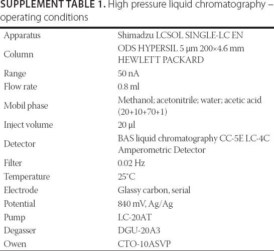
Hihg pressor liquid chromatography (HPLC)
Qualitative and quantitative analyses of the coumarin derivatives in methanol extract was performed by high pressure liquid chromatography with an electrochemical detector. Conditions for HPLC was given in Supplement Table 1.
Principles of the anticoagulant activity test
Human blood was sampled into a 4 ml test tube containing 3.2% sodium citrate. The blood was mixed and then centrifuged for 15 min. Obtained plasma was analyzed on coagulation analyzer (Diamed CDX, Diamond Diagnostics, USA) at 37°C. The principle of the test was based on re-enabling the blood to clot by adding calcium. For accurate results, a proportion of blood and citrate in the test tube must be fixed (1:10), before being placed in a centrifuge. The sample was visuali verified in order to determine if there were any clot formation within the tube, because in that case the test results would not be valid. All samples were stored at 25°C until the time of testing.
In order to determine the protrombin time (PT) a liquid calcium thromboplastin, also known as tissue factor III (Diaplastin-E, Pentapharm Ltd., Basel, Switzerland) was used. Diaplastin-E is stable, standardized to British corporate thromboplastin, and has the International sensitivity index of 1.1. Time to coagulation was measured optically. An INR value of normal plasma and solvent DMSO were determined for each test.
Tested extracts
Methanol extract, chloroform extract, water extract after essential oil distillation (extract E) and decoction of tarragon leaves were investigated for their anticoagulant activity. Dilution of the extracts was performed in DMSO in a way that the total concentration of DMSO does not exceed 30%. Only DMSO at a concentration of 30% and less, gave no problems with turbidity and it was possible to measure the PT.
Methanol and chloroform extracts were prepared in concentrations of 5%, 1.5%, 0.75%, and 0.09%. Extract E was prepared in concetrations of 1.5%, 0.75%, 0.375%, and 0.09%, due to its poor solubility in dimethyl sulfoxide. The decoction was prepared at a ratio of 1:10 and subsequently dilutions of 1:50, 1:100, and 1:500.
As a blank, for the first three extracts, pure solvent dimethyl sulfoxide (DMSO) was used. For the samples of decoction dilutions, distilled water was used as a blank. The anticoagulant activity was evaluated by measuring the effect of the extract on the total coagulation assay by determining PT. All the measurements were done in triplicate.
Statistical analysis
SPSS Statistics 17.0 software package (SPSS Inc., 233 South Wacker Drive, 11th Floor, Chicago, IL) was used for descriptive statistical analysis.
RESULTS
Extraction of coumarin derivatives
The qualitative composition of three extracts obtained by successive extractions with hexane, chloroform and methanol, was obtained by thin layer chromatography (TLC). Since the coumarin compounds are the objects of the study, the coumarin standards were used for their identification in different extracts.
Analysis of the TLC chromatograms showed that extraction conducted with three solvents failed to satisfy, since clear separation of coumarin compounds in the leaves of tarragon was not achieved. Thin layer chromatography partly confirmed the presence of coumarin compounds in chloroform and methanol extracts, but the coumarin derivatives spots were more intense in the methanol extract. Further separation of the methanol extract was carried out using a different solubility of the components in function of their pKa values. The results of separated coumarin compounds in the methanol extract, visible in the form of blue fluorescing spots observed under UV light at 365 nm and 254 nm, are shown in the chromatogram presented in Figure 1.
Figure 1.
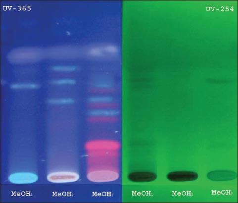
Methanol extract fractions separated according to pKa values and observed under UV-365 and 254 nm. MeOH1=organic fraction: Et2O/H2O (pH 5.3); MeOH2= organic fraction: Et2O/H2O (pH 11.5); MeOH3= water fraction Et2O/H2O (pH 11.5).
High pressure liquid chromatography
By comparing the retention time of the coumarin standards with retention times obtained in the tested methanol extract, five coumarin compounds were identified and quantified (Figure 2, Table 1).
Figure 2.
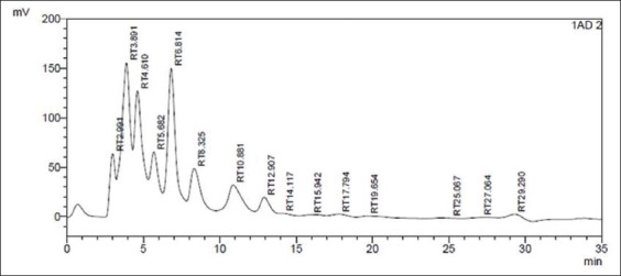
Chromatogram of pretreated methanol extract of tarragon leaves. *The five peaks corresponding to the coumarin derivatives are: RT 4.610, RT 5.682, RT 8.325, RT 12.907, RT 14.117.
Table 1.
Coumarin derivatives identified in tarragon leaves
Extraction of coumarin derivatives from plant residue after essential oil distillation
Plant residue remaining after distillation was subjected to further extraction in order to check whether coumarin compounds are lagging behind in plant material after the extraction of essential oil. The obtained extracts and fractions were applied to the chromatographic plate presented in Figure 3 in order to identify fractions with a major amount of coumarin derivatives.
Figure 3.

Chromatogram of separated spots obtained from different extracts of plant residue after essential oil destilation with water steam. A, chloroform extract of plant residue; B, diethyl ether extract of plant residue; C = diethyl ether fraction of water residue; D = water extract of plant residue - pH 9; E =water extract of plant residue - pH 11.5; F = water fraction of water residue.
The results show that coumarin compounds are lagging behind in plant material after steam distillation. Clearly visible blue fluorescing spots from coumarin compounds, particularly accentuated in extracts D and E, indicate that this compounds can be extracted from plant residue by varying the aqueous pH value.
Anticoagulant activity
The PT was used for estimating the total amount of prothrombin in the blood. The diagrams presented in Figure 4 show the results of anticoagulant activity of investigated extracts, expressed as INR values. Each extract was diluted in four different concentrations in order to monitor its effects on PT of healthy individuals and to see whether there is a correlation between different concentrations and INR values.
Figure 4.
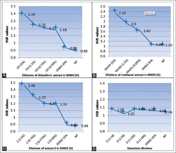
The relationship between different concentrations of test extracts and INR value. (A) Influence of chloroform extract on PT; (B) Influence of methanol extract on PT; (C) Influence of extract E on PT; (D) Influence of decoction on PT.
PT results were expressed as INR values but also as percentages and seconds for different extracts and their respective dilutions (Tables 2, 3, 4 and 5).
Table 2.
PT results and INR values for different chloroform extract concentrations
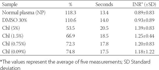
Table 3.
PT results and INR values for different methanol extract concentrations
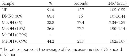
Table 4.
PT results and INR values for different extract E concentrations
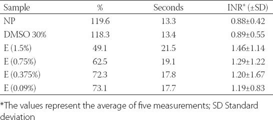
Table 5.
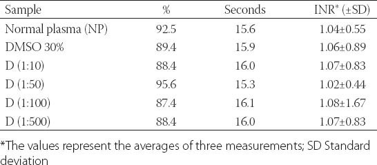
Prothrombin time and INR values for different decoction concentrations
Relationship diagrams between different extract concentration and INR values show that chloroform, methanol and extract E influence PT. There is a notable progressive linear increase in INR values with the increase of the concentration of extract.
A previously treated methanol extract showed the highest anticoagulant activity. The maximum INR value of added methanol extract at a concentration of 5% was 2.34. INR value of normal plasma in this case was 1.05. (Figure 4, Table 3).
The attached results show that the INR value of normal plasma (1.04) was almost identical with INR values in plasma that were added with different dilutions of tarragon decoction. Small differences are attributed to variations in the testing procedure. Decoction has no effect on PT, given that this pharmaceutical form does not contain coumarin derivatives (Figure 4, Table 5).
DISCUSSION
Many compounds present in diets may affect coagulation processes. Many of them are bioactive compounds, such as phenolic and polyphenolic compounds, vitamins and carotenoids, found in vegetables and fruits. Studies have shown these compounds have inhibitory effects on platelet function, thus having beneficial effects on heart disease and atherosclerosis. These reports encourage many studies on anticoagulant activity of medicinal plants with the hope that new and more effective agents will be found [14].
Artemisia dracunculus L., Asteraceae, owing to its phytochemical composition, has become very intriguing in the possible preparation of medicinal products against diseases caused by thromboembolic events. Among other natural polyphenolic compounds, this plant species contains coumarin derivatives with potential therapeutic effect [15]. For example, some coumarins inhibit the activity of vitamin K-dependent γ-carboxylase involved in the activation of coagulation factors [16]. Coumarins may inhibit platelet functions through multiple mechanisms including scavenging of reactive oxygen species, inhibiting cyclic nucleotide phosphodiesterase and prostaglandin syntheses [16].
Previous studies on the anticoagulant activity of various plant extracts of Artemisia dracunculus were performed in vitro, on isolated platelets [17, 13], while anticoagulant activity in this study was performed directly on samples of human blood by determining the PT. The PT is one of the most important tests to monitor coagulation and anticoagulant therapy and for the detection of blood-clotting disorders. The study (Yazdanparast R., et al) on tarragon leaf extract demonstrated the inhibition of platelet aggregation, adhesion to lamina coated plates by 60%, as well as decreased protein secretion by 50% [18].
Our experiments proved the presence of coumarin compounds in tarragon leaves and the method of their extraction has proven to be satisfactory. The extracts in which coumarin derivatives were concentrated showed anticoagulant activity and among them, methanol extracts had the highest effect on increase of the PT (INR 2.34). In our experiments all of the three extracts showed positive relation between the anticoagulant effect (INR value) and the concentration of applied extracts. Since the decoction is most frequently used in pharmaceutical herbal drug preparation, it was very important to investigate the anticoagulant activity of the tarragon leaves decoction. Unfortunately, decoction does not contain coumarin compounds subsquently it was not observed anticoagulant activity of this extract.
Our results support the data on anticoagulant effects of the tarragon leaf extracts and provide a scientific basis for the traditional use of this plant as an anticoagulant.
CONCLUSION
Artemisia dracunculus L., Asteraceae, contains coumarin compounds and is important plant species from a pharmacological point of view. Its anticoagulant activity confirmed in vitro and its use in folk medicine suggests the need for further research in this field.
ACKNOWLEDGMENTS
Authors would like to thank Professor Samir Ðug form the Faculty of Science, Department of Botany, University of Sarajevo, for his contribution and help in identification of plant material used in this study.
Appendix
Supplement Table 1.
High pressure liquid chromatography – operating conditions
DECLARATION OF INTERESTS
The authors declare no conflict of interests.
REFERENCES
- [1].Rajaram S. The effect of vegetarian diet, plant foods, and phytochemical on hemostasis and thrombosis. Am J Clin Nutr. 2003;78:552–8. doi: 10.1093/ajcn/78.3.552S. [DOI] [PubMed] [Google Scholar]
- [2].Bruneton J. 2nd ed. Paris: Tec&Doc; 1999. Pharmacognosy, Phytochemistry medicinal plants. [Google Scholar]
- [3].Golfakhrabadi F, Abdollahi M, Ardakani MR, Saeidnia S, Akbarzadeh T, Ahmadabadi AN, et al. Anticoagulant activity of isolated coumarins and toxicity evaluation of Ferulago carduchorum in rats. Pharm Biol. 2014;52(10):1335–40. doi: 10.3109/13880209.2014.892140. http://dx.doi.org/10.3109/13880209.2014.892140 . [DOI] [PubMed] [Google Scholar]
- [4].Olas B, Wachowicz B, Stochmal A, Oleszek W. Inhibition of blood platelet adhesion and secretion by different phenolics from Yucca schidigera Roezl. Bark. Nutrition. 2005;21:199–206. doi: 10.1016/j.nut.2004.03.024. http://dx.doi.org/10.1016/j.nut.2004.03.024 . [DOI] [PubMed] [Google Scholar]
- [5].Aglarova AM, Zilfikarov IN, Severtseva OV. Biological characteristics and useful properties of tarragon (Artemisia dracunculus L.) Pharm Chem J. 2008;42(2):31–5. http://dx.doi.org/10.1007/s11094-008-0064-3 . [Google Scholar]
- [6].Obolskiy D, Pischel I, Fiestel B, Glotov N, Heinrich M. Artemisia dracunculus L. (Tarragon): A Critical Review of Its Traditional Use, Chemical Composition, Pharmacology, and Safety. J Agric Food Chem. 2011;59(21):11367–84. doi: 10.1021/jf202277w. http://dx.doi.org/10.1021/jf202277w . [DOI] [PubMed] [Google Scholar]
- [7].Lopez-Lutz D, Alviano DS, Alviano CS, Kolodziejczyk PP. Screening of chemical composition, antimicrobial and antioxidant activities of Artemisia essential oils. Phytochemistry. 2008;69(8):1732–8. doi: 10.1016/j.phytochem.2008.02.014. http://dx.doi.org/10.1016/j.phytochem.2008.02.014 . [DOI] [PubMed] [Google Scholar]
- [8].Govorko D, Logendra S, Wang Y, Esposito D, Komarnytsky S, Ribnicky D. Polyphenolic compounds from Artemisia dracunculus L. inhibit PEPCK gene expression and gluconeogenesis in an H4IIE hepatoma cell line. Am J Physiol Endocrinol Metab. 2007;293(6):1503–10. doi: 10.1152/ajpendo.00420.2007. http://dx.doi.org/10.1152/ajpendo.00420.2007 . [DOI] [PubMed] [Google Scholar]
- [9].Ribnicky DM, Kuhn P, Poulev A, Logendra S, Zuberi A, Cefalu WT, Raskin I. Improved absorbtion and bioactivity of active compounds from an anti-diabetic extract of Artemisia dracunculus L. Int J Pharm. 2009;370(1-2):87–92. doi: 10.1016/j.ijpharm.2008.11.012. http://dx.doi.org/10.1016/j.ijpharm.2008.11.012 . [DOI] [PMC free article] [PubMed] [Google Scholar]
- [10].Sayyah M, Nadjafnia L, Kamalinejad M. Anticonvulsant activity and chemical composition of Artemisia dracunculus L. essential oil. J Ethnopharmacol. 2004;94(2-3):283–7. doi: 10.1016/j.jep.2004.05.021. http://dx.doi.org/10.1016/j.jep.2004.05.021 . [DOI] [PubMed] [Google Scholar]
- [11].Benli M, Kaya I, Yigit N. Screening antimicrobial activity of various extracts of Artemisia dracunculus L. Cell Biochem Funct. 2007;25:681–686. doi: 10.1002/cbf.1373. http://dx.doi.org/10.1002/cbf.1373 . [DOI] [PubMed] [Google Scholar]
- [12].Wang ZQ, Ribnicky D, Zhang XH, Raskin I, Yu Y, Cefalu WT. Bioactives of Artemisia dracunculus L. enhance cellular insulin signaling in primary human skeletal muscle culture. Metabolism. 2008;57(1):S58–S64. doi: 10.1016/j.metabol.2008.04.003. http://dx.doi.org/10.1016/j.metabol.2008.04.003 . [DOI] [PMC free article] [PubMed] [Google Scholar]
- [13].Yazdan Parast R, Saei A. Effects of aqueous tarragon, Artemisia dracunculus, extract on lipid and coagulatory parameters in rats. Biomed Lett. 1999;59(233):137–41. [Google Scholar]
- [14].Visioli F, Borsani L, Galli C. Diet and prevention of coronary heart disease: the potential role of phytochemicals. Cardiovasc Res. 2000;47:419–25. doi: 10.1016/s0008-6363(00)00053-5. http://dx.doi.org/10.1016/S0008-6363(00)00053-5 . [DOI] [PubMed] [Google Scholar]
- [15].Wright CW. New York: Taylor & Francis; 2002. Artemisia. [Google Scholar]
- [16].Garro HA, Garcia C, Martin VS, Tonn CE, Pungitore CR. Chemistry and biological activity of coumarins at molecular level. Nat Prod Commun. 2014;8:1091–4. [PubMed] [Google Scholar]
- [17].Okazaki K, Nakayama S, Kawazoe K, Takaishi Y. Antiaggregant effects on human platelets of culinary herbs. Phytother Res. 1998;12(8):603–5. http://dx.doi.org/10.1002/(SICI)1099-1573(199812)12:8<603:AID-PTR372>3.0.CO;2-G . [Google Scholar]
- [18].Yazdanparast R, Shahriyary L. Comparative effects of Artemisia dracunculus, Satureja hortensis and Origanum majorana on inhibition of blood platelet adhesion, aggregation and secretion. Vasc Pharmacol. 2008;48(1):32–7. doi: 10.1016/j.vph.2007.11.003. http://dx.doi.org/10.1016/j.vph.2007.11.003 . [DOI] [PubMed] [Google Scholar]