Abstract
We identified Tf2, the first β-scorpion toxin from the venom of the Brazilian scorpion Tityus fasciolatus. Tf2 is identical to Tb2-II found in Tityus bahiensis. We found that Tf2 selectively activates human (h)Nav1.3, a neuronal voltage-gated sodium (Nav) subtype implicated in epilepsy and nociception. Tf2 shifts hNav1.3 activation voltage to more negative values, thereby opening the channel at resting membrane potentials. Seven other tested mammalian Nav channels (Nav1.1-1.2; Nav1.4-1.8) expressed in Xenopus oocytes are insensitive upon application of 1 μM Tf2. Therefore, the identification of Tf2 represents a unique addition to the repertoire of animal toxins that can be used to investigate Nav channel function.
Introduction
The Tityus fasciolatus scorpion is found in the Cerrado biome in Brazil and is phylogenetically related to Tityus serrulatus, a scorpion responsible for many envenomation cases [1,2]. In contrast to T. serrulatus however, getting stung by T. fasciolatus is less likely since this scorpion is predominantly found in termite hills and rarely moves into human habitats. The venom toxicity of T. fasciolatus is considered moderate with a median lethal dose (LD50) of 56.65μg/mouse [3]. So far, Tf4 is the only peptide purified from this venom and based on its amino acid sequence and activity in sucrose gap assays in frog nerves, it is classified as an α-scorpion toxin acting on voltage-gated sodium (Nav) channels [4]. Since their pivotal role in cellular excitability makes them an ideal target to incapacitate prey or predators, scorpion venoms have evolved to contain a plethora of toxins targeting Nav channels [5].
α-Scorpion toxins primarily interfere with the voltage-drive activation process of the domain IV voltage sensor within the channel [6–8]. Since the primary role of this region is to inactivate the channel a few milliseconds after it opens (i.e. fast inactivation) [9–17], α-scorpion toxins slow down fast inactivation resulting in a persistent inward Na+ current when the channel should no longer conduct. In contrast to α-scorpion toxins, β-scorpion toxins activate Nav channels at voltages where they should normally be closed without affecting fast inactivation [18,19]. In general, the working mechanism underlying β-scorpion toxin activity involves stabilizing the voltage sensor in domains I, II, or III in an activated state to facilitate Nav channel activation [9,18–23]. Given their potency and exquisite selectivity, both toxin groups have been used extensively to investigate Nav channel function [5]. However, very few toxins have been identified that target just one of the nine Nav channel isoforms found in mammals. Such a tool would be beneficial to probe the physiological role of a particular isoform in an organism [24].
While testing the venom of T. fasciolatus on an array of Nav channel isoforms, we came across a fraction that selectively activated human (h)Nav1.3, a neuronal subtype believed to be involved in epilepsy as well as pain perception after channel upregulation due to spinal cord injury [25–27]. Further fraction purification resulted in the identification of Tf2, the first β-scorpion toxin isolated from the venom of T. fasciolatus. Tf2 can shift hNav1.3 activation voltage to much more negative values, effectively opening the channel at resting membrane potentials. Remarkably, seven other tested mammalian Nav channels (Nav1.1–1.2; Nav1.4–1.8) are insensitive to Tf2 upon application of an identical quantity. As such, the identification of Tf2 represents a distinct addition to the range of animal toxins that can be used to investigate hNav1.3 function.
Materials and Methods
Animal and venom collection
Specimens of T. fasciolatus were collected in Brasilia, Federal District, Brazil (15°46’S 47°50’W), under license n° 19138–1 (IBAMA- Instituto Brasileiro do Meio Ambiente e dos Recursos Naturais). They were kept at appropriate facilities at the University of Brasilia with food and water ad libitum. Scorpions were submitted to electrical stimulation close to the telson, respecting a 30 day interval. The venom was collected, solubilized in a watery trifluoroacetic acid (TFA) 0.12% solution, and centrifuged at 15,000 × g for 15 min. The supernatant was collected and quantified at 280 nm and dried. Hereafter, the material was kept at -20°C.
Toxin purification
Dried T. fasciolatus venom was dissolved in purified water and separated by RP-HPLC (reverse-phase high performance liquid chromatography) using a C18 analytical column (Synergi Fusion RP 4μ 80 Å 250 × 4.6 mm Phenomenex, Inc., USA), using a linear gradient from 0% solvent A (0.12% TFA in water) to 60% solvent B (0.10% TFA in acetonitrile) over 60 min at a flow rate of 1 mL/min, with detection at a wavelength of 216 and 280nm. The optimized chromatography protocols used to purify Tf2 consisted of three extra steps with a gradient from 25% to 45% solvent B in 40 min with the two last steps performed at 45°C. The purity of the toxin was verified by MALDI-TOF mass spectrometry and by the area under curve on HPLC and was higher than 97%.
Molecular mass and amino acid sequence determination
The molecular mass and purity of the sample was checked on an Ultraflex III MALDI TOF mass spectrometer (Bruker Daltonics, Germany). The sample was dissolved in an α-cyano-4-hydroxycinnamic acid matrix (1:3, v:v), spotted onto the MALDI target plate and dried at room temperature for 15 min and analyzed in positive reflected and linear modes. External calibration was preceded by a Peptide Calibration Standard for mass spectrometry (Bruker Daltonics) for reflector mode and Protein 1 mixture (Bruker Daltonics) for linear mode. Spectra were analyzed with FlexAnalysis 3.3 (Bruker Daltonics, Germany). In order to check the accurate monoisotopic molecular mass of Tf2, a micrOTOF-QII (Bruker Daltonics) equipped with an orthogonal electrospray ionization source (ESI) operated in the positive mode was used. Sample was diluted in variable concentrations of 1% formic acid in a water/acetonitrile mixture (1:1, v:v) and applied to the mass spectrometer source by direct infusion.
The complete amino acid sequence was obtained from a transcriptomic library performed with RNA isolated from the T. fasciolatus venom gland (Hiseq, Illumina, USA). The signal peptide was identified using SignalP 4.1 server [28], and the amidation signal site was determined using insights gained from previous characterized toxins. To verify the peptide sequence, the first 15 amino acid residues were sequenced by automated Edman degradation on a Beckman (Palo Alto, CA) LF 3000 protein peptide sequencer. Similarity searches were performed using blastp (http://www.ncbi.nlm.nih.gov/blast) with an e value cutoff set to <10–5 to identify putative functions. ClustalW version 2.0 [29] was used for sequence alignments and calculation of the percentage of identity between paired sequences. The consensus sequence of alignment was colored using Chroma software [30]. The protein reported in this work is deposited in UniProt Knowledgebase as entry C0HJM9 and nucleotide sequence is deposited in EMBL-EBI as entry LN606597.
Molecular modeling
Model construction of Tf2, Ts2 and CssIV was performed by the fold recognition approach for identifying potential templates using the Phyre2 server [31]. This server is able to identify homologs using distant sequences obtained from algorithms such as PSI-BLAST and HMM [32,33]. The template with the highest score, Ts1 (Protein Data Bank ID 1NPI, 1.16 Å resolution), was chosen for both Tf2 and Ts2 model construction, with 100% coverage and 75% / 74% identity, respectively [34]; and Cn2 template (PDB ID 1CN2, 2.21 Å resolution) [35] was chosen for CssIV modeling, with 100% coverage and 83% identity. Additionally, CssII and AaHII three-dimensional structures (PDB ID 2LI7 [36] and 1PTX [37], respectively), were used in the structural and electrostatic potential surface comparison with Tf2. The alpha toxin AaHII was used in order to emphasize the structural differences between alpha and beta sodium scorpion toxins. Structural alignment of all proteins (Tf2, Ts2, Ts1, CssIV, CssII, and AaHII) was performed using the plugin MultiSeq by Visual Molecular Dynamics (VMD) [38] and the electrostatic potential surfaces were calculated using CCP4 Molecular Graphics Program [39] by the built-in Poisson-Boltzmann method.
Electrophysiology using Xenopus oocytes
Human (h)Nav1.4, hNav1.5, and rat (r)Nav1.8 were a gift from Peter Ruben (Simon Fraser University, Canada), Chris Ahern (University of Iowa, USA), and John Wood (UCL, UK), respectively. hNav1.1–1.3, hNav1.6–1.7 were obtained from Origene Technologies, Inc. (MD, USA). Accession numbers are NM_001165963.1 (hNav1.1), NM_021007.2 (hNav1.2), NM_006922.3 (hNav1.3), NM_000334 (hNav1.4), NM_198056 (hNav1.5), NM_014191.2 (hNav1.6), NM_002977.2 (hNav1.7), and NM_017247.1 (rNav1.8). cRNA of hNav1.1–1.7 and rNav1.8 was synthesized using T7 polymerase (mMessage mMachine kit, Life technologies, USA) after linearizing the fully-sequenced DNA with appropriate restriction enzymes. Channels were expressed together with the hβ1 subunit (Origene Technologies, Inc., NM_001037.4) (1:5 molar ratio) in Xenopus laevis oocytes (acquired from Xenopus one, USA) that were incubated at 17°C in 96 mM NaCl, 2 mM KCl, 5 mM Hepes, 1 mM MgCl2, 1.8mMCaCl2, and 50 g/mL gentamycin (pH 7.6 with NaOH) for 1–4 days after cRNA injection, and then were studied using two-electrode voltage-clamp recording techniques (OC-725C; Warner Instruments) with a 150-μl recording chamber. Data were filtered at 4 kHz and digitized at 20 kHz using pClamp10 software (Molecular Devices, USA). Microelectrode resistances were 0.5–1 MΩ when filled with 3 M KCl. The external recording solution contained 100 mM NaCl, 5 mM Hepes, 1 mM MgCl2, and 1.8 mM CaCl2 (pH 7.6 with NaOH). All experiments were performed at room temperature (22°C). Leak and background conductance, identified by blocking the channel with tetrodotoxin (Alomone Laboratories, Israel), were subtracted for all Nav channel currents. The G-V curves were plotted with OriginPro8 (3<n<6), normalized, and fitted with the Boltzmann equation. A t-test was carried out to calculate the statistical relevance of each experiment with p<0.05.
Results
Toxin purification and sequence determination
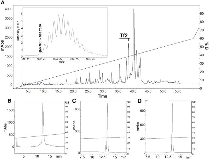
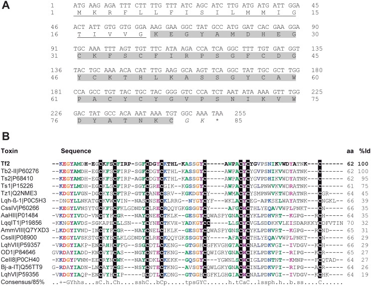
1 mg of T. fasciolatus venom was divided into 60 fractions using reversed-phase HPLC with a linear acetonitrile gradient and Tf2 was collected at 38.5% of acetonitrile (Fig 1A). Next, three additional purification steps with varying degrees of acetonitrile gradients were performed to obtain the final pure toxin (Fig 1B–1D). The amino acid sequence was deduced by a combination of Edman sequencing and a RNA library (unpublished data) obtained from RNA extracted from the venom gland. The nucleotide sequence that codes for Tf2 contains 255 nucleotides, including the stop codon, and the translated peptide contains a signal peptide with 20 amino acid residues, and a 62 residue mature peptide (Fig 2A). Nav channel scorpion toxins containing a GK at the C-terminal end are frequently amidated [40], an important feature for toxins such as Ts1 and CssII since this post translational modification increases their biological activity [41,42]. Taking protein amidation into account (-0.98 Da), as well as the presence of four disulfide bridges, the theoretical monoisotopic molecular mass is [M+H]+ 6,950.0302 Da, which corresponds with the experimental monoisotopic molecular mass of [M+H]+ 6,949.9350 Da (or [M+7H]7+ = 993.7050) obtained by using the micrOTOF QII (see inset Fig 1A). The calculated error between theoretical and experimental molecular mass, using the fifth isotope of the isotopic series, was 0.6 ppm.
Fig 1. Purification and molecular mass determination of Tf2.
(A) Chromatography by RP-HPLC of 1 mg of T. fasciolatus crude venom. Fractionation was performed on a C18 analytical column, using a linear gradient from 0% solvent A (0.12% TFA in water) to 60% solvent B (0.10% TFA in acetonitrile) over 60 min at a flow rate of 1 mL/min, with detection at a wavelength of 216 and 280nm. The component eluted at 38.5% of acetonitrile corresponds to Tf2. (B-D) Three additional chromatographic protocols performed to obtain pure Tf2. (B) Linear gradient of B solvent, from 25 to 45% B in 40 minutes, at room temperature (22°C). (C) Linear gradient of B solvent, from 25 to 45% of B in 40 minutes, at 45°C. (D) Linear gradient of B solvent, from 30 to 40% of B in 40 minutes, at 45°C. Inset on (A) shows mass spectrometry analysis of Tf2 by micrOTOF-Q II, presenting the monoisotopic distribution of the +7 charged ion ([M+7H]7+ = 993.7050), which is equivalent to [M+H]+ 6949.9350 Da.
Fig 2. Tf2 sequence and alignment with scorpion Nav channel toxins.
(A) The nucleotide sequence of Tf2 was obtained by HiSeq (Illumina, USA). Signal peptide is underlined, mature peptide is highlighted in gray, and the amidation set point is marked in italic. (B) Multiple sequence alignment of Tf2 with other Nav channel toxins. Toxins are presented by their short names and UniProt KB codes. Capital letters denote amino acids. Lower-case letters denote: h, hydrophobic; s, small; b, big; p, polar; t, tiny; a, aromatic; l, aliphatic. Positive (+) and negative (-) amino acid residues that are part of the consensus sequence are also colored. Cys residues are shaded in black. aa means amino acid residues, and %Id is the percentage of sequence identity with Tf2.
Aligning Tf2 with other toxins found in databases (Fig 2B) reveals 100% identity with Tb2-II, a toxin from the South American scorpion Tityus bahiensis that is lethal upon injection into mammals and insects [43]. However, the molecular target of Tb2-II has not been identified yet. Tf2 is also 95% identical (3 amino acid difference) to Ts2, a toxin from T. serrulatus venom which inhibits fast inactivation of Nav1.2, Nav1.3, Nav1.5, Nav1.6, and Nav1.7, but does not affect Nav1.4 or NaV1.8 [44,45]. Interestingly, Ts2 also shifts the voltage-dependence of activation of Nav1.3 to more negative potentials suggesting that multiple voltage sensors may be targeted [9]. Sequence alignments were conducted with toxins that were tested on heterologously expressed-voltage gated ion channels as well as representative examples of toxins of which 3D structures have been determined. Such an alignment could provide insights into the features that are important for the biological activity of Tf2. For example, in Tf2, Tb2-II, and Ts2, a conserved cluster of aromatic residues formed by Y4, Y37, Y44, Y46, W40 and W55 was observed (Fig 2B). This is known to be as an important feature for the activity of β-scorpion toxins [46,47].
Homology modeling
For structural analysis, the NMR structures of Ts1, CssII, and AaHII, were compared with the homology models of Tf2, Ts2, and CssIV. Tf2 and Ts2 models were constructed using Ts1 as template whereas the CssIV model was obtained using Cn2 as template. Since the effect of different amino acid mutations is well studied in the β-scorpion toxins CssII and CssIV, we chose these as reference molecules to better understand the structure-function relationship of Ts1, Ts2 and Tf2 [48,49]. The α-toxin AaHII was chosen as a representative example of α-scorpion toxins to evaluate the differences with this particular family [42,50].
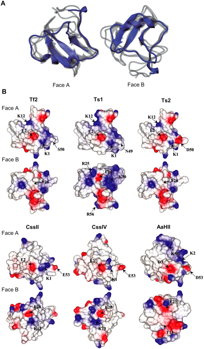
Resulting structural alignments reveal a superposition of secondary structures, an observation that is typical for scorpion toxins targeting Nav channels (Fig 3A) [46]. However, the electrostatic potential diverges when comparing the structures of toxins Ts1, Ts2 and Tf2 with the structures of CssII, CssIV and AaHII (Fig 3A and 3B). This feature has been suggested before to be responsible for the difference in target sensitivity [51–55]. All toxins possess a centrally-located residue with negative electrostatic potential (Fig 3B, face A), corresponding to a glutamic acid (E2, present in all β-scorpion toxins, including Ts2) or an aspartic acid (D3, present in the α-scorpion toxin AaHII). Another feature observed in all analyzed structures is the presence of a positive region close to the negative core, i.e. a lysine residue situated at position 1 for the β-scorpion toxins, and position 2 for the α-scorpion toxin AaHII. Comparing Tf2 to other scorpion toxins suggests that positively charged groups at positions 1 and 12 (K12 to Tf2, Ts1 and Ts2, K13 to CssII and CssIV), as well a negative charge at position 2 are likely determinants of β-scorpion toxin specificity [47]. In α-scorpion toxins, the positively charged residue is commonly found in position 58 instead of 12 (K58 in AaH2, Fig 3) [47], whereas with Ts2 has a K12 and A58 (Fig 2B) [44]. However, all β-scorpion toxins analyzed here possess these characteristics, yet Tf2 acts primarily on hNav1.3 whereas the others do not.
Fig 3. Structural comparison between Tf2 and other Nav channel toxins.
(A) Structural alignment between Tf2 (in blue) and five other Nav channel scorpion toxins—Ts1, Ts2, CssII, CssIV, and AaHII (in gray). (B) Comparison of electrostatic potentials between the toxins Tf2, Ts1, Ts2, CssII, CssIV, and AaHII. The figure shows charge distribution along the toxin surface, divided into faces A and B. Shown in red are acidic residues whereas blue represents basic residues; in white, neutral regions are shown.
The primary sequences of Tf2 and Ts2 differ in three residues (S20A, S50D and N51H) (Fig 2B), among which only the substitution at position 50 induces a significant change in the electrostatic potential of the molecule. The presence of this aspartic acid generates a negative potential in Ts2, which is also absent in Ts1 (N49 residue in the equivalent position) (Fig 3B, face A). When comparing the structures of Ts1 to those from Tf2 and Ts2, we observe an increase of positively charged regions, originating from an arginine at positions 25/56 and a lysine at position 30 (Fig 2B, face B). The high degree of sequence and structural conservation of charged groups observed in Tf2, Ts1 and Ts2 at face A suggests that this structural pattern may have an important role in the recognition and specificity towards particular Nav channel isoforms.
CssII, CssIV, and AaHII also have an acid residue in a similar acidic region of D50 found in Ts2 (E53, E53 and D53, respectively) (Figs 2B and 3B). Although presenting a different electrostatic potential in comparison to Ts2 and Tf2, all these toxins have small amino acid residues in equivalent positions, as observed in the consensus alignment (Fig 2B) and at face A of structures (Fig 3B). As expected by their high degree of similarity, face B of Tf2 and Ts2 have similar electrostatic potentials. In comparison, face B of Ts1 is more positively charged due to the presence of residues R25, K30 and R56. Additionally, an acidic region, usually at position E28, can be visualized at face B in almost all these proteins, which is known as a key pharmacophore region in scorpion toxins targeting Nav channels [46]. However, both Tf2 and Ts2 present an aromatic residue at this position (Y26; see Fig 3B, face B).
Tf2 activity on mammalian Nav channels expressed in oocytes
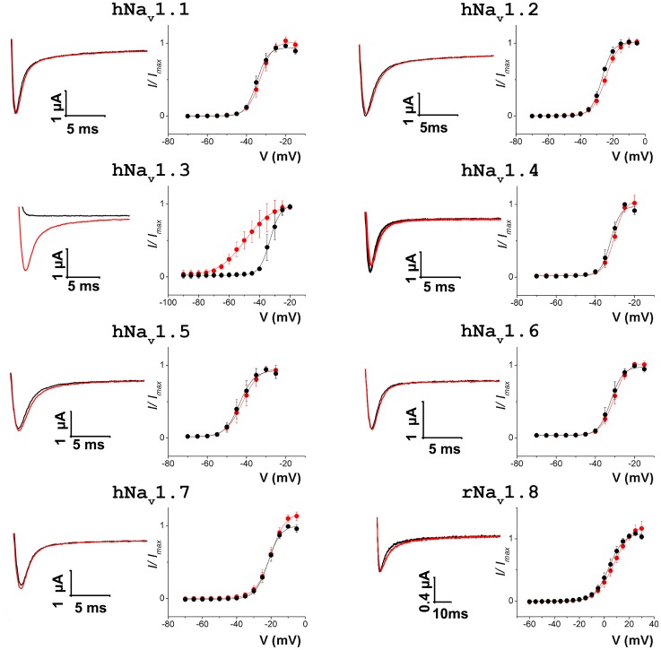
To examine the biological activity of Tf2, we used the two-electrode voltage-clamp technique to measure sodium currents from 8 mammalian Nav channel isoforms expressed in X. laevis oocytes before and after toxin addition. At 1 μM, Tf2 does not influence currents generated by hNav1.1–1.2, 1.4–1.7, and rNav1.8 (Fig 4); however, hNav1.3 activation is drastically influenced. At 1μM, Tf2 shifts the voltage-dependence of activation by ~16 mV (V1/2 from -33.1 ± 0.2mV to -49.3 ± 0.5mV; slopes 3.4 ± 0.2 and 8.9 ± 0.5). To a certain extent, this effect resembles that of Ts2, which promotes opening of rNav1.3 when applying 1 μM toxin [44]. However, Ts2 also hampers fast inactivation of this and other rNav channel isoforms whereas Tf2 does not appear to influence activation or fast inactivation of other Nav channel isoforms. Thus, Tf2 activity seems to be unique among β-scorpion toxins and may be valuable to further hNav1.3 research.
Fig 4. Effect of Tf2 on Nav channel isoforms expressed in X. laevis oocytes.
Shown on the left in each column is a representative trace of experimental Na+ currents obtained by depolarizing the membrane to a suitable voltage from a holding potential of -90mV, at -25mV for Nav 1.1–1.2, 1.4–1.7, at -40mV for Nav1.3, and at 20mV for Nav1.8. Shown on the right in each column is a deduced conductance (G)—voltage (V) relationship before (black) and after (red) the application of 1μM Tf2. This concentration only influences the activation of hNav1.3. Data is shown as mean ± SEM of n > 3.
Discussion
In this work, we report the discovery and characterization of Tf2, the first β-scorpion toxin from the venom of T. fasciolatus. In contrast to related β-scorpion toxins [34,36,44,45,49], Tf2 preferentially potentiates hNav1.3 opening, resulting in a dramatic shift in activation voltage. As a result, delivery of 1 μM Tf2 to Nav1.3 expressed in neurons may cause the channel to open at resting membrane voltages, an effect that would be detrimental to the envenomed organism.
Given its exquisite selectivity pattern, it is interesting to consider the mechanism underlying Tf2 activity. When comparing the amino acid sequence of Tf2 and Ts2, only three substitutions can be found (S20A, S50D, and N51H). As a result of the substitution S50D, Ts2 has a lower overall charge that may affect toxin ability to interact with its binding site, which may be located within the lipid membrane. In Ts1, the residue W50, equivalent to the residues H51 in Ts2 or N51 in Tf2, is crucial for toxin activity [47]. Thus, it is possible that the different effects between Tf2 and Ts2 on Nav channels are due to this substitution or due to the charge difference caused by S50D. Hence, in order to elucidate the different effects of Tf2 and Ts2, it will be interesting to perform site-directed mutagenesis and test both Tf2 and Ts2 under similar electrophysiological conditions.
Overall, Tf2 possess all the important characteristics mentioned by Polikarpov et al., [47] that are relevant for the activity of Nav channel toxins, such as a positive electrostatic potential at position 1 (K1), a negative potential at position 2 (E2), a positively charged group at position 12 (K12) and a solvent-exposed aromatic core (Y4, Y37, Y44, Y46, W40 and W55). In all presented structures, the central negative residue (E2 or D3) is surrounded by the aromatic core. At face B, Tf2 and Ts2 possess an Y28 instead of the E28. As discussed by Cohen et al., [49], the charge neutralization of this acidic residue resulted in a three order decrease of CssIV binding affinity. Maybe this neutralization could represent a clue for the high specificity of Tf2 to hNav1.3. CssII and CssIV also possess these characteristics, but the basic residue at position 12 is actually at position 13, which is located at face B. Despite of the different face, these peptides maintain the β-scorpion toxin activity. Instead of a K12, the toxin AaHII has a lysine residue at position 58, as a proposed conserved feature in α-scorpion toxins.
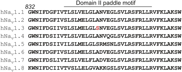
In general, β-scorpion toxins interact primarily with the S3b-S4 paddle motif within the domain II voltage sensor in Nav channels [9,18–23]. However, a sequence alignment of this region reveals a high degree of amino acid conservation between hNav1.1-r1.8 (Fig 5). Even though the Ser at position 842 in hNav1.3 is unique among most Nav channel isoforms and may therefore contribute to subtype selectivity of the toxin, a Ser is also found in hNav1.5 which is insensitive to Tf2. However, hNav1.5 differs in the four ensuing residues (Arg/Met/Ser/Asn) from hNav1.3. In contrast to α-scorpion toxins for which an interaction with the paddle motif is the domain IV voltage sensor is sufficient to exert their effect [9,56–59], it is important to mention that residues outside of the domain II paddle motif may be required for activity of β-scorpion toxins [8,18,19,21,51,60–62]. As such, the Nav channel isoform selectivity of Tf2 may be determined by amino acids in regions outside of the anchoring point in the domain II paddle motif. Further mutagenesis studies will therefore help delineate the complete binding site of Tf2. Finally, it is also worth considering that the extent of the effect on hNav1.3 as observed in the X. laevis system, may differ in mammalian neurons due to a different membrane lipid composition or the presence of β-subunits [63,64]. Altogether, the distinct selectivity pattern of Tf2 may be a powerful tool to investigate the role of hNav1.3 in epilepsy or nociception after spinal cord injury [25–27]. Moreover, the dramatic shift in channel activation voltage in the presence of Tf2 could be exploited in drug screening assays (e.g. FLIPR) in which voltage-control is impractical to open the channel in the presence of therapeutics [24,65].
Fig 5. Sequence alignment of the domain II paddle motif in 8 mammalian Nav channel isoforms.
Figure shows a sequence alignment of the domain II paddle motif as found in 8 mammalian Nav channel isoforms. As a reference, the number in italic indicates the coordinates of the first Gly residue in hNav1.1. Although the Ile in hNav1.3 (position 830 according to hNav1.3 coordinates, 840 according to hNav1.1 coordinates) differs from the Phe found in other neuronal isoforms, this residue is not present within the paddle motif and may not be accessible to Tf2. The Ser at position 842 (hNav1.3 coordinates—indicated in red) is unique among hNav1.1–1.3.
Acknowledgments
We also like to thank Tainá Raiol, João Victor Araújo Oliveira, Rafel Costa and Dr. Marcelo Brígido from the Laboratory of Molecular Biology, University of Brasilia, for bioinformatics analysis of the T. fasciolatus transcriptome library. We kindly thank Dr. Carlos Bloch and Maura Vianna Prates, from Mass Spectrometry Laboratory—Embrapa Recursos Genéticos e Biotecnologia, for mass spectrometry and Edman sequencing, respectively.
Data Availability
All relevant data are within the paper.
Funding Statement
Parts of the research in this publication were supported by the National Institute of Neurological Disorders and Stroke (NINDS) of the National Institutes of Health (NIH) under award number R00NS073797 (F.B.) and by CNPq (564.223/2010-7)/FAPDF (193.000.471/2011) (INOVATOXIN project) (E.F.S). T.S.C. is a graduate student financed by CNPq and CAPES Foundation. S.C.R. and C.B.F.M. are graduate students supported by CNPq. E.F.S. receives support from CNPq. The funders had no role in study design, data collection and analysis, decision to publish, or preparation of the manuscript.
References
- 1. Chippaux JP, Goyffon M (2008) Epidemiology of scorpionism: a global appraisal. Acta Trop 107: 71–79. 10.1016/j.actatropica.2008.05.021 [DOI] [PubMed] [Google Scholar]
- 2. Lourenço WR, Cloudsley-Thompson JL, Cuellar O, Von Eickstedt VRD, Barravieira B, Knox MB (1996) The evolution of scorpionism in Brazil in recent years. Journal of Venomous Animals and Toxins 2: 121–134. [Google Scholar]
- 3. Guimarães PTC (2009) Caracterização Molecular e Imunológica do veneno de Tityus fasciolatus e sua ação sobre camundongos. Belo Horizonte: Universidade Federal de Minas Gerais; 130 p. 10.1080/713663671 [DOI] [Google Scholar]
- 4. Wagner S, Castro MS, Barbosa JA, Fontes W, Schwartz EN, Sebben A, et al. (2003) Purification and primary structure determination of Tf4, the first bioactive peptide isolated from the venom of the Brazilian scorpion Tityus fasciolatus. Toxicon 41: 737–745. [DOI] [PubMed] [Google Scholar]
- 5. Gilchrist J, Olivera BM, Bosmans F (2014) Animal toxins influence voltage-gated sodium channel function. Handbook of experimental pharmacology 221: 203–229. 10.1007/978-3-642-41588-3_10 [DOI] [PMC free article] [PubMed] [Google Scholar]
- 6. Campos F, Coronas F, Beirão P (2004) Voltage-dependent displacement of the scorpion toxin Ts3 from sodium channels and its implication on the control of inactivation. British journal of pharmacology 142: 1115–1122. [DOI] [PMC free article] [PubMed] [Google Scholar]
- 7. Rogers J, Qu Y, Tanada T, Scheuer T, Catterall W (1996) Molecular determinants of high affinity binding of alpha-scorpion toxin and sea anemone toxin in the S3-S4 extracellular loop in domain IV of the Na+ channel alpha subunit. The Journal of biological chemistry 271: 15950–15962. [DOI] [PubMed] [Google Scholar]
- 8. Martin-Eauclaire MF, Ferracci G, Bosmans F, Bougis PE (2015) A surface plasmon resonance approach to monitor toxin interactions with an isolated voltage-gated sodium channel paddle motif. The Journal of general physiology 145: 155–162. 10.1085/jgp.201411268 [DOI] [PMC free article] [PubMed] [Google Scholar]
- 9. Bosmans F, Martin-Eauclaire MF, Swartz KJ (2008) Deconstructing voltage sensor function and pharmacology in sodium channels. Nature 456: 202–208. 10.1038/nature07473 [DOI] [PMC free article] [PubMed] [Google Scholar]
- 10. Campos FV, Chanda B, Beirão PS, Bezanilla F (2008) Alpha-scorpion toxin impairs a conformational change that leads to fast inactivation of muscle sodium channels. J Gen Physiol 132: 251–263. 10.1085/jgp.200809995 [DOI] [PMC free article] [PubMed] [Google Scholar]
- 11. Capes DL, Arcisio-Miranda M, Jarecki BW, French RJ, Chanda B (2012) Gating transitions in the selectivity filter region of a sodium channel are coupled to the domain IV voltage sensor. Proceedings of the National Academy of Sciences of the United States of America 109: 2648–2653. 10.1073/pnas.1115575109 [DOI] [PMC free article] [PubMed] [Google Scholar]
- 12. Cha A, Ruben PC, George AL Jr., Fujimoto E, Bezanilla F (1999) Voltage sensors in domains III and IV, but not I and II, are immobilized by Na+ channel fast inactivation. Neuron 22: 73–87. [DOI] [PubMed] [Google Scholar]
- 13. Chanda B, Bezanilla F (2002) Tracking voltage-dependent conformational changes in skeletal muscle sodium channel during activation. J Gen Physiol 120: 629–645. [DOI] [PMC free article] [PubMed] [Google Scholar]
- 14. Horn R, Ding S, Gruber HJ (2000) Immobilizing the moving parts of voltage-gated ion channels. J Gen Physiol 116: 461–476. [DOI] [PMC free article] [PubMed] [Google Scholar]
- 15. Sheets MF, Kyle JW, Hanck DA (2000) The role of the putative inactivation lid in sodium channel gating current immobilization. J Gen Physiol 115: 609–620. [DOI] [PMC free article] [PubMed] [Google Scholar]
- 16. Sheets MF, Kyle JW, Kallen RG, Hanck DA (1999) The Na channel voltage sensor associated with inactivation is localized to the external charged residues of domain IV, S4. Biophys J 77: 747–757. [DOI] [PMC free article] [PubMed] [Google Scholar]
- 17. Bezanilla F (2008) How membrane proteins sense voltage. Nature reviews Molecular cell biology 9: 323–332. 10.1038/nrm2376 [DOI] [PubMed] [Google Scholar]
- 18. Cestèle S, Qu Y, Rogers J, Rochat H, Scheuer T, Catterall WA (1998) Voltage sensor-trapping: enhanced activation of sodium channels by beta-scorpion toxin bound to the S3-S4 loop in domain II. Neuron 21: 919–931. [DOI] [PubMed] [Google Scholar]
- 19. Cestèle S, Yarov-Yarovoy V, Qu Y, Sampieri F, Scheuer T, Catterall WA (2006) Structure and function of the voltage sensor of sodium channels probed by a beta-scorpion toxin. The Journal of biological chemistry 281: 21332–21344. [DOI] [PMC free article] [PubMed] [Google Scholar]
- 20. Leipold E, Borges A, Heinemann SH (2012) Scorpion beta-toxin interference with Nav channel voltage sensor gives rise to excitatory and depressant modes. The Journal of general physiology 139: 305–319. 10.1085/jgp.201110720 [DOI] [PMC free article] [PubMed] [Google Scholar]
- 21. Leipold E, Hansel A, Borges A, Heinemann S (2006) Subtype specificity of scorpion beta-toxin Tz1 interaction with voltage-gated sodium channels is determined by the pore loop of domain 3. Molecular pharmacology 70: 340–347. [DOI] [PubMed] [Google Scholar]
- 22. Marcotte P, Chen LQ, Kallen RG, Chahine M (1997) Effects of Tityus serrulatus scorpion toxin gamma on voltage-gated Na+ channels. Circ Res 80: 363–369. [DOI] [PubMed] [Google Scholar]
- 23. Campos FV, Chanda B, Beirão PS, Bezanilla F (2007) b-Scorpion toxin modifies gating transitions in all four voltage sensors of the sodium channel. The Journal of general physiology 130: 257–268. [DOI] [PMC free article] [PubMed] [Google Scholar]
- 24. Kalia J, Milescu M, Salvatierra J, Wagner J, Klint JK, King GF, et al. (2014) From foe to friend: Using animal toxins to investigate ion channel function. J Mol Biol. [DOI] [PMC free article] [PubMed] [Google Scholar]
- 25. Estacion M, Gasser A, Dib-Hajj SD, Waxman SG (2010) A sodium channel mutation linked to epilepsy increases ramp and persistent current of Nav1.3 and induces hyperexcitability in hippocampal neurons. Experimental neurology 224: 362–368. 10.1016/j.expneurol.2010.04.012 [DOI] [PubMed] [Google Scholar]
- 26. Hains BC, Waxman SG (2007) Sodium channel expression and the molecular pathophysiology of pain after SCI. Progress in brain research 161: 195–203. [DOI] [PubMed] [Google Scholar]
- 27. Vanoye CG, Gurnett CA, Holland KD, George AL Jr., Kearney JA (2014) Novel SCN3A variants associated with focal epilepsy in children. Neurobiology of disease 62: 313–322. 10.1016/j.nbd.2013.10.015 [DOI] [PMC free article] [PubMed] [Google Scholar]
- 28. Petersen TN, Brunak S, von Heijne G, Nielsen H (2011) SignalP 4.0: discriminating signal peptides from transmembrane regions. Nat Methods 8: 785–786. 10.1038/nmeth.1701 [DOI] [PubMed] [Google Scholar]
- 29. Larkin MA, Blackshields G, Brown NP, Chenna R, McGettigan PA, McWillian H, et al. (2007) Clustal W and Clustal X version 2.0. Bioinformatics 23: 2947–2948. [DOI] [PubMed] [Google Scholar]
- 30. Goodstadt L, Ponting CP (2001) CHROMA: consensus-based colouring of multiple alignments for publication. Bioinformatics 17: 845–846. [DOI] [PubMed] [Google Scholar]
- 31. Kelley LA, Sternberg MJ (2009) Protein structure prediction on the Web: a case study using the Phyre server. Nat Protoc 4: 363–371. 10.1038/nprot.2009.2 [DOI] [PubMed] [Google Scholar]
- 32. Altschul SF, Gish W, Miller W, Myers EW, Lipman DJ (1990) Basic local alignment search tool. J Mol Biol 215: 403–410. [DOI] [PubMed] [Google Scholar]
- 33. Soding J (2005) Protein homology detection by HMM-HMM comparison. Bioinformatics 21: 951–960. [DOI] [PubMed] [Google Scholar]
- 34. Pinheiro CB, Marangoni S, Toyama MH, Polikarpov I (2003) Structural analysis of Tityus serrulatus Ts1 neurotoxin at atomic resolution: insights into interactions with Na+ channels. Acta Crystallogr D Biol Crystallogr 59: 405–415. [DOI] [PubMed] [Google Scholar]
- 35. Pintar A, Possani LD, Delepierre M (1999) Solution structure of toxin 2 from Centruroides noxius Hoffmann, a beta-scorpion neurotoxin acting on sodium channels. J Mol Biol 287: 359–367. [DOI] [PubMed] [Google Scholar]
- 36. Saucedo AL, del Rio-Portilla F, Picco C, Estrada G, Prestipino G, Possani LD, et al. (2012) Solution structure of native and recombinant expressed toxin CssII from the venom of the scorpion Centruroides suffusus suffusus, and their effects on Nav1.5 sodium channels. Biochim Biophys Acta 1824: 478–487. 10.1016/j.bbapap.2012.01.003 [DOI] [PubMed] [Google Scholar]
- 37. Housset D, Habersetzer-Rochat C, Astier JP, Fontecilla-Camps JC (1994) Crystal structure of toxin II from the scorpion Androctonus australis Hector refined at 1.3 A resolution. J Mol Biol 238: 88–103. [DOI] [PubMed] [Google Scholar]
- 38. Humphrey W, Dalke A, Schulten K (1996) VMD: visual molecular dynamics. J Mol Graph 14: 33–38, 27–38. [DOI] [PubMed] [Google Scholar]
- 39. McNicholas S, Potterton E, Wilson KS, Noble ME (2011) Presenting your structures: the CCP4mg molecular-graphics software. Acta Crystallogr D Biol Crystallogr 67: 386–394. 10.1107/S0907444911007281 [DOI] [PMC free article] [PubMed] [Google Scholar]
- 40. Guerrero-Vargas JA, Mourao CB, Quintero-Hernandez V, Possani LD, Schwartz EF (2012) Identification and phylogenetic analysis of Tityus pachyurus and Tityus obscurus novel putative Na+-channel scorpion toxins. PLoS One 7: e30478 10.1371/journal.pone.0030478 [DOI] [PMC free article] [PubMed] [Google Scholar]
- 41. Coelho VA, Cremonez CM, Anjolette FA, Aguiar JF, Varanda WA, Arantes EC (2014) Functional and structural study comparing the C-terminal amidated beta-neurotoxin Ts1 with its isoform Ts1-G isolated from Tityus serrulatus venom. Toxicon 83: 15–21. 10.1016/j.toxicon.2014.02.010 [DOI] [PubMed] [Google Scholar]
- 42. Estrada G, Garcia BI, Schiavon E, Ortiz E, Cestele S, Wanke E, et al. (2007) Four disulfide-bridged scorpion beta neurotoxin CssII: heterologous expression and proper folding in vitro. Biochim Biophys Acta 1770: 1161–1168. [DOI] [PubMed] [Google Scholar]
- 43. Pimenta AM, Martin-Eauclaire M, Rochat H, Figueiredo SG, Kalapothakis E, Afonso LC, et al. (2001) Purification, amino-acid sequence and partial characterization of two toxins with anti-insect activity from the venom of the South American scorpion Tityus bahiensis (Buthidae). Toxicon 39: 1009–1019. [DOI] [PubMed] [Google Scholar]
- 44. Cologna CT, Peigneur S, Rustiguel JK, Nonato MC, Tytgat J, Arantes EC (2012) Investigation of the relationship between the structure and function of Ts2, a neurotoxin from Tityus serrulatus venom. FEBS J 279: 1495–1504. 10.1111/j.1742-4658.2012.08545.x [DOI] [PubMed] [Google Scholar]
- 45. Sampaio SV, Arantes EC, Prado WA, Riccioppo Neto F, Giglio JR (1991) Further characterization of toxins T1IV (TsTX-III) and T2IV from Tityus serrulatus scorpion venom. Toxicon 29: 663–672. [DOI] [PubMed] [Google Scholar]
- 46. Pedraza Escalona M, Possani LD (2013) Scorpion beta-toxins and voltage-gated sodium channels: interactions and effects. Frontiers in bioscience 18: 572–587. [DOI] [PubMed] [Google Scholar]
- 47. Polikarpov I, Junior MS, Marangoni S, Toyama MH, Teplyakov A (1999) Crystal structure of neurotoxin Ts1 from Tityus serrulatus provides insights into the specificity and toxicity of scorpion toxins. J Mol Biol 290: 175–184. [DOI] [PubMed] [Google Scholar]
- 48. Estrada G, Restano-Cassulini R, Ortiz E, Possani LD, Corzo G (2011) Addition of positive charges at the C-terminal peptide region of CssII, a mammalian scorpion peptide toxin, improves its affinity for sodium channels Nav1.6. Peptides 32: 75–79. 10.1016/j.peptides.2010.11.001 [DOI] [PubMed] [Google Scholar]
- 49. Cohen L, Karbat I, Gilles N, Ilan N, Benveniste M, Gordon D, et al. (2005) Common features in the functional surface of scorpion beta-toxins and elements that confer specificity for insect and mammalian voltage-gated sodium channels. J Biol Chem 280: 5045–5053. [DOI] [PubMed] [Google Scholar]
- 50. Karbat I, Frolow F, Froy O, Gilles N, Cohen L, Turkov M, et al. (2004) Molecular basis of the high insecticidal potency of scorpion alpha-toxins. J Biol Chem 279: 31679–31686. [DOI] [PubMed] [Google Scholar]
- 51. Alami M, Vacher H, Bosmans F, Devaux C, Rosso JP, Bougis PE, et al. (2003) Characterization of Amm VIII from Androctonus mauretanicus mauretanicus: a new scorpion toxin that discriminates between neuronal and skeletal sodium channels. Biochem J 375: 551–560. [DOI] [PMC free article] [PubMed] [Google Scholar]
- 52. Darbon H, Jover E, Couraud F, Rochat H (1983) Photoaffinity labeling of alpha- and beta- scorpion toxin receptors associated with rat brain sodium channel. Biochem Biophys Res Commun 115: 415–422. [DOI] [PubMed] [Google Scholar]
- 53. el Ayeb M, Bahraoui EM, Granier C, Rochat H (1986) Use of antibodies specific to defined regions of scorpion alpha-toxin to study its interaction with its receptor site on the sodium channel. Biochemistry 25: 6671–6678. [DOI] [PubMed] [Google Scholar]
- 54. Kharrat R, Darbon H, Granier C, Rochat H (1990) Structure-activity relationships of scorpion alpha-neurotoxins: contribution of arginine residues. Toxicon 28: 509–523. [DOI] [PubMed] [Google Scholar]
- 55. Possani LD, Martin BM, Svendsen I, Rode GS, Erickson BW (1985) Scorpion toxins from Centruroides noxius and Tityus serrulatus. Primary structures and sequence comparison by metric analysis. Biochem J 229: 739–750. [DOI] [PMC free article] [PubMed] [Google Scholar]
- 56. Rogers JC, Qu Y, Tanada TN, Scheuer T, Catterall WA (1996) Molecular determinants of high affinity binding of alpha-scorpion toxin and sea anemone toxin in the S3-S4 extracellular loop in domain IV of the Na+ channel alpha subunit. Journal of Biological Chemistry 271: 15950–15962. [DOI] [PubMed] [Google Scholar]
- 57. Wang J, Yarov-Yarovoy V, Kahn R, Gordon D, Gurevitz M, Scheuer T, et al. (2011) Mapping the receptor site for alpha-scorpion toxins on a Na+ channel voltage sensor. Proceedings of the National Academy of Sciences of the United States of America 108: 15426–15431. 10.1073/pnas.1112320108 [DOI] [PMC free article] [PubMed] [Google Scholar]
- 58. Benzinger GR, Kyle JW, Blumenthal KM, Hanck DA (1998) A specific interaction between the cardiac sodium channel and site-3 toxin anthopleurin B. Journal of Biological Chemistry 273: 80–84. [DOI] [PubMed] [Google Scholar]
- 59. Gur M, Kahn R, Karbat I, Regev N, Wang JT, Catterall W, et al. (2011) Elucidation of the Molecular Basis of Selective Recognition Uncovers the Interaction Site for the Core Domain of Scorpion alpha-Toxins on Sodium Channels. Journal of Biological Chemistry 286: 35209–35217. 10.1074/jbc.M111.259507 [DOI] [PMC free article] [PubMed] [Google Scholar]
- 60. Karbat I, Ilan N, Zhang JZ, Cohen L, Kahn R, Benveniste M, et al. (2010) Partial agonist and antagonist activities of a mutant scorpion beta-toxin on sodium channels. Journal of Biological Chemistry 285: 30531–30538. 10.1074/jbc.M110.150888 [DOI] [PMC free article] [PubMed] [Google Scholar]
- 61. Zhang JZ, Yarov-Yarovoy V, Scheuer T, Karbat I, Cohen L, Gordon D, et al. (2012) Mapping the interaction site for a beta-scorpion toxin in the pore module of domain III of voltage-gated Na(+) channels. Journal of Biological Chemistry 287: 30719–30728. 10.1074/jbc.M112.370742 [DOI] [PMC free article] [PubMed] [Google Scholar]
- 62. Bende NS, Dziemborowicz S, Mobli M, Herzig V, Gilchrist J, Wagner J, et al. (2014) A distinct sodium channel voltage-sensor locus determines insect selectivity of the spider toxin Dc1a. Nature communications 5: 4350 10.1038/ncomms5350 [DOI] [PMC free article] [PubMed] [Google Scholar]
- 63. Mihailescu M, Krepkiy D, Milescu M, Gawrisch K, Swartz KJ, White S (2014) Structural interactions of a voltage sensor toxin with lipid membranes. Proceedings of the National Academy of Sciences of the United States of America 111: E5463–5470. 10.1073/pnas.1415324111 [DOI] [PMC free article] [PubMed] [Google Scholar]
- 64. Gilchrist J, Das S, Van Petegem F, Bosmans F (2013) Crystallographic insights into sodium-channel modulation by the beta4 subunit. Proceedings of the National Academy of Sciences of the United States of America 110: E5016–5024. 10.1073/pnas.1314557110 [DOI] [PMC free article] [PubMed] [Google Scholar]
- 65. Felix JP, Williams BS, Priest BT, Brochu RM, Dick IE, Warren VA, et al. (2004) Functional assay of voltage-gated sodium channels using membrane potential-sensitive dyes. Assay and drug development technologies 2: 260–268. [DOI] [PubMed] [Google Scholar]
Associated Data
This section collects any data citations, data availability statements, or supplementary materials included in this article.
Data Availability Statement
All relevant data are within the paper.


