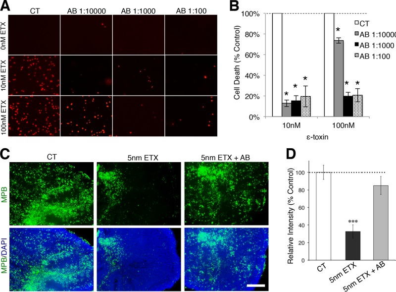
FIG 7 .
Neutralizing ε-toxin antibody protects oligodendrocytes from cell death and prevents myelin loss. (A) Primary mixed glial cells were treated with the indicated ε-toxin (ETX) doses and ε-toxin neutralizing antibody (AB) dilutions for 24 h. Cell viability was evaluated by PI exclusion. (B) Quantification of cell death in mixed culture treated with the indicated doses of ε-toxin and neutralizing antibody dilutions. The percent cell death was calculated by enumerating the number of PI-positive nuclei and normalizing the values against the values for the controls (100%). Values are means ± SD (n = 3). Values that are significantly different (P < 0.001) by ANOVA from the values for the control are indicated by an asterisk. Similar results were obtained in two independent experiments. (C) MBP immunostaining (green) of untreated cerebellar slices (control [CT]), slices treated with 5 nM ε-toxin (5nM ETX), or slices treated with 5 nM ε-toxin plus neutralizing ε-toxin antibody (5nM ETX + AB) for 20 h. DAPI (blue) is counterstained to identify cell nuclei. Bar, 500 µm. (D) Quantification of MBP staining normalized to control (100%). Values are means ± SEM. There were 5 or 6 slices for each condition. Values that are significantly different (P < 0.001) from the value for the control by two-tailed Student’s t test are indicated (***). Similar results were obtained in three independent experiments.

