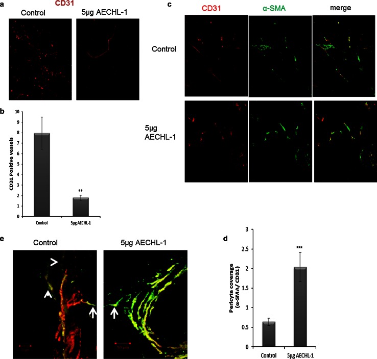
Fig. 9.
Effect of AECHL-1 on tumor angiogenesis. MCF-7 cells were injected in the right flank of SCID mice (n = 5 per group) as described in “Materials and methods.” Eight-micrometer cryosections were stained for CD-31 to a visualize and b quantify vessel density (×20), or CD-31 and α-SMA to c visualize and d quantify pericyte coverage of tumor vasculature. e Organization of mural cells in AECHL-1-treated and control tumors as analyzed in the 30-µm cryosections doublestained for CD-31 and α-SMA (×40, projected stacks).Control vessels show decreased mural cell support with treated vessels showing higher mural cell investment. Arrows, SMA+ cells extending away from vessel. Arrowheads, SMA+ cells without any associated vessels. Columns, mean from five mice per group; bars, SE. **P < 0.01; ***P < 0.001 versus control

