

Abstract
Sporadic human infections by a novel H7N9 virus occurred over a large geographic region in China. In this study, we show that Newcastle disease virus (NDV)-vectored H7 (NDV-H7) and NDV-H5 vaccines are able to induce antibodies with high hemagglutination inhibition (HI) titers and completely protect chickens from challenge with the novel H7N9 or highly pathogenic H5N1 viruses, respectively. Notably, a baculovirus-expressed H7 protein failed to protect chickens from H7N9 virus infection.
TEXT
A novel avian H7N9 influenza virus which emerged in poultry in China around March 2013 has caused more than 300 human infections and more than 100 deaths (1). More than 160 of these cases were reported in the first month of 2014. Because of the lack of an existing immunity against H7 subtype influenza viruses in the human population and domestic poultry and because of the absence of an available vaccine, there is a great concern that H7N9 virus may emerge as a potential pandemic virus for humans. In addition, the possible evolution of this low-pathogenicity H7N9 virus into a highly pathogenic virus for chickens is of concern (1, 3, 4). Sporadic human infections occurred over a large geographic region in China, suggesting a possible wide spread of H7N9 virus in poultry and at live poultry markets (5, 6). To date, no licensed commercial vaccine is available for the novel H7N9 virus in both avian species and humans. Vaccination could be a critical tool to prevent infection of domestic poultry and to prepare for a potential pandemic in humans.
In this study, two H7 and two H5 vaccine candidates were investigated in chickens. The Newcastle disease virus (NDV)-vectored H7 (NDV-H7) vaccine was generated using reverse genetics to insert the ectodomain gene of the H7 hemagglutinin (HA) from Anhui/1/2013 H7N9 influenza virus between the P and M genes of an NDV vaccine strain (Lasota). To be recognized as an additional viral gene, the inserted sequence contained NDV's gene end (GE), intergenic (IS), and gene start (GS) sequences, as well as a Kozak sequence for efficient translation, preceding the H7 initiation codon (Fig. 1A). To improve the incorporation of the hemagglutinin ectodomain protein in the NDV, the transmembrane and cytoplasmic tail of the NDV F protein were fused to the C terminus of the ectodomain of the H7 protein (Fig. 1A). The ectodomain (amino acids 1 to 515) of the hemagglutinin protein of the novel H7N9 virus (A/Anhui/1/13) was expressed in a baculovirus system featuring a C-terminal trimerization domain as described before (7) and also evaluated in chickens. Additionally, NDV-H5 of a highly pathogenic avian influenza (HPAI) H5N1 virus (A/chicken/Bali/U8661/2009, clade 2.1.3.2) and baculovirus-expressed recombinant H5 protein (from A/Vietnam/1203/04, clade 1) were tested in this study as well. The NDV-H5 and H5 subunit vaccine candidates were generated using the same strategy as that for the H7 vaccines (Fig. 1A). The H5 ectodomain sequence inserted in the NDV vector was modified to replace the multiple basic cleavage site (ESRRKKR/GLF) with a monobasic cleavage site (ESR/GLF). To check the expression of hemagglutinin proteins in NDV-H5- and NDV-H7-infected cells, immunofluorescence assays were conducted on Vero cells infected with NDV-H5 or NDV-H7 by using monoclonal antibodies against either H5 or H7. Both the H5 and H7 hemagglutinin proteins were expressed successfully in infected Vero cells, and the results were further confirmed by flow cytometry (Fig. 1B). Protein expression levels in chicken cells were analyzed by Western blotting (Fig. 1C). Chicken embryo fibroblast (CEF) primary cultures were infected with NDV-H5, NDV-H7, and wild-type NDV at a multiplicity of infection of 1 PFU/cell. At 20 h postinfection (p.i.), total cell extracts were analyzed by Western blotting using murine H5- and H7-specific antibodies. Under the conditions used (1:4,000 dilution), the two antibodies recognize identical amounts of the corresponding purified hemagglutinin (HA) with similar sensitivities (data not shown). A commercial antibody against the NDV glycoprotein HN was used to confirm similar viral loads. To analyze the incorporation of the chimeric hemagglutinins in the NDV particle, we compared the amounts of H5 in recombinant NDVs expressing the chimeric H5 or a full-length H5 (unpublished data). Viral particles from allantoic fluid were purified by ultracentrifugation through a 30% sucrose cushion and analyzed by Western blotting with H5- and HN-specific monoclonal antibodies as described above. As shown in Fig. 1D, replacing the original transmembrane and cytoplasmic domains with those of the NDV F protein resulted in increased incorporation of the chimeric protein in the viral particle.
FIG 1.

Construction of NDV-H7 and NDV-H5 vaccines and detection of hemagglutinin expression by immunofluorescence assay, flow cytometry, and Western blotting. (A) Construction strategy for NDV-H5 and NDV-H7. (B) Immunofluorescence and flow cytometry analysis of Vero cells 48 h after infection with either NDV-H5 or NDV-H7. (C) Western blotting and expression levels of HA and HN proteins in chicken embryo fibroblast cells infected with NDV-H5, NDV-H7, and wild-type (wt) NDV at a multiplicity of infection (MOI) of 1. Relative protein amounts were calculated and normalized using ratios to the NDV-HN protein. (D) Western blotting of H5 proteins in NDV-chimeric H5 (chimeric H5) and NDV-wt-H5 (wild-type H5) virus particles. Relative protein amounts were calculated and normalized using ratios to the NDV-HN protein in the viral particle.
Six-week-old specific-pathogen-free (SPF) White Leghorn chickens were used. The chicken studies were approved by the Institutional Animal Care and Use Committee at Kansas State University (IACUC 3018). Groups of 10 chickens were vaccinated with NDV-H7 (5 × 10⁶ PFU/bird) and NDV-H5 (5 × 10⁶ PFU/bird) vaccine virus by either the intramuscular (i.m.) or oculonasal (o.n.) route. None of the vaccinated chickens showed clinical signs after i.m. or o.n. vaccination with NDV-H7 or NDV-H5. Another two groups of 10 chickens were immunized with either recombinant H7 (10 μg/bird) or H5 (10 μg/bird) proteins adjuvanted with Montanide ISA 70VG (Seppic, USA). Two groups of chickens (n = 20 per group), vaccinated with phosphate-buffered saline (PBS), served as mock-vaccinated controls. Booster vaccination was conducted 2 weeks after the first vaccination using the same procedure as that for the first vaccination. Blood samples were collected before vaccination and 2 weeks after each vaccination. All birds were challenged 2 weeks after boost with either H5N1 (A/chicken/Bali/U8661/2009) or H7N9 (A/Anhui/1/2013) virus intranasally at a dose of 106 50% tissue culture infective doses (TCID50). Cloacal and oropharyngeal swabs were collected at 0, 1, 3, 6, 10, and 14 days postinfection (dpi) for further evaluation. All chickens were observed twice a day for clinical disease with clinical scores from 0 to 3 which reflected the severity of disease (0, healthy; 1, sick; 2, severely ill; 3, dead). Two or three chickens from the vaccinated groups and five birds from the mock groups were necropsied at 2 or 4 days postinfection. Lung, brain, intestine, and bursa of Fabricius were collected for virus titration. A complete set of tissues was collected for histopathological analysis. All surviving chickens were necropsied at 14 dpi. Clinical scores, antibody titers, and survival rates were analyzed by using analysis of variance (ANOVA) in GraphPad Prism version 5.0 (GraphPad Software Inc., CA). Those response variables were subjected to comparisons for all pairs by using the Tukey-Kramer test. Pairwise mean comparisons between vaccinated and mock groups were made using the Student t test. A P value of <0.05 was considered statistically significant.
NDV-H7 induced low-level hemagglutination inhibition (HI) antibodies after the first vaccination; only a few chickens seroconverted in both the i.m.- and o.n.-vaccinated groups. No detectable antibody was found in the H7 protein-vaccinated groups after the first immunization (Fig. 2A). After the booster, high HI antibody titers (1:40 to 1:160) were detected in chickens vaccinated intramuscularly with NDV-H7 (Fig. 2B and C). Similarly, all chickens except one seroconverted in the NDV-H7-oculonasally vaccinated group, although the HI titers were lower (1:10 to 1:40) than those in the NDV-H7 i.m.-vaccinated group (Fig. 2C). However, only four out of 10 H7 protein-vaccinated chickens seroconverted, with low levels of HI antibody titers (1:10 to 1:20) after booster (Fig. 2B). These data indicate that the NDV-H7 live vaccines induce higher HI antibody levels than does the H7 protein vaccine at the applied doses after two rounds of immunization.
FIG 2.

Hemagglutination inhibition antibody titers of chickens vaccinated with different H7 vaccines and virus shedding after challenge. (A) HI antibody titer in serum of individual chicken at 2 weeks after first vaccination with NDV-H7 or baculovirus-expressed H7 proteins. ON, oculonasal immunization; IM, intramuscular immunization. (B) HI antibody titer in serum of individual chickens at 2 weeks after booster. (C) Geometric means of HI titers in serum of chicken at 2 weeks after booster; the dashed line represents the detection limit. (D) Percentages of chickens shedding virus by oropharyngeal (solid lines) or cloacal (dashed lines) routes. Results are shown as the number of chickens shedding virus/total numbers. (E) Virus titers (mean ± standard error of the mean of log10 TCID50) in oropharyngeal swabs collected at different time points. (F) Virus titers (mean ± standard error of the mean of log10 TCID50) in cloacal swabs collected at different time points. *, P < 0.05.
After challenge with the novel H7N9 (A/Anhui/1/2013) virus, no clinical signs were observed in the mock-vaccinated groups and all mock-vaccinated chickens survived through the entire observation period of 14 days. This confirmed that the H7N9 virus is a low-pathogenicity avian influenza virus in chicken. Similarly, all vaccinated chickens survived without showing any clinical signs. More than half of the mock-vaccinated chickens shed virus, with titers ranging from 102 to 103 TCID50, via the oropharynx at 1 and 3 dpi (Fig. 2D and E). Four or five out of 10 H7 protein-vaccinated chickens also shed virus efficiently via the oropharynx at 1 and 3 dpi, respectively (Fig. 2D and E). However, the four seroconverted chickens in the H7 protein-vaccinated group did not shed virus in the entire study. Virus was also detected (102 to 105 TCID50/ml) in cloacal swabs from a small percentage of mock-vaccinated chickens and H7 protein-vaccinated chickens after challenge (Fig. 2D, dashed line, and F). In contrast, none of the NDV-H7-intramuscularly or -oculonasally vaccinated chickens shed virus during the entire observation period (Fig. 2D). During necropsies at 2 and 4 dpi, no clear gross lesion was observed; virus titration results revealed that virus was present in 2 out of 5 bursas of Fabricius of mock-vaccinated H7N9-infected control chickens at 4 dpi (102 to 103 TCID50/ml). No virus was detected in the tissues collected from vaccinated birds, including the H7 protein-vaccinated chickens. Also, no virus antigen (NP-specific antibody; GenScript, USA) was detected in lung and bursa of Fabricius by immunohistochemical staining in both mock- and H7-vaccinated chickens at the respective necropsy days after infection. Collectively, the novel H7N9 virus is able to infect chickens which shed virus via both oropharynx and cloaca, although without causing clinical signs and gross lesions. The NDV-H7 vaccine completely protects chickens from H7N9 virus shedding independently of the routes of immunization.
Next, we analyzed the immunogenicity and efficacy of the H5 vaccines. All NDV-H5-vaccinated chickens except one from the o.n.-vaccinated group seroconverted (1:10 to 1:80) after a single immunization with NDV-H5 (Fig. 3A). However, only three chickens seroconverted in the H5 protein-vaccinated group after the first round of vaccination (Fig. 3A). High-HI-titer antibodies were detected in all NDV-H5-i.m.-vaccinated chickens after booster (Fig. 3B and C). All chickens in the NDV-H5-o.n.-vaccinated group and all but one from the H5 protein-vaccinated group seroconverted after booster (Fig. 3B). After challenge with an HPAI H5N1 virus, as shown in Fig. 3D, the mock-vaccinated chickens showed typical clinical signs of highly pathogenic influenza virus, e.g., depression, skin cyanosis, facial edema, and central nervous system (CNS) signs; 60% of these chickens died (Fig. 3E); in contrast, all vaccinated chickens independent of vaccine formulation and immunization route survived without showing any clinical signs during the observation period of 14 days (Fig. 3D and E). Virus was detected in lung tissues of four out of five mock-vaccinated chickens necropsied at 4 dpi; one chicken also showed virus replication additionally in brain, intestine, and bursa of Fabricius at 4 dpi (Fig. 3F). In contrast, no virus was detected in the tissues of NDV-H5- and H5 protein-vaccinated chickens. Immunohistochemical staining showed that NP antigen was detected in the lung and bursa of Fabricius of mock-vaccinated chickens after infection (Fig. 4); no antigen was observed in NDV-H5- and H5 subunit-vaccinated groups. Although the mortality rate of the H5N1 virus (harboring the RRKKR motif at the cleavage site of the HA protein) used in this study is only 60%, it caused a systemic infection in all mock-vaccinated birds. Both the NDV-H5 and the H5 subunit vaccine can protect chickens from systemic infection and/or death with H5N1, suggesting that the vaccines should be able to inhibit virus replication and provide protection against more virulent homologous H5N1 strains. In addition, a few mock-vaccinated chickens (3 out of 15) shed virus at 3 dpi via the oropharynx, whereas no virus shedding was detected in vaccinated chickens during the entire observation period (data not shown). Taken together, both NDV-H5 and H5 subunit vaccines induced influenza virus-specific antibody titers after two rounds of vaccination and protected chickens from clinical disease, virus replication, and mortality associated with H5N1 virus infection.
FIG 3.

Hemagglutination inhibition antibody titers of chickens vaccinated by different H5 vaccines and survival rates of chickens after challenge. (A) HI antibody titers in serum of individual chicken at 2 weeks after first vaccination with NDV-H5 or baculovirus-expressed H5 proteins. ON, oculonasal immunization; IM, intramuscular immunization. (B) HI antibody titer in serum of individual chickens at 2 weeks after booster. (C) Geometric mean of HI titers in serum of chickens at 2 weeks after booster; dashed line represents detection limit. (D) Clinical sign scores of chickens after challenge. (E) Survival rates of mock and vaccinated chickens after challenge with H5N1. (F) Virus replication in tissues of chickens necropsied at 4 dpi after challenge with H5N1. *, P < 0.05; **, P < 0.01.
FIG 4.
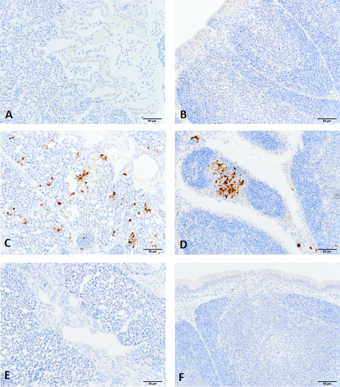
Immunohistochemical staining of microscopic lung and bursa of Fabricius sections from chickens infected with H5N1 virus at 4 days postinfection. No positive immunohistochemical reaction was seen in lung and bursa of Fabricius sections of control chickens (A and B) and NDV-vaccinated chickens infected with H5N1 virus (E and F). Positive reactions were seen in lung and bursa of Fabricius sections of mock-vaccinated chickens after infection with the H5N1 virus (C and D). No antigen was found in sections of H5 protein-vaccinated chickens after infection (data not shown). Anti-influenza A virus antibody to nucleoprotein (GenScript, USA) was used for the immunohistochemical staining. Bar, 50 μm in all photographs.
Although the antibody levels of chickens vaccinated oculonasally are lower than those of chickens vaccinated intramuscularly for both the NDV-H5 and NDV-H7 vaccines (Fig. 2C and 3C), both immunization routes are successful in inducing seroconversion and protection of chicken from challenge. Previous studies showed that oculonasal immunization with NDV-based H5N1 vaccine could elicit immune responses in chickens which were able to protect against highly pathogenic H5N1 infection (9). The NDV-H5 vaccine induced a better antibody response than the NDV-H7 vaccine in chickens. The H5 subunit vaccine also completely protected chickens from clinical signs and death after challenge with a highly pathogenic H5N1 influenza virus. In contrast, the H7 subunit vaccine induced only low-level antibody responses and failed to protect chickens from virus shedding, suggesting that the H7 HA protein might be less immunogenic in chickens than the H5 protein, as has been described previously in mice and humans (10). It should be noted that the two NDV vectors express similar amounts of the recombinant HA proteins (Fig. 1C), and the same amounts of baculovirus-expressed proteins and vaccine adjuvant were used for the immunizations. The observation that an H7 subunit vaccine did not protect all animals against H7N9 challenge might be due to a lower induction of CD4 help. It has been reported that the H7 protein contains 14 to 24% fewer T-cell epitopes than the HA proteins of other subtypes, which may explain the low immunogenicity of H7 in humans (11, 12); whether this also applies in chickens remains unknown at this time. Low immunogenicity of the H7 HA implies that the production of an effective H7N9 vaccine will be more difficult than that of previously produced influenza vaccines and might require improvement of the H7 immunogenicity or potent vaccine platforms/formulations.
The H7 subunit vaccine induced only a limited immune response and failed to protect chickens from H7N9 infection. To determine whether this low level of antibody response can drive H7N9 virus antigen drift, we sequenced the HA genes of viruses shed from the cloaca and oropharynx in both mock- and H7 protein-vaccinated groups. In total, five amino acid substitutions were found in the H7 protein of viruses isolated from the vaccinated group and six substitutions were observed in the H7 protein of viruses isolated from the mock group (Table 1). Interestingly, four of these substitutions (N141D, A143T, N167D, and L235Q) were found in both groups; the L235Q (L226Q, H3 numbering) substitution was observed in both groups already at 3 days postinfection, which suggests that the continuous virus circulation in chickens results in rapid adaptation of this human H7N9 isolate to birds. One substitution, R316K, which is located underneath the ectodomain of H7 protein, was observed only in the H7 protein-vaccinated group. All sequence data suggest that the low level of antibody produced in H7 protein-vaccinated chickens did not stimulate a significant antigenic drift compared to the mock-vaccinated group.
TABLE 1.
Amino acid substitutions occurring in HA protein of shed viruses of both H7 protein-vaccinated group and mock-vaccinated group
| Swab | Substitutions (dpi of occurrence) in vaccination groupa: |
|
|---|---|---|
| H7 Baculo-IMb | Mock | |
| Oropharyngeal | A143T (3), L235Q (3), R316K (3) | N141D (3), A143T (3), N167D (3), G214E (3), L235Q (3) |
| Cloacal | N141D (10), N167D (10), L235Q (10) | E114Q (6), N141D (6), A143T (3), N167D (6), L235Q (6) |
Amino acid substitutions shown in bold were observed in both the mock- and protein-vaccinated groups. H7 numbering is used.
Baculo-IM, intramuscular vaccination with baculovirus-expressed protein.
A few H7-based live vaccines have been studied in mammalian models (e.g., mice or ferrets) or in vitro for potential protection against the novel H7N9 virus (13–15). H7-based or H3 stalk subunit and modified live vaccines have been shown to protect mice from challenge with the novel H7N9 virus (16–20). However, none of these H7 vaccine candidates were evaluated in chickens against the novel H7N9 virus. Importantly, the use of live attenuated influenza viruses as H7 vaccines in avian or mammalian species raises a major biosafety concern, because of possible reassortment with other circulating influenza virus strains (e.g., H3N2, H1N1, H5N1, and H10N8), which may generate new strains with different phenotypes and pandemic potential. NDV LaSota strain is well established as an attenuated vaccine for Newcastle disease; it has also been shown to be a good vector to express foreign proteins (21, 22). Various NDV-based vaccine candidates have been evaluated in different animal models (e.g., mouse, chicken, pig, dog, etc.), showing that NDV-vectored vaccine candidates work well in various animal species (9, 23–28). NDV-vectored vaccines are safer than live attenuated influenza virus vaccines because they do not harbor the risk of reassortment with other influenza virus strains. Additionally, an NDV-vectored influenza virus vaccine can function as a bivalent vaccine against both influenza virus and NDV infections in chickens (10). This conclusion is based on the HI antibody titers against NDV in both NDV-H5- and NDV-H7-vaccinated groups (Table 2), since both vaccines elicited good immune responses against NDV in oculonasally and intramuscularly vaccinated chickens, with HI titers ranging from 1:40 to 1:320 after booster; these HI titers should be able to provide protection against NDV infection based on previous observations (29). In the field, although the seropositive prevalence of NDV in chickens is high, based on our data describing specific HI titers against NDV (Table 2), an anamnestic booster response was found in chickens which had HI titers of 1:10 to 1:40 against NDV after the first vaccination. This suggests that both the NDV-H5 and NDV-H7 vaccines could work in chickens containing HI titers of 1:40 or lower against NDV. However, vaccination studies in chickens younger than 6 weeks would need to be performed to investigate the possible use of this vaccine right after birth.
TABLE 2.
NDV-specific antibody responses in serum of chickens after vaccination
| Vaccination | No. of seropositive chickens/total no.a | NDV HI titerb |
|
|---|---|---|---|
| First vaccination | Boost | ||
| Mock | 0/20 | 0 | 0 |
| NDV-H5 o.n. | 10/10 | 3.5 ± 0.20 | 6.4 ± 0.35 |
| NDV-H5 i.m. | 10/10 | 4.4 ± 0.53 | 7.1 ± 0.64 |
| Baculo-H5 i.m.c | 0/10 | 0 | 0 |
| NDV-H7 o.n. | 10/10 | 4.6 ± 0.55 | 6.1 ± 0.50 |
| NDV-H7 i.m. | 10/10 | 4.3 ± 0.47 | 7.4 ± 0.71 |
| Baculo-H7 i.m. | 0/10 | 0 | 0 |
Two weeks after boost.
The hemagglutination inhibition (HI) titer is expressed as log2 mean ± standard deviation.
Baculo-, expressed in baculovirus.
In summary, the sporadic appearance of human cases of H7N9 infection continues in various provinces in China, which suggests a wide geographic spread of the novel H7N9 virus. This indicates that an effective H7N9 vaccine is needed to control the spread of this virus in avian species and to avoid cross-species transmission to humans. Based on the present study, both NDV-vectored H7 and H5 vaccines could be potential vaccine candidates to mitigate infections in chickens and consequently protect public health.
ACKNOWLEDGMENTS
We thank Richard J. Webby from St. Jude Children's Research Hospital for supplying us with the A/Anhui/1/2013 virus stock. We also thank Haixia Liu, Chester McDowell, Daniel Madden, Jinhwa Lee, and Michael Duff for providing technical support. All work was done in the Biosecurity Research Institute (BSL3/BSL3Ag) located at Kansas State University.
This work was partially funded by the National Institute of Allergy and Infectious Diseases, National Institutes of Health, Department of Health and Human Services, under contract number HHSN266200700005C, and by the U.S. Department of Homeland Security under grant award number 2010-ST-AG0001.
A.G.-S. is an inventor of patents encompassing NDV-vectored vaccines owned by the Icahn School of Medicine at Mount Sinai.
REFERENCES
- 1.Zhou J, Wang D, Gao R, Zhao B, Song J, Qi X, Zhang Y, Shi Y, Yang L, Zhu W, Bai T, Qin K, Lan Y, Zou S, Guo J, Dong J, Dong L, Zhang Y, Wei H, Li X, Lu J, Liu L, Zhao X, Li X, Huang W, Wen L, Bo H, Xin L, Chen Y, Xu C, Pei Y, Yang Y, Zhang X, Wang S, Feng Z, Han J, Yang W, Gao GF, Wu G, Li D, Wang Y, Shu Y. 2013. Biological features of novel avian influenza A (H7N9) virus. Nature 499:500–503. doi: 10.1038/nature12379. [DOI] [PubMed] [Google Scholar]
- 2.Reference deleted.
- 3.Richard M, Schrauwen EJ, de Graaf M, Bestebroer TM, Spronken MI, van Boheemen S, de Meulder D, Lexmond P, Linster M, Herfst S, Smith DJ, van den Brand JM, Burke DF, Kuiken T, Rimmelzwaan GF, Osterhaus AD, Fouchier RA. 2013. Limited airborne transmission of H7N9 influenza A virus between ferrets. Nature 501:560–563. doi: 10.1038/nature12476. [DOI] [PMC free article] [PubMed] [Google Scholar]
- 4.To KK, Chan JF, Chen H, Li L, Yuen KY. 2013. The emergence of influenza A H7N9 in human beings 16 years after influenza A H5N1: a tale of two cities. Lancet Infect Dis 13:809–821. doi: 10.1016/S1473-3099(13)70167-1. [DOI] [PMC free article] [PubMed] [Google Scholar]
- 5.Lam TT, Wang J, Shen Y, Zhou B, Duan L, Cheung CL, Ma C, Lycett SJ, Leung CY, Chen X, Li L, Hong W, Chai Y, Zhou L, Liang H, Ou Z, Liu Y, Farooqui A, Kelvin DJ, Poon LL, Smith DK, Pybus OG, Leung GM, Shu Y, Webster RG, Webby RJ, Peiris JS, Rambaut A, Zhu H, Guan Y. 2013. The genesis and source of the H7N9 influenza viruses causing human infections in China. Nature 502:241–244. doi: 10.1038/nature12515. [DOI] [PMC free article] [PubMed] [Google Scholar]
- 6.Zhu H, Wang D, Kelvin DJ, Li L, Zheng Z, Yoon SW, Wong SS, Farooqui A, Wang J, Banner D, Chen R, Zheng R, Zhou J, Zhang Y, Hong W, Dong W, Cai Q, Roehrl MH, Huang SS, Kelvin AA, Yao T, Zhou B, Chen X, Leung GM, Poon LL, Webster RG, Webby RJ, Peiris JS, Guan Y, Shu Y. 2013. Infectivity, transmission, and pathology of human-isolated H7N9 influenza virus in ferrets and pigs. Science 341:183–186. doi: 10.1126/science.1239844. [DOI] [PubMed] [Google Scholar]
- 7.Margine I, Palese P, Krammer F. 2013. Expression of functional recombinant hemagglutinin and neuraminidase proteins from the novel H7N9 influenza virus using the baculovirus expression system. J Vis Exp 2013(81):e51112. doi: 10.3791/51112. [DOI] [PMC free article] [PubMed] [Google Scholar]
- 8.Reference deleted.
- 9.Ge J, Deng G, Wen Z, Tian G, Wang Y, Shi J, Wang X, Li Y, Hu S, Jiang Y, Yang C, Yu K, Bu Z, Chen H. 2007. Newcastle disease virus-based live attenuated vaccine completely protects chickens and mice from lethal challenge of homologous and heterologous H5N1 avian influenza viruses. J Virol 81:150–158. doi: 10.1128/JVI.01514-06. [DOI] [PMC free article] [PubMed] [Google Scholar]
- 10.Mao H, Yen HL, Liu Y, Lau YL, Malik Peiris JS, Tu W. 2014. Conservation of T cell epitopes between seasonal influenza viruses and the novel influenza A H7N9 virus. Virol Sin 29:170–175. doi: 10.1007/s12250-014-3473-3. [DOI] [PMC free article] [PubMed] [Google Scholar]
- 11.De Groot AS, Ardito M, Terry F, Levitz L, Ross T, Moise L, Martin W. 2013. Low immunogenicity predicted for emerging avian-origin H7N9: implication for influenza vaccine design. Hum Vaccin Immunother 9:950–956. doi: 10.4161/hv.24939. [DOI] [PMC free article] [PubMed] [Google Scholar]
- 12.De Groot AS, Moise L, Liu R, Gutierrez AH, Terry F, Koita OA, Ross TM, Martin W. 2014. Cross-conservation of T-cell epitopes: now even more relevant to (H7N9) influenza vaccine design. Hum Vaccin Immunother 10:256–262. doi: 10.4161/hv.28135. [DOI] [PMC free article] [PubMed] [Google Scholar]
- 13.Rudenko L, Isakova-Sivak I, Donina S. 2013. H7N3 live attenuated influenza vaccine has a potential to protect against new H7N9 avian influenza virus. Vaccine 31:4702–4705. doi: 10.1016/j.vaccine.2013.08.040. [DOI] [PubMed] [Google Scholar]
- 14.Rudenko L, Isakova-Sivak I, Rekstin A. 2014. H7N9: can H7N3 live-attenuated influenza vaccine be used at the early stage of the pandemic? Expert Rev Vaccines 13:1–4. doi: 10.1586/14760584.2014.864564. [DOI] [PubMed] [Google Scholar]
- 15.Xu Q, Chen Z, Cheng X, Xu L, Jin H. 2013. Evaluation of live attenuated H7N3 and H7N7 vaccine viruses for their receptor binding preferences, immunogenicity in ferrets and cross reactivity to the novel H7N9 virus. PLoS One 8:e76884. doi: 10.1371/journal.pone.0076884. [DOI] [PMC free article] [PubMed] [Google Scholar]
- 16.Goff PH, Krammer F, Hai R, Seibert CW, Margine I, Garcia-Sastre A, Palese P. 2013. Induction of cross-reactive antibodies to novel H7N9 influenza virus by recombinant Newcastle disease virus expressing a North American lineage H7 subtype hemagglutinin. J Virol 87:8235–8240. doi: 10.1128/JVI.01085-13. [DOI] [PMC free article] [PubMed] [Google Scholar]
- 17.Klausberger M, Wilde M, Palmberger D, Hai R, Albrecht RA, Margine I, Hirsh A, Garcia-Sastre A, Grabherr R, Krammer F. 2014. One-shot vaccination with an insect cell-derived low-dose influenza A H7 virus-like particle preparation protects mice against H7N9 challenge. Vaccine 32:355–362. doi: 10.1016/j.vaccine.2013.11.036. [DOI] [PMC free article] [PubMed] [Google Scholar]
- 18.Krammer F, Albrecht RA, Tan GS, Margine I, Hai R, Schmolke M, Runstadler J, Andrews SF, Wilson PC, Cox RJ, Treanor JJ, Garcia-Sastre A, Palese P. 2014. Divergent H7 immunogens offer protection from H7N9 virus challenge. J Virol 88:3976–3985. doi: 10.1128/JVI.03095-13. [DOI] [PMC free article] [PubMed] [Google Scholar]
- 19.Krammer F, Margine I, Hai R, Flood A, Hirsh A, Tsvetnitsky V, Chen D, Palese P. 2014. H3 stalk-based chimeric hemagglutinin influenza virus constructs protect mice from H7N9 challenge. J Virol 88:2340–2343. doi: 10.1128/JVI.03183-13. [DOI] [PMC free article] [PubMed] [Google Scholar]
- 20.Smith GE, Flyer DC, Raghunandan R, Liu Y, Wei Z, Wu Y, Kpamegan E, Courbron D, Fries LF III, Glenn GM. 2013. Development of influenza H7N9 virus like particle (VLP) vaccine: homologous A/Anhui/1/2013 (H7N9) protection and heterologous A/chicken/Jalisco/CPA1/2012 (H7N3) cross-protection in vaccinated mice challenged with H7N9 virus. Vaccine 31:4305–4313. doi: 10.1016/j.vaccine.2013.07.043. [DOI] [PubMed] [Google Scholar]
- 21.Nakaya T, Cros J, Park MS, Nakaya Y, Zheng H, Sagrera A, Villar E, Garcia-Sastre A, Palese P. 2001. Recombinant Newcastle disease virus as a vaccine vector. J Virol 75:11868–11873. doi: 10.1128/JVI.75.23.11868-11873.2001. [DOI] [PMC free article] [PubMed] [Google Scholar]
- 22.Park MS, Steel J, Garcia-Sastre A, Swayne D, Palese P. 2006. Engineered viral vaccine constructs with dual specificity: avian influenza and Newcastle disease. Proc Natl Acad Sci U S A 103:8203–8208. doi: 10.1073/pnas.0602566103. [DOI] [PMC free article] [PubMed] [Google Scholar]
- 23.DiNapoli JM, Nayak B, Yang L, Finneyfrock BW, Cook A, Andersen H, Torres-Velez F, Murphy BR, Samal SK, Collins PL, Bukreyev A. 2010. Newcastle disease virus-vectored vaccines expressing the hemagglutinin or neuraminidase protein of H5N1 highly pathogenic avian influenza virus protect against virus challenge in monkeys. J Virol 84:1489–1503. doi: 10.1128/JVI.01946-09. [DOI] [PMC free article] [PubMed] [Google Scholar]
- 24.DiNapoli JM, Yang L, Suguitan A Jr, Elankumaran S, Dorward DW, Murphy BR, Samal SK, Collins PL, Bukreyev A. 2007. Immunization of primates with a Newcastle disease virus-vectored vaccine via the respiratory tract induces a high titer of serum neutralizing antibodies against highly pathogenic avian influenza virus. J Virol 81:11560–11568. doi: 10.1128/JVI.00713-07. [DOI] [PMC free article] [PubMed] [Google Scholar]
- 25.Ge J, Wang X, Tao L, Wen Z, Feng N, Yang S, Xia X, Yang C, Chen H, Bu Z. 2011. Newcastle disease virus-vectored rabies vaccine is safe, highly immunogenic, and provides long-lasting protection in dogs and cats. J Virol 85:8241–8252. doi: 10.1128/JVI.00519-11. [DOI] [PMC free article] [PubMed] [Google Scholar]
- 26.Kong D, Wen Z, Su H, Ge J, Chen W, Wang X, Wu C, Yang C, Chen H, Bu Z. 2012. Newcastle disease virus-vectored Nipah encephalitis vaccines induce B and T cell responses in mice and long-lasting neutralizing antibodies in pigs. Virology 432:327–335. doi: 10.1016/j.virol.2012.06.001. [DOI] [PubMed] [Google Scholar]
- 27.Lee DH, Park JK, Kwon JH, Yuk SS, Erdene-Ochir TO, Jang YH, Seong BL, Lee JB, Park SY, Choi IS, Song CS. 2013. Efficacy of single dose of a bivalent vaccine containing inactivated Newcastle disease virus and reassortant highly pathogenic avian influenza H5N1 virus against lethal HPAI and NDV infection in chickens. PLoS One 8:e58186. doi: 10.1371/journal.pone.0058186. [DOI] [PMC free article] [PubMed] [Google Scholar]
- 28.Nayak B, Rout SN, Kumar S, Khalil MS, Fouda MM, Ahmed LE, Earhart KC, Perez DR, Collins PL, Samal SK. 2009. Immunization of chickens with Newcastle disease virus expressing H5 hemagglutinin protects against highly pathogenic H5N1 avian influenza viruses. PLoS One 4:e6509. doi: 10.1371/journal.pone.0006509. [DOI] [PMC free article] [PubMed] [Google Scholar]
- 29.Zhao W, Spatz S, Zhang Z, Wen G, Garcia M, Zsak L, Yu Q. 2014. Newcastle disease virus (NDV) recombinants expressing infectious laryngotracheitis virus (ILTV) glycoproteins gB and gD protect chickens against ILTV and NDV challenges. J Virol 88:8397–8406. doi: 10.1128/JVI.01321-14. [DOI] [PMC free article] [PubMed] [Google Scholar]