Abstract
Management of the open abdomen has advanced significantly in recent years with the increasing use of vacuum assisted closure (VAC) techniques leading to increased rates of fascial closure. We present the case of a patient who suffered two complete abdominal wall dehiscences after an elective laparotomy, meaning primary closure was no longer possible. She was treated successfully with a VAC system combined with continuous medial traction using a Prolene® mesh. This technique has not been described before in the management of patients following wound dehiscence.
Keywords: Open abdomen, Laparostomy, Vacuum assisted wound closure
Case History
We describe the case of a 57-year-old woman with a background of asthma and obesity who was a heavy smoker. She presented to our clinic requesting a reversal of her ileostomy following a convoluted surgical history.
The patient was originally referred to our hospital in December 2011 with a large umbilical hernia. On examination, however, a pelvic mass was found and urgent computed tomography (CT) was organised. Two weeks later she presented as an emergency with an acutely ischaemic leg and was transferred to the regional vascular centre. Revascularisation was unsuccessful and she underwent an above-knee amputation. Following the procedure, she developed peritonitis. CT showed a large ovarian mass and free gas so a laparotomy was performed. The findings were a right ovarian tumour adherent to the right colon, which was necrotic and perforated. A hysterectomy, bilateral salpingo-oophorectomy and right hemicolectomy were carried out. Primary anastomosis was felt to be too high risk so an end ileostomy and transverse colon mucous fistula were formed at separate sites. Histology showed a T1a ovarian tumour.
The patient re-presented to our clinic requesting an ileostomy reversal. She was having significant problems with prolapse of the mucous fistula was unable to manage this. On examination, the prolapse was over 20cm. She also had a symptomatic incisional hernia. Owing to her ongoing symptoms and difficulty in managing the prolapse, the decision was made to go ahead with surgery.
In September 2012 the patient was admitted for surgery. Her laparotomy wound was reopened, both stomas were mobilised and a handsewn ileocolic anastomosis was performed. A mass closure was carried out with size 1 nylon sutures and the sheath was approximated well with no tension. A superficial vacuum drain was left in the wound and this was removed on day 2. She was discharged home on day 9. However, two days later she was readmitted with purulent discharge from her wound. On examination, the upper part of her wound had dehisced, with small bowel visible through the wound. She was taken back to theatre immediately. A lateral release was carried out but it was still difficult to approximate the rectus sheath so a 28cm × 18cm Permacol™ biological mesh (Covidien, Dublin, Ireland) was inserted and sutured in with interrupted Prolene® (Ethicon, Somerville, NJ, US).
The patient made good progress on the ward initially but 15 days later, she had a further wound dehiscence and was taken back to theatre for a second time. The finding on this occasion was a complete failure of the mesh; the edges were still sutured to the sheath but the middle of the mesh had split. There was no option of closing the abdomen at this point so it was left open as a laparostomy and she was taken to the intensive care unit. An intra-abdominal vacuum assisted closure (VAC) system (ABThera™ OA Negative Pressure Therapy Unit; KCI, San Antonio, TX, US) was inserted 48 hours later.
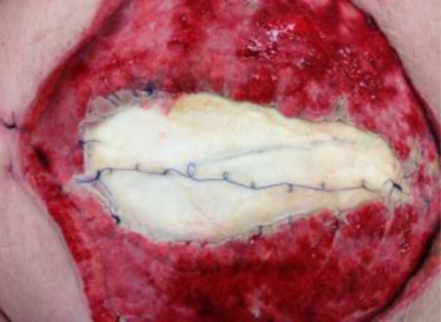
The patient was taken back to theatre every 3–4 days over the next month (8 returns to theatre in total) and, in combination with the VAC system, a Prolene® mesh was sutured to the sheath to provide medial traction (Fig 1). Mepitel® (Mölnlycke, Dunstable, UK) was placed between the mesh and the bowel. On each return to theatre, the central portion of the mesh was excised and sutured back together to provide ongoing traction to narrow the defect and facilitate eventual closure.
Figure 1.

Prolene® mesh sutured to fascia to provide medial traction. The middle portion of the mesh was excised on each return to theatre and sutured again to tighten it.
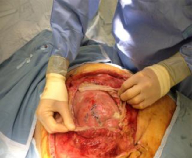
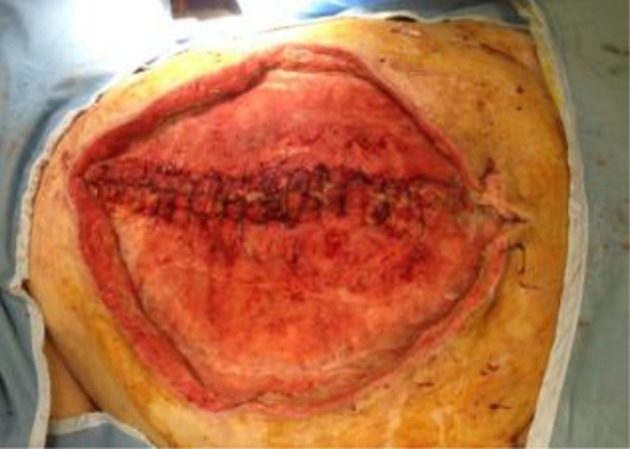
During this time, the patient was noted to have small bowel content in the intra-abdominal drains and, on return to theatre, she was found to have two small bowel fistulas. These were repaired with Vicryl® sutures (Ethicon) (Fig 2). On reinspection at subsequent returns to theatre, this appeared to have closed the fistulas successfully. Initially, she required parenteral feeding owing to the concern about further fistulation but enteral feeding was later introduced with no evidence of feed or small bowel content in the drains. The sheath was eventually closed six weeks later (Fig 3) and a standard VAC sponge was placed over this.
Figure 2.

Fistula sites in small bowel that have been oversewn with Vicryl®
Figure 3.

The fascia was closed with interrupted nylon sutures.
During the subsequent weeks, the wound granulated well and there were no further specific surgical problems. Respiratory weaning via a tracheostomy was complicated by multiple episodes of chest sepsis with Stenotrophomonas maltophilia. This required treatment with several courses of antibiotics and a 65-day stay on the intensive care unit with a tracheostomy before decannulation was possible. The patient was referred to the regional plastic surgery unit in December 2012 and transferred for skin grafting later that month. This was successful and she was discharged to a community hospital for ongoing rehabilitation in January 2013 after a total hospital stay of 115 days.
Discussion
This case describes the successful use of a mesh to provide medial traction during progressive fascial closure in the open abdomen. This technique has been described previously in small numbers of patients.1,2 In these cases, however, the abdomen was either left open primarily or reopened owing to abdominal compartment syndrome. A similar technique using a Wittmann Patch® (Starsurgical, Burlington, WI, US), which consists of two overlapping hook and burr sheets attached to the fascia, has also been used primarily in trauma patients.3
No previous cases have been reported in patients where the abdomen could not be closed owing to wound dehiscence where primary closure was not possible. Despite many similarities, the burst abdomen presents a slightly different challenge as there is often a concomitant wound infection and the abdomen may have been open for some time in an unsterile environment. Using a VAC system in combination with the mesh allowed the exposed fascia to begin to granulate and heal earlier as well as enabling easier wound management and nursing care on the ward.
Small bowel fistula formation is a recognised complication of VAC in the open abdomen. The reported incidence of this varies from 4.5%4 to 20%,5 leading some authors to advocate caution in its use, especially in patients with intra-abdominal sepsis.6 This case illustrates the steps involved in the procedure photographically and also describes successful management of the potential complications of negative pressure wound management.
References
- 1. Petersson U, Acosta S, Björck M. Vacuum-assisted wound closure and mesh-mediated fascial traction – a novel technique for late closure of the open abdomen. World J Surg 2007; 31: 2,133–2,137. [DOI] [PubMed] [Google Scholar]
- 2. Acosta S, Bjarnason T, Petersson U et al. Multicentre prospective study of fascial closure rate after open abdomen with vacuum and mesh-mediated fascial traction. Br J Surg 2011; 98: 735–743. [DOI] [PubMed] [Google Scholar]
- 3. Hadeed JG, Staman GW, Sariol HS et al. Delayed primary closure in damage control laparotomy: the value of the Wittmann patch. Am Surg 2007; 73: 10–12. [PubMed] [Google Scholar]
- 4. Barker DE, Kaufman HJ, Smith LA et al. Vacuum pack technique of temporary abdominal closure: a 7-year experience with 112 patients. J Trauma 2000; 48: 201–206. [DOI] [PubMed] [Google Scholar]
- 5. Rao M, Burke D, Finan PJ, Sagar PM. The use of vacuum-assisted closure of abdominal wounds: a word of caution. Colorectal Dis 2007; 9: 266–268. [DOI] [PubMed] [Google Scholar]
- 6. Starr-Marshall K. Vacuum-assisted closure of abdominal wounds and entero-cutaneous fistulae; the St Marks experience. Colorectal Dis 2007; 9: 573. [DOI] [PubMed] [Google Scholar]


