

Abstract
We report a rare case of ingestion of a large stone in a male patient with a known psychiatric disorder. Failure of endoscopic removal necessitated retrieval of the impacted stone by an open oesophagotomy. This case highlights an important yet unusual presentation and management of an oesophageal foreign body.
Keywords: Oesophagus, Foreign body, Endoscopy, Oesophagotomy
Case History
A 29-year-old man presented to the emergency department with absolute dysphagia 6 hours after having swallowed a stone. He complained of a dull pain in the throat and the sensation of an obstruction at the level of the thyroid cartilage. There were no associated symptoms of difficulty in breathing. He had a background of treated bipolar affective disorder and admitted to swallowing a stone on impulse after listening to a song by the musician Joss Stone.
On examination, the patient was retching but maintaining his own airway. Examination of the oropharynx and chest was unremarkable, and there was no evidence of surgical emphysema. He declined flexible nasoendoscopy. A lateral soft tissue neck x-ray demonstrated a smooth, large opacity in the cervical oesophagus (Fig 1). A subsequent chest x-ray confirmed the position of the stone (Fig 2).
Figure 1.
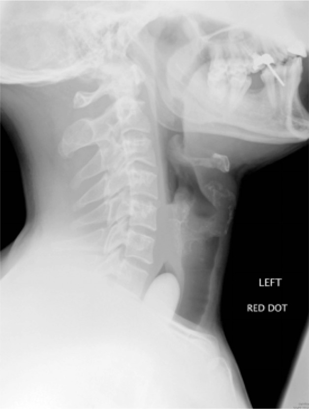
Lateral soft tissue neck radiography showing radio-opaque foreign body extending from the upper level of the C7 vertebra. The foreign body was extending downwards into the thoracic inlet and the distal aspect of the foreign body was not visible.
Figure 2.

Posteroanterior chest radiography showing foreign body at the thoracic inlet.
A rigid oesophagoscopy was performed under general anaesthesia to reveal a large stone distal to the cricopharyngeus. Owing to its size and the surrounding mucosal oedema, the foreign body could not be dislodged or delivered retrogradely into the pharynx. Direct attempts to remove the stone using Foley catheters and a Dormia basket proved futile, and the decision was taken to convert to an open approach via a cervical oesophagotomy.
A left transverse skin incision was made and subplatysmal flaps were elevated. The sternocleidomastoid muscle was retracted laterally and the omohyoid muscle was divided. The carotid sheath was exposed medially and retracted. Dissection was performed down to the prevertebral fascia and the proximal oesophagus containing the foreign body was identified. Further attempts to milk the stone into the pharynx failed and an incision was made at the site of the impacted foreign body. A stone measuring 3.6cm × 3.1cm × 1.8cm was impacted tightly in the oesophagus. Following manual removal of the stone, the oesophagotomy was repaired in two layers. A Blake® drain (Ethicon, Somerville, NJ, US) was sited and skin closure was performed. A nasogastric tube was inserted and secured in position. Postoperatively, the patient remained nil by mouth for a week prior to a soluble contrast swallow being performed, which showed no evidence of a leak.
Discussion
Oesophageal foreign body impaction is a common otolaryngological emergency and it is usually treated successfully by endoscopic intervention. Foreign body ingestion is more often an accidental rather than an intentional occurrence.1 The majority of ingested foreign bodies resolve spontaneously without causing complications.2,3 However, it is a potentially serious condition that can result in oesophageal perforation and mediastinitis if left untreated.4
Most cases of foreign body ingestion occur in the paediatric population (peak incidence 6 months – 6 years). Children often present with inorganic foreign bodies such as coins or toys. Adults generally present with impacted food (eg meat without bone) and this may be associated with underlying pathology such as stenosis or oesophageal atresia. Repeated, multiple and deliberate foreign body ingestion is a well recognised presentation among patients with psychiatric disorders.
Most patients present with sensation of a foreign body, choking or respiratory distress.4 Typical symptoms include dysphagia, drooling and chest pain although clinical presentation may be delayed. History may be vague in young children, mentally impaired or psychiatric patients.
Clinical evaluation should include assessment of the patient’s airway and respiratory status. Examination of the oropharynx and assessment of the neck for swelling, tenderness or presence of surgical emphysema should then be performed. Subsequent flexible nasoendoscopic examination of the larynx and hypopharynx may detect the foreign body but pooling of saliva is the most common finding.
The most common site of impaction is at the level of the cricoid cartilage in the cervical oesophagus at the cricopharyngeal junction as this is the narrowest part of the oesophagus. Foreign body impaction also commonly occurs at the indentation from the aortic arch and the gastro-oesophageal junction.4
In spite of the mostly benign natural course, prolonged impaction of an oesophageal foreign body can cause mucosal ischaemia and inflammation. This may result in perforation, mediastinitis, fistula formation,2 development of a deep neck abscess or a pleural empyema. Consequently, there is high associated morbidity and mortality.2,4
Radiographic evaluation can aid confirmation of an impacted foreign body in the oesophagus.2 Plain films (lateral soft tissue neck and chest radiography) can facilitate localisation of a radio-opaque foreign body and can also demonstrate the presence of free mediastinal air. Computed tomography can confirm the presence of an oesophageal foreign body as well as provide more information with regard to neighbouring structures at risk from the obstruction.
Most objects that successfully traverse the oesophagus pass uneventfully (80–90%).2–4 The remaining patients (10–20%) require conservative or surgical intervention.2 Endoscopic or surgical treatment is reserved for those where medical management has proven to be ineffective.
The choice between flexible or rigid endoscopy remains controversial and is often dependent on the specialty to which the patient has presented.5 The use of rigid oesophagoscopy is more prevalent in otorhinolaryngology and its use is particularly advocated for the removal of sharp foreign bodies or batteries as it is a more controlled technique allowing improved visibility.2 Flexible endoscopy may be useful if the obstruction is distal to the upper cervical oesophagus or if the patient is an unsuitable candidate for general anaesthesia.
Various non-endoscopic techniques for the removal of oesophageal foreign bodies have been described.2,4 The use of these methods, however, cannot be applied to a large impacted foreign object, such as in the case presented here. When endoscopic retrieval is not possible, immediate open surgical extraction should be performed. A few rare cases have been reported in which oesophagotomy was required to remove an impacted oesophageal foreign body, and this approach is indicated if signs of life-threatening complications occur or appear imminent in view of the size and the shape of the object.
Conclusions
Prompt diagnosis and treatment are essential in the management of ingested foreign bodies to prevent the occurrence of complications. Impacted oesophageal foreign bodies are usually treated endoscopically. When this approach is either unfeasible or unsuccessful, as in the case presented here, open surgical removal may be mandatory.
References
- 1.Abraham B, Alao AO. An unusual foreign body ingestion in a schizophrenic patient: case report. Int J Psychiatry Med 2005; 35: 313–318. [DOI] [PubMed] [Google Scholar]
- 2.Balasubramaniam SK, Bray D, Black MI et al A review of the current management of impacted foreign bodies in the oesophagus in adults. Eur Arch Otorhinolaryngol 2008; 265: 951–956. [DOI] [PubMed] [Google Scholar]
- 3.Ikenberry SO, Jue TL, Anderson MA et al Management of ingested foreign bodies and food impactions. Gastrointest Endosc 2011; 73: 1,085–1,091. [DOI] [PubMed] [Google Scholar]
- 4.Pelucchi S, Bianchini C, Ciorba A, Pastore A. Unusual foreign body in the upper cervical oesophagus: case report. Acta Otorhinolaryngol Ital 2007; 27: 38–40. [PMC free article] [PubMed] [Google Scholar]
- 5.Leopard D, Fishpool S, Winter S. The management of oesophageal soft food bolus obstruction: a systematic review. Ann R Coll Surg Engl 2011; 93: 441–444. [DOI] [PMC free article] [PubMed] [Google Scholar]