Abstract
Axonal sorting and transport of fully assembled pseudorabies virus (PRV) virions is dependent on the viral protein Us9. Here we identify a Us9-independent mechanism for axonal localization of viral glycoprotein M (gM). We detected gM-mCherry assemblies transporting in the anterograde direction in axons. Furthermore, unlabeled gM, but not glycoprotein B, was detected by Western blotting in isolated axons during Us9-null PRV infection. These results suggest that gM differs from other viral proteins regarding axonal transport properties.
TEXT
Pseudorabies virus (PRV) is a member of the family Herpesviridae and infects the peripheral nervous system (PNS) (1). Spread of PRV infections within the nervous system is directional, with virions moving from peripheral sites to neuronal cell bodies (retrograde) or from the neuronal cell body to the periphery (anterograde) (2, 3). Anterograde spread requires the sorting of virions from the cell body into axons with subsequent transport away from the cell body. The PRV protein Us9, a nonglycosylated viral type II membrane protein, is essential for anterograde transport and spread. In the absence of Us9 expression, PRV virions are excluded from the axon (4, 5). Us9 interacts with the molecular motor Kif1A to direct sorting and transport (6). While Us9 is known to mediate transport of infectious virions, its role in axonal transport of other viral assemblies is unclear.
Alphaherpesvirus assembly is a complex multistep process involving interactions of virus proteins with cellular membranes as well as several membrane budding and fusion events that produce distinct structures besides infectious virions (7, 8). Herpesviral proteins can assemble into noninfectious particles called light particles, or L particles (9, 10). These noninfectious particles, which have an envelope and tegument proteins but lack a capsid, can be detected in axons in vitro and in vivo (11–14). Furthermore, in HSV-infected neurons, glycoproteins C and D have been detected in axons on structures devoid of capsid protein during infection by Us9-null mutants (15). While distinct structures can be sorted into axons and transported, their functional relevance to anterograde spread of infection and the requirement of Us9 for their transport is unknown. In this study, we focused on the Us9-independent axonal transport of viral glycoprotein M (gM).
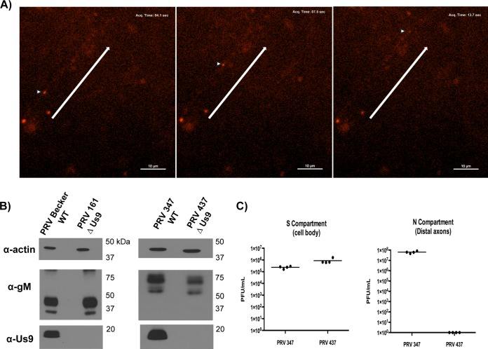
We constructed a plasmid encoding mCherry-tagged glycoprotein M (gM) through de novo synthesis, designated pML124 (11). Two single fluorescent PRV strains expressing this fusion were then derived: PRV 347 (gM-mCherry/wild-type Becker) and PRV 437 (gM-mCherry/Us9-null). PRV 347 and 437 were isolated following cotransfection of linearized pML124 and nucleocapsid DNA from PRV Becker (WT) or PRV 161 (Us9-null) (16), as previously described (11). We performed live-cell imaging of PRV 347 and PRV 437 in dissociated rat superior cervical ganglion (SCG) cultures under conditions previously described (11). As for other PRV membrane proteins (6, 17), substantially more fluorescent signal was detected in neuronal cell bodies than on puncta in axons. As expected, mCherry-labeled puncta trafficked in the anterograde direction after infection with PRV 347, similar to the previously characterized gM-mCherry/GFP-Us9 dually labeled puncta (11). However, we also observed anterograde transport of gM-mCherry puncta after infection with PRV 437 (Us9-null) (see Movie S1 in the supplemental material). Puncta moving in an anterograde direction were detected beginning at 8 h postinfection (Fig. 1A). The motility of these structures was qualitatively different between PRV 347 and PRV 437, with Us9-null mCherry puncta appearing less motile with an apparently higher stall frequency due to the high number of immobile mCherry puncta. To our knowledge, this is the first observation of definitive anterograde transport of a PRV membrane protein in the absence of Us9 expression.
FIG 1.
Characterization of a Us9-independent pathway for anterograde axonal transport of gM. (A) Live cell imaging stills of anterograde transport of gM-mCherry in rat SCG neurons at 8 h postinfection with PRV 437. Triangles indicate punctate structures moving in an antergrade direction. Arrows indicates anterograde directionality. (B) WB assessment of Us9 and gM expression in PK15 whole-cell extracts infected with PRV Becker, PRV 161 (Us9-null parental strain), PRV 347 (gM-mCherry), and PRV 437 (gM-mCherry, Us9-null). (C) Anterograde spreading capacities of PRV 347 and PRV 437 in chambered neuronal cultures at 24 h postinfection. Point estimates reflect viral titers in the N compartment for infections performed in quadruplicate for each viral strain. Lines denote median titers.
Previous work indicated that glycoproteins gB, gC, and gE as well as bulk virion envelope epitopes could not be detected in axons during Us9-null infections by immunofluorescence (4, 18, 19). To reconcile these observations with our finding of Us9-independent transport of gM, we verified the Us9-null phenotype and Us9 expression levels of PRV 437. We measured expression of Us9 by PRV 347, 437, and 161 during infection of PK15 cells by Western blotting (WB) using a polyclonal rabbit Us9 antiserum (5). No Us9 was detected for the Us9-null mutants PRV 437 and PRV 161 (Fig. 1B). The gM-mCherry fusion protein was detected using a polyclonal rabbit gM antiserum (20) as two distinct bands at 70 and 50 kDa for PRV 347 and PRV 437, showing the expected increase in size over the untagged gM protein. We then assessed the anterograde-spread capacity of PRV 347 and PRV 437 by infecting primary cultures of rat SCG neurons in modified Campenot chambers as described previously (21). No anterograde spread into the isolated N compartment was observed at 24 h postinfection with PRV 437, compared to the robust spread of PRV 347 (Fig. 1C), confirming the Us9-null phenotype.
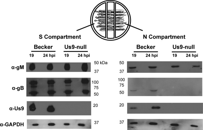
Our results suggest that a vesicle containing gM-mCherry enters and is transported in axons without Us9. Moreover, the live-cell imaging assay confirmed that gM-mCherry labeled puncta were mobile and therefore not extracellular inoculum or stalled particles. To exclude confounding effects on gM localization due to the mCherry fluorophore tag, we analyzed native untagged proteins in PRV-infected SCG cultures using the modified Campenot chamber system as described above. Cultures were infected with PRV Becker or PRV 161, and the contents of each compartment were harvested separately for WB analysis. The S compartment samples demonstrated expression of the viral membrane proteins gB and gM, indicative of productive infection and new protein synthesis above input virion material (Fig. 2). Us9 was detected in PRV Becker- but not PRV 161-infected chambers. Robust gM signal was detected in the N compartment axons at 18 and 24 h postinfection with both PRV Becker and PRV 161, confirming our observations with the live-cell imaging assay. These results verified the Us9-independent entry of untagged, native gM protein into axons. Furthermore, the viral glycoprotein gB was not detected in axons after PRV 161 (Us9-null) infection, while the mature cleaved gB form was detected in axons after wild-type infection (Fig. 2). These findings confirmed the dependence of glycoproteins like gB on Us9 for transport (18) and further suggested that Us9-independent transport of gM constitutes a process that does not involve all viral membrane proteins.
FIG 2.
Western blot assessment of axonal entry of viral membrane proteins during PRV infection of chambered rat SCG cultures. Samples from chambered neuronal cultures infected with PRV Becker or PRV 161 were assessed by WB to determine the axonal targeting properties of various membrane proteins in the presence/absence of Us9 expression. S and N compartment samples were extracted separately at 18 or 24 h postinfection. Results are representative of two independent biological replicates.
It remains unclear if gM is unique in its ability to undergo Us9-independent axonal sorting and transport. gM is a type III transmembrane protein expressed as a heterodimer with gN (22) that mediates secondary envelopment through interactions with gE and tegument (23–25). gM also facilitates endocytic retrieval and relocalization of both viral and host proteins from the plasma membrane (20, 26). It is possible that the membrane topology of gM targets it to trans-Golgi-derived vesicles that form part of the secretory pathway and are sorted constitutively into axons by host adaptor proteins such as AP3 (27, 28). The host secretory pathway has already been implicated in transport and egress of infectious virions (14, 29, 30). It is also possible that gM actively directs the axonal targeting of these currently uncharacterized vesicles to modulate the viral life cycle. However, it is difficult to establish a more precise functionality for gM in anterograde transport, as deletion of this glycoprotein severely impacts overall viral replication/assembly (31), resulting in a pleiotropic effect on other steps of the viral life cycle. Future experiments are needed to detail the kinesin motor that moves these vesicles in axons and to characterize the cohort of gM binding partners.
Supplementary Material
ACKNOWLEDGMENTS
We thank Tal Kramer for scientific advice and support, as well as Orkide Koyuncu and Ian Hogue for helpful commentary on the manuscript.
L.W.E. and R.K. are supported by U.S. National Institutes of Health grants R37 NS033506-16 and R01 NS060699-03. M.P.T. is supported by a National Institutes of Health Career Development Award (K22106948-01A1) and by IDeA Program grant GM110732 (8P20GM103500-10).
Footnotes
Supplemental material for this article may be found at http://dx.doi.org/10.1128/JVI.00625-15.
REFERENCES
- 1.Pomeranz LE, Reynolds AE, Hengartner CJ. 2005. Molecular biology of pseudorabies virus: impact on neurovirology and veterinary medicine. Microbiol Mol Biol Rev 69:462–500. doi: 10.1128/MMBR.69.3.462-500.2005. [DOI] [PMC free article] [PubMed] [Google Scholar]
- 2.Granstedt AE, Bosse JB, Thiberge SY, Enquist LW. 2013. In vivo imaging of alphaherpesvirus infection reveals synchronized activity dependent on axonal sorting of viral proteins. Proc Natl Acad Sci U S A 110:E3516–E3525. doi: 10.1073/pnas.1311062110. [DOI] [PMC free article] [PubMed] [Google Scholar]
- 3.Smith G. 2012. Herpesvirus transport to the nervous system and back again. Annu Rev Microbiol 66:153–176. doi: 10.1146/annurev-micro-092611-150051. [DOI] [PMC free article] [PubMed] [Google Scholar]
- 4.Tomishima MJ, Enquist LW. 2001. A conserved alpha-herpesvirus protein necessary for axonal localization of viral membrane proteins. J Cell Biol 154:741–752. doi: 10.1083/jcb.200011146. [DOI] [PMC free article] [PubMed] [Google Scholar]
- 5.Brideau AD, Banfield BW, Enquist LW. 1998. The Us9 gene product of pseudorabies virus, an alphaherpesvirus, is a phosphorylated, tail-anchored type II membrane protein. J Virol 72:4560–4570. [DOI] [PMC free article] [PubMed] [Google Scholar]
- 6.Kramer T, Greco TM, Taylor MP, Ambrosini AE, Cristea IM, Enquist LW. 2012. Kinesin-3 mediates axonal sorting and directional transport of alphaherpesvirus particles in neurons. Cell Host Microbe 12:806–814. doi: 10.1016/j.chom.2012.10.013. [DOI] [PMC free article] [PubMed] [Google Scholar]
- 7.Johnson DC, Baines JD. 2011. Herpesviruses remodel host membranes for virus egress. Nat Rev Microbiol 9:382–394. doi: 10.1038/nrmicro2559. [DOI] [PubMed] [Google Scholar]
- 8.Mettenleiter TC, Klupp BG, Granzow H. 2009. Herpesvirus assembly: an update. Virus Research 143:222–234. doi: 10.1016/j.virusres.2009.03.018. [DOI] [PubMed] [Google Scholar]
- 9.Szilágyi JF, Cunningham C. 1991. Identification and characterization of a novel non-infectious herpes simplex virus-related particle. J Gen Virol 72:661–668. doi: 10.1099/0022-1317-72-3-661. [DOI] [PubMed] [Google Scholar]
- 10.McLauchlan J, Rixon FJ. 1992. Characterization of enveloped tegument structures (L particles) produced by alphaherpesviruses: integrity of the tegument does not depend on the presence of capsid or envelope. J Gen Virol 73:269–276. doi: 10.1099/0022-1317-73-2-269. [DOI] [PubMed] [Google Scholar]
- 11.Taylor MP, Kramer T, Lyman MG, Kratchmarov R, Enquist LW. 2012. Visualization of an alphaherpesvirus membrane protein that is essential for anterograde axonal spread of infection in neurons. mBio 3:e00063-12. doi: 10.1128/mBio.00063-12. [DOI] [PMC free article] [PubMed] [Google Scholar]
- 12.Liu WW, Goodhouse J, Jeon NL, Enquist LW. 2008. A microfluidic chamber for analysis of neuron-to-cell spread and axonal transport of an alpha-herpesvirus. PLoS One 3:e2382. doi: 10.1371/journal.pone.0002382. [DOI] [PMC free article] [PubMed] [Google Scholar]
- 13.Alemañ N, Quiroga MI, López-Peña M, Vázquez S, Guerrero FH, Nieto JM. 2003. L-particle production during primary replication of pseudorabies virus in the nasal mucosa of swine. J Virol 77:5657–5667. doi: 10.1128/JVI.77.10.5657-5667.2003. [DOI] [PMC free article] [PubMed] [Google Scholar]
- 14.Antinone SE, Zaichick SV, Smith GA. 2010. Resolving the assembly state of herpes simplex virus during axon transport by live-cell imaging. J Virol 84:13019–13030. doi: 10.1128/JVI.01296-10. [DOI] [PMC free article] [PubMed] [Google Scholar]
- 15.LaVail JH, Tauscher AN, Sucher A, Harrabi O, Brandimarti R. 2007. Viral regulation of the long distance axonal transport of herpes simplex virus nucleocapsid. Neuroscience 146:974–985. doi: 10.1016/j.neuroscience.2007.02.010. [DOI] [PMC free article] [PubMed] [Google Scholar]
- 16.Brideau AD, Card JP, Enquist LW. 2000. Role of pseudorabies virus Us9, a type II membrane protein, in infection of tissue culture cells and the rat nervous system. J Virol 74:834–845. doi: 10.1128/JVI.74.2.834-845.2000. [DOI] [PMC free article] [PubMed] [Google Scholar]
- 17.Kratchmarov R, Kramer T, Greco TM, Taylor MP, Ch'ng TH, Cristea IM, Enquist LW. 2013. Glycoproteins gE and gI are required for efficient KIF1A-dependent anterograde axonal transport of alphaherpesvirus particles in neurons. J Virol 87:9431–9440. doi: 10.1128/JVI.01317-13. [DOI] [PMC free article] [PubMed] [Google Scholar]
- 18.Lyman MG, Feierbach B, Curanovic D, Bisher M, Enquist LW. 2007. Pseudorabies virus Us9 directs axonal sorting of viral capsids. J Virol 81:11363–11371. doi: 10.1128/JVI.01281-07. [DOI] [PMC free article] [PubMed] [Google Scholar]
- 19.Lyman MG, Curanovic D, Enquist LW. 2008. Targeting of pseudorabies virus structural proteins to axons requires association of the viral Us9 protein with lipid rafts. PLoS Pathog 4:e1000065. doi: 10.1371/journal.ppat.1000065. [DOI] [PMC free article] [PubMed] [Google Scholar]
- 20.Crump CM, Bruun B, Bell S, Pomeranz LE, Minson T, Browne HM. 2004. Alphaherpesvirus glycoprotein M causes the relocalization of plasma membrane proteins. J Gen Virol 85:3517–3527. doi: 10.1099/vir.0.80361-0. [DOI] [PubMed] [Google Scholar]
- 21.Curanovic D, Ch'ng TH, Szpara M, Enquist L. 2009. Compartmented neuron cultures for directional infection by alpha herpesviruses. Curr Protoc Cell Biol Chapter 26:Unit 26.4. doi: 10.1002/0471143030.cb2604s43. [DOI] [PMC free article] [PubMed] [Google Scholar]
- 22.Jöns A, Dijkstra JM, Mettenleiter TC. 1998. Glycoproteins M and N of pseudorabies virus form a disulfide-linked complex. J Virol 72:550–557. [DOI] [PMC free article] [PubMed] [Google Scholar]
- 23.Leege T, Fuchs W, Granzow H, Kopp M, Klupp BG, Mettenleiter TC. 2009. Effects of simultaneous deletion of pUL11 and glycoprotein M on virion maturation of herpes simplex virus type 1. J Virol 83:896–907. doi: 10.1128/JVI.01842-08. [DOI] [PMC free article] [PubMed] [Google Scholar]
- 24.Nixdorf R, Klupp BG, Mettenleiter TC. 2001. Role of the cytoplasmic tails of pseudorabies virus glycoproteins B, E and M in intracellular localization and virion incorporation. J Gen Virol 82:215–226. [DOI] [PubMed] [Google Scholar]
- 25.Brack AR, Klupp BG, Granzow H, Tirabassi R, Enquist LW, Mettenleiter TC. 2000. Role of the cytoplasmic tail of pseudorabies virus glycoprotein E in virion formation. J Virol 74:4004–4016. doi: 10.1128/JVI.74.9.4004-4016.2000. [DOI] [PMC free article] [PubMed] [Google Scholar]
- 26.Ren Y, Bell S, Zenner HL, Lau S.-YK, Crump CM. 2012. Glycoprotein M is important for the efficient incorporation of glycoprotein H-L into herpes simplex virus type 1 particles. J Gen Virol 93:319–329. doi: 10.1099/vir.0.035444-0. [DOI] [PubMed] [Google Scholar]
- 27.Robinson MS. 2004. Adaptable adaptors for coated vesicles. Trends Cell Biol 14:167–174. doi: 10.1016/j.tcb.2004.02.002. [DOI] [PubMed] [Google Scholar]
- 28.Newell-Litwa K, Seong E, Burmeister M, Faundez V. 2007. Neuronal and non-neuronal functions of the AP-3 sorting machinery. J Cell Sci 120:531–541. doi: 10.1242/jcs.03365. [DOI] [PubMed] [Google Scholar]
- 29.Hogue IB, Bosse JB, Hu J.-R, Thiberge SY, Enquist LW. 2014. Cellular mechanisms of alpha herpesvirus egress: live cell fluorescence microscopy of pseudorabies virus exocytosis. PLoS Pathog 10:e1004535. doi: 10.1371/journal.ppat.1004535. [DOI] [PMC free article] [PubMed] [Google Scholar]
- 30.Miranda-Saksena M, Boadle RA, Aggarwal A, Tijono B, Rixon FJ, Diefenbach RJ, Cunningham AL. 2009. Herpes simplex virus utilizes the large secretory vesicle pathway for anterograde transport of tegument and envelope proteins and for viral exocytosis from growth cones of human fetal axons. J Virol 83:3187–3199. doi: 10.1128/JVI.01579-08. [DOI] [PMC free article] [PubMed] [Google Scholar]
- 31.Dijkstra JM, Visser N, Mettenleiter TC, Klupp BG. 1996. Identification and characterization of pseudorabies virus glycoprotein gM as a nonessential virion component. J Virol 70:5684–5688. [DOI] [PMC free article] [PubMed] [Google Scholar]
Associated Data
This section collects any data citations, data availability statements, or supplementary materials included in this article.


