

Abstract
Background
Pathway analysis has been widely used to gain insight into essential mechanisms of the response to myocardial infarction (MI). Currently, there exist multiple pathway databases that organize molecular datasets and manually curate pathway maps for biological interpretation at varying forms of organization. However, inconsistencies among different databases in pathway descriptions, frequently due to conflicting results in the literature, can generate incorrect interpretations. Furthermore, although pathway analysis software provides detailed images of interactions among molecules, it does not exhibit how pathways interact with one another or with other biological processes under specific conditions.
Methods
We propose a novel method to standardize descriptions of enriched pathways for a set of genes/proteins using Gene Ontology terms. We used this method to examine the relationships among pathways and biological processes for a set of condition-specific genes/proteins, represented as a functional biological pathway-process network. We applied this algorithm to a set of 613 MI-specific proteins we previously identified.
Results
A total of 96 pathways from Biocarta, KEGG, and Reactome, and 448 Gene Ontology Biological Processes were enriched with these 613 proteins. The pathways were represented as Boolean functions of biological processes, delivering an interactive scheme to organize enriched information with an emphasis on involvement of biological processes in pathways. We extracted a network focusing on MI to demonstrate that tyrosine phosphorylation of Signal Transducer and Activator of Transcription (STAT) protein, positive regulation of collagen metabolic process, coagulation, and positive/negative regulation of blood coagulation have immediate impacts on the MI response.
Conclusions
Our method organized biological processes and pathways in an unbiased approach to provide an intuitive way to identify biological properties of pathways under specific conditions. Pathways from different databases have similar descriptions yet diverse biological processes, indicating variation in their ability to share similar functional characteristics. The coverages of pathways can be expanded with the incorporation of more biological processes, predicting involvement of protein members in pathways. Further, detailed analyses of the functional biological pathway-process network will allow researchers and scientists to explore critical routes in biological systems in the progression of disease.
Background
The emergence of publicly available pathway databases has provided biologists excellent resources to attain a deeper understanding of biological mechanisms by providing organization to a large list of differentially expressed genes and proteins. Knowledge of molecular-level interactions and reactions has been curated in many knowledge databases, forming biological pathways. These knowledge databases include BioCarta (http://biocarta.com/), Kyoto Encyclopedia of Genes and Genomes (KEGG), Reactome, Protein Analysis Through Evolutionary Relationships (PANTHER), and MetaCyc [1-5]. Most often, pathways are organized as directed graphs of interacting molecules and often are accompanied by visualizations that demonstrate relationships among gene products, gene function types (e.g., regulation, activation, and inhibition) and translated protein locations (e.g., extracellular matrix, cell membrane, or nucleus). Recently, the integration of various omics data such as proteomics, genomics, transcriptomics, and metabolomics for knowledge discovery has drawn much attention [6-9]. In addition to the aforementioned pathway knowledge databases, the Gene Ontology (GO) Consortium pursues approaches to standardize the representation of gene products across different species and databases [10]. GO consists of a controlled vocabulary of terms, covering three domains: cellular components, molecular functions and biological processes. A GO Biological Process (GOBP) is a series of molecular events, with a defined beginning and end. However, a biological process is not equivalent to a pathway; GOBPs are assumed to be independent and do not represent the interactions among molecules.
Despite manual curation and careful revision, different knowledge databases could have different descriptions, participating molecules, interacting diagrams, and supporting literature for similar pathways. For example, considering the Transforming Growth Factors Beta (TGF-beta) signaling pathway in human, KEGG reported as hsa04350: TGF-beta signaling pathway, Reactome reported as REACT_111102.4: Signaling by TGF-beta Receptor Complex, and Biocarta reported as h_tgfbpathway. In detail, KEGG annotated 80 genes/proteins, Reactome annotated 120 genes/proteins, and Biocarta annotated 17 genes/proteins with TGF-beta signaling pathway. Descriptions of TGF-beta signaling pathway in the nucleus were excerpted to show related yet distinctive contents among KEGG, Reactome and Biocarta databases (Material in quote marks and italic type represents verbatim quotation from the knowledge databases):
KEGG - "Once phosphorylated, R-Smads associate with the co-mediator Smad, Smad4, and the heteromeric complex then translocates into the nucleus. In the nucleus, Smad complexes activate specific genes through cooperative interactions with other DNA-binding and coactivator (or co-repressor) proteins".
(http://www.genome.jp/kegg-bin/show_pathway?hsa04350)
Reactome - "The general signaling scheme is rather simple: upon binding of a ligand, an activated plasma membrane receptor complex is formed, which passes on the signal towards the nucleus through a phosphorylated receptor SMAD (R-SMAD). In the nucleus, the activated R-SMAD promotes transcription in complex with a closely related helper molecule termed Co-SMAD (SMAD4)".
(http://www.reactome.org/PathwayBrowser/#DIAGRAM = 170834&PATH = 162582)
Biocarta - "The activated TGF-beta R1 phosphorylates SMAD2 and SMAD3, which bind to the SMAD4 mediator to move into the nucleus and form complexes that regulate transcription. SMADs regulate transcription in several ways, including binding to DNA, interacting with other transcription factors, and interacting with transcription corepressors and coactivators like p300 and CBP".
(http://www.biocarta.com/pathfiles/h_tgfbpathway.asp).
These variations in knowledge representation among different databases prompt an urgent need for standard pathway representations. For a set of proteins or genes with enriched pathways and GOBPs, we propose a method that integrates molecular interaction, biological pathways and GOBP to standardize descriptions of pathways using GOBPs through the establishment of the functional biological pathway-process network. We demonstrated with the set of 613 proteins related to myocardial infarction (MI) from the MI-specific protein-protein interaction network [11].
Methods
In this study, we started with 613 MI-specific proteins to find enriched pathways and GOBPs [11]. We performed analyses to statistically examine the similarities between pathways and biological processes and identify the hierarchical structures for the GOBPs. Based on the similarity score matrix and the structure of GOBPs, we established the logical circuitry between GOBPs and pathways, and visualize the circuitry with networks.
Selection of condition-specific genes/proteins
We previously identified 613 proteins specific to MI in an MI-specific protein-protein interaction network (MIPIN); the network and its protein members were used here to demonstrate the developed method [11].
Functional annotation analysis
Many tools are available to provide gene-annotation enrichment analysis and pathway mapping. We performed functional annotation analysis using DAVID Functional Annotation Tool, with the parameters Count to be 2 and EASE to be 0.05, to obtain enriched GOBP terms, KEGG and Reactome pathways [12].
Statistical measure of inter-annotator agreement
We evaluated the pairwise similarity between different annotation terms, including GO terms and pathways using Kappa statistics because annotation terms sharing common members might be related to one another [13]. Considering a set of all annotated genes/proteins G, two annotation terms Ti and Tj annotated by two set of genes Gi and Gj (i≠j; i,j = 1, 2, ..., N), we denoted the number of proteins annotated by both terms as aij, the number of proteins annotated by Ti but not Tj as bij, the number of proteins annotated by Tj but not Ti as cij, and the number of proteins not annotated by neither terms among the union of proteins annotated by N annotation terms as dij.
Thus, we have,
The Kappa score κij was defined as,
where Pr(agreeij) was the observed percentage agreement and Pr(randomij) was the overall probability of random agreement for annotation terms Ti and Tj. The observed percentage agreement Pr(agreeij) could be calculated as follows,
Out of total number of associated proteins, Ti annotates and Tj annotates Thus, the probability that both annotation terms randomly annotate the same proteins wasSimilarly, the probability that neither pathway randomly annotate the same protein was As a result, the overall probability of random agreement Pr(randomij) could be calculated as,
A high Kappa score indicated that two annotation terms share many common proteins.
Construction of undirected GOBP graph
An undirected GOBP graph GraphGOBPenriched was constructed to describe the relationships among NenrichedGOBP enriched GOBP terms, i.e., GraphGOBPenriched = (VGOBP, EGOBP), |VGOBP|= NenrichedGOBP, and EGOBP defines the set of edges in the graph. The relationships between GOBP terms, represented by edges connecting them, were evaluated based on the ancestor/offspring relationships in the complete directed acyclic graph of all GOBP terms from the Gene Ontology Consortium. We mapped NenrichedGOBP enriched GOBP terms to the corresponding vertices of the complete directed acyclic graph of all GOBP terms from the Gene Ontology Consortium using the package "GO.db" from Bioconductor [14]. Let GraphGOBPComplete = (VcompleteGOBP, EcompleteGOBP) be the complete directed acyclic graph of all GOBP terms. Then, VGOBP is mapped to VcompleteGOBP (VGOBP ⊂ V' and V' ⊂ VcompleteGOBP). Two GOBP terms would be connected if there existed a link between this pair of vertices in the complete graph of GOBP. All networks and graphs in this study were constructed and analyzed with the assistance of the package 'igraph' in R [15].
Construction of undirected Boolean bipartite pathway and GOBP graph
The relationships between pathways and GOBP terms were represented as an undirected graph where edges between pathways and GOBP terms were evaluated based on Kappa statistics. We computed the Kappa similarity matrix of size NtotalGOBP x NtotalPathway, where NtotalPathway is the total number of pathways including Biocarta, KEGG and Reactome pathways. Each row of the similarity matrix represents a GOBP term, and each column represents a pathway. Top 1% of the most similar pairs of pathway and biological process were selected and connected based on the Kappa similarity scores. Figure 1 showed that choosing the top 1% of the most similar pairs allowed the selection of a reasonable number of edges with high similarity scores (the average of Kappa scores was 0.025, and the chose cut-off value was 0.27). The set of pairs of pathway and GOBP terms satisfying such condition as was denoted as EPathwayGOBP. We then established the pathway and GOBP graph as an undirected bipartite graph BipartiteGraphPathwayGOBP = {VPathway, VGOBP, EPathwayGOBP} where VPathway is the set of pathways and VGOBP is the set of GOBP terms included in EPathwayGOBP (|VPathway| ≤ NtotalPathway and |VB| ≤ NtotalGOBP). Thus, the graph BipartiteGraphPathwayGOBP would consist of pathways that could be well represented by GOBP terms.
Figure 1.
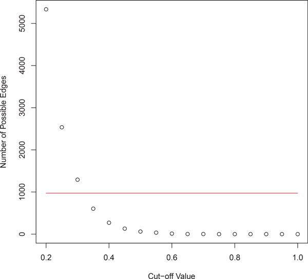
The graph showing Number of possible edges vs. Cut-off value, and the selected number of edges. Choosing top 1% of the most similar pairs of pathway and biological process considered a reasonable number of pairs of pathways and biological processes with high similarity scores.
We further introduced Boolean rules to BipartiteGraphPathwayGOBP to represent pathways as Boolean functions of biological processes, assuming that connected biological processes have direct impacts on the pathways. Since a pathway contains dynamics and dependencies among participating molecules, which are annotated by biological processes, we assume that different combinations of biological process states can affect the state of the pathway, which is either 'active' (binary state 1) or 'inactive' (binary state 0). For every pathway VPathwayi in the graph BipartiteGraphPathwayGOBP, let VPathwayGOBPi be the set of GOBP terms connected to that pathway and VGOBP = ∪VPathwayGOBPi-, we performed Boolean mapping such that the pathway VPathwayi could be described as a Boolean algebra functions of its connected GOBP terms, VPathwayi = f(VPathwayGOBPi).
The Boolean rules were derived from the relationships between GOBP terms connected to the pathway. If two GOBP terms were connected, then the Boolean relationship between these GOBP terms would be "OR." Such assumption arose from the fact two connected GOBP terms would share a significant amount of protein; thus, if a biological process was active, then its connected process must be simultaneously active as well. The relationship between two unconnected GOBP terms would be "AND." For example, considering a small network with 3 GOBP terms, GOBP1, GOBP2 and GOBP3, and a pathway P, where GOBP1 and GOBP2 were connected, GOPB3 was not connected with GOBP1 and GOBP2, and all GOBP terms were connected to pathway P. Then, the Boolean function for P could be written as, VP = (VGOBP1 ∪ VGOBP2) ∩ VGOBP3.
The functional biological pathway-process network and the extracted MI network
We combined the GOBP graph GraphGOBPenriched from section 2.4 and the bipartite graph BipartiteGraphPathwayGOBP from section 2.5 to have a complete functional biological pathway-process network, where there were connections among GOBPs, and pathways communicated with each other through biological processes. As the complete network had many vertices and edges, we presented the MI pathway, h_amiPathway, from Biocarta, to illustrate the result. We retained important GOBP terms which were crossed by the shortest paths among other pathways to the MI pathway. Shortest paths were calculated using the un-weighted breadth-first search method. The extracted network allowed us to identify how the MI pathway could lead to other pathways and vice versa, initiating cardiac remodeling post-MI.
Results
Undirected GOBP graph
Using DAVID Functional Annotation Tool, we obtained 993 enriched GOBP terms from the list of 613 MI-specific proteins. From the ancestor/offspring relationships, the graph GraphGOBPenriched was constructed, resulting in a network of 993 vertices and 4284 edges. GraphGOBPenriched had 16 connected sub-graphs having more than 1 vertex and 46 isolated vertices. The largest connected sub-graph consisted of 885 vertices and 4199 edges.
It is interesting to note that GOBP terms with the highest degree, measuring the number of direct links incident on a vertex in a graph, were related to phosphorylation, phosphate, phosphorus, and kinase activity (Table 1). Since phosphorus and phosphate metabolic processes have the highest connections, this could mean that the chemical reactions and pathways involving intracellular signaling might initiate the cascade of events post-MI. In fact, serum phosphorus has been shown to serve as a sensitive indicator of MI and is linked to all-cause mortality and heart failure in patients after MI [16,17]. Hypophosphatemia in MI is associated with a greater degree of dysfunction of the left ventricle (LV), resulting in increased 30 days mortality [18]. In patients with MI, plasma sphingosine-1-phosphate concentration is reduced, leading to decreases protective action on cardiomyocyte viability [19].
Table 1.
Top 20 GO Biological Processes ranked by degree measurements.
| GOBP ID | Name | Degree |
|---|---|---|
| GO:0006793 | phosphorus metabolic process* | 55 |
| GO:0006796 | phosphate metabolic process* | 52 |
| GO:0006955 | immune response | 47 |
| GO:0010033 | response to organic substance | 47 |
| GO:0016310 | phosphorylation* | 45 |
| GO:0048584 | positive regulation of response to stimulus | 45 |
| GO:0051174 | regulation of phosphorus metabolic process* | 44 |
| GO:0019220 | regulation of phosphate metabolic process* | 43 |
| GO:0043507 | positive regulation of JUN kinase activity* | 43 |
| GO:0042325 | regulation of phosphorylation* | 39 |
| GO:0043406 | positive regulation of MAP kinase activity* | 38 |
| GO:0000187 | activation of MAPK activity* | 38 |
| GO:0032268 | regulation of cellular protein metabolic process | 37 |
| GO:0031659 | positive regulation of cyclin-dependent protein kinase activity during G1/S* | 37 |
| GO:0006468 | protein amino acid phosphorylation* | 36 |
| GO:0010604 | positive regulation of macromolecule metabolic process | 34 |
| GO:0001932 | regulation of protein amino acid phosphorylation* | 34 |
| GO:0001775 | cell activation | 34 |
| GO:0045860 | positive regulation of protein kinase activity* | 33 |
| GO:0006952 | defense response | 33 |
*GOBPs related with phosphorus, phosphate, phosphorylation, and kinase activity.
In addition, biological processes involved with phosphorylation accounted for 4 GOBP terms while there were 5 kinase-activity-related GOBPs in Table 1. Phosphorylation is a major post-translational modification to regulate protein function. In a phosphorylation process, a protein kinase modifies target proteins, or substrates, by chemically adding phosphate groups to them. This result corresponded well with our previous work which identified Kinase Pathways as one of the major groups of pathways significantly enriched following MI [11].
Network of biological pathways and GOBP showed similarities and differences among pathways in regard to GOBP annotation
At selected parameters, we retrieved 98 pathways, including 37 KEGG, 13 Reactome, and 48 Biocarta pathways using DAVID Functional Annotation Tool. Analysing statistical measures of inter-annotator agreement between 98 pathways and 993 GOBP terms, we established a graph BipartiteGraphPathwayGOBP with 544 vertices, containing 96 pathways 448 associated GOBPs, and 973 edges. These edges represented the most significantly enriched pairs of pathways and GOBP in the context of MI. This graph consisted of 8 sub-graphs, with the largest connected component having 76 pathways and 396 GOBP terms.
Earlier, we mentioned the TGF-beta signaling pathway and how it was defined differently among the KEGG, Reactome, and Biocarta pathway databases. We further examined the associated GOBP terms to compare these 3 pathways (Figure 2). The variations were due to different literature being used to construct the pathways: REACT_6844: Signaling by TGF beta were involved with 56 GOBP terms, hsa04350: TGF-beta Signaling Pathway was associated with 14 GOBP terms, and the h_tgfbPathway was linked to 27 GOBP terms. Nonetheless, the common biological processes among these pathways included phosphorylation of SMAD proteins, serine/threonine kinase signaling pathway, epithelial-mesenchymal transition, and response to cholesterol and cell morphogenesis involved in differentiation (Figure 2: Box 6). It can be seen that the REACT_6844 provided a more complete description of TGFβ signaling pathway (Figure 2: Box 1-2&5), hsa04350 mainly focused on protein transport, transcription, gene expression and cell development (Figure 2: Box 2-3), whereas h_tgfbPathway emphasized organ development (Figure 2: Box 4-5). As a result, we can understand the different characteristics assigned for each pathway under the different circumstances. Individually, TGF-beta signaling pathways from KEGG, Biocarta, and Reactome annotated 21, 12, and 7 proteins, respectively, from the initial 613 MI-specific proteins. Thus, by incorporating the signaling pathways from different sources, we updated the knowledge of TGF-beta signaling pathways with more biological processes, and identified additional proteins participating in the pathway. Using this approach, the total number of proteins annotated with TGF-beta signaling pathways, by combining proteins from KEGG, Biocarta and Reactome, was expanded to 25 proteins.
Figure 2.
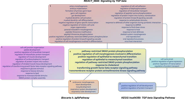
Representations of TGF-beta signalling pathway from Biocarta, KEGG and RACTOME in terms of Gene Ontology biological processes in the condition of MI. Box 1: GOBP exclusive to REACTOME REACT_6844: Signaling by TGF beta. Box 2: Common GOBP between REACTOME and KEGG. Box 3: GOBP exclusive to KEGG has04350: TGF-beta Signaling Pathway. Box 4: GOBP exclusive to BioCarta h_tgfbPathway. Box 5: Common GOBP between BioCarta and REACTOME. Box 6: Common GOBP between BioCarta, KEGG and REACTOME.
Additionally, we investigated how this system acts using three other cardiovascular disease processes, namely hsa05412: Arrhythmogenic Right Ventricular Cardiomyopathy (ARVC), hsa05410: Hypertrophic Cardiomyopathy (HCM), and hsa05414: Dilated Cardiomyopathy (DCM). These analyses provide additional examples to demonstrate how representing pathways in terms of biological processes helped us to quickly understand the characteristics of such conditions under specific circumstances (Figure 3). ARVC is an inherited disease that results in fat and fibrous tissues replacing the heart muscle of the right ventricle and subepicardial region of the left ventricle. With HCM, a portion of the myocardium is hypertrophied, forcing the heart to work harder to pump blood because of the thickened heart muscle. DCM is a condition in which the heart weakens and becomes dilated, resulting in inefficient blood pumping to other organs. All three aforementioned cardiomyopathy pathways involve integrin-mediated signaling pathway, cell-matrix adhesion, and cell-substrate adhesion. However, HCM and DCM are specifically related to leukocyte adhesion. It has been confirmed that human leukocyte antigens are associated with HCM and DCM [20-23].
Figure 3.
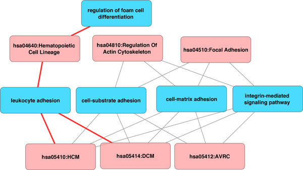
Sub-network of Cardiomyopathy. Pathways were represented in red while GOBPs were represented in blue. Pathways of Hypertrophic, Dilated and Arrhythmogenic Right Ventricular Cardiomyopathy were shown to be connected to biological processes including leukocyte adhesion, cell-substrated adhesion, and cell-matrix adhesion. Integrin-ECM interactions are required for cell adhesion.
We showed a visualization of a sub-graph consisting of 7 pathways and 34 GOBP terms that intersected with the MI response (Figure 4). Two pathways having the largest number of associated GOBP terms were hsa04610: Complement And Coagulation Cascades (characterized by 17 GOBP terms) and h_fibrinolysisPathway (characterized by 22 GOBP terms). The center of this sub-network is the MI pathway from Biocarta, h_amiPathway. Altogether, 3 pathways were represented by 32 out of 34 GOBP terms in this sub-network, and there were 8 common GOBP terms, including coagulation, regulation of coagulation, negative regulation of coagulation, blood coagulation, regulation of blood coagulation, negative regulation of blood coagulation, homeostasis and regulation of body fluid levels (Table 2). As a result, we noticed that blood coagulation, coagulation, homeostasis and regulation of body fluid levels were the underlying processes in these pathways. Table 2 and Figure 4 also pointed out the differences among these pathways: hsa04610 was associated with activation of proteins involved in acute inflammatory response and wound healing, whereas the fibrinolysis pathway was specifically involved with fibrinolysis, platelet activation, protein phosphorylation, collagen process and tissue regeneration.
Figure 4.
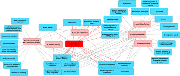
Sub-network of MI. Pathways were represented in while GOBPs were represented in blue. The major underlying processes for MI included coagulation, homeostasis, collagen metabolic/biosynthetic process, calcium ion transport, tissue regeneration, and wound healing.
Table 2.
Pathways and GOBP in the MI functional pathway-process network.
| GOBP names | Pathways | F | C | A | E | P | I | H |
|---|---|---|---|---|---|---|---|---|
| activation of plasma proteins involved in acute inflammatory response | √ | |||||||
| acute inflammatory response | √ | |||||||
| blood coagulation | √ | √ | √ | √ | √ | √ | ||
| blood coagulation, extrinsic pathway | √ | √ | ||||||
| coagulation | √ | √ | √ | √ | √ | √ | ||
| complement activation | √ | |||||||
| developmental growth | √ | √ | ||||||
| fibrinolysis | √ | √ | ||||||
| growth | √ | √ | ||||||
| hemostasis | √ | √ | √ | √ | √ | √ | ||
| negative regulation of blood coagulation | √ | √ | √ | √ | √ | |||
| negative regulation of coagulation | √ | √ | √ | √ | √ | |||
| negative regulation of multicellular organismal process | √ | |||||||
| platelet activation | √ | √ | √ | |||||
| positive regulation of blood coagulation | √ | √ | √ | √ | ||||
| positive regulation of calcium ion transport | √ | √ | ||||||
| positive regulation of coagulation | √ | √ | √ | √ | ||||
| positive regulation of collagen biosynthetic process | √ | √ | √ | |||||
| positive regulation of collagen metabolic process | √ | √ | √ | |||||
| protein maturation | √ | |||||||
| protein maturation by peptide bond cleavage | √ | |||||||
| protein processing | √ | |||||||
| regeneration | √ | |||||||
| regulation of blood coagulation | √ | √ | √ | √ | √ | |||
| regulation of body fluid levels | √ | √ | √ | √ | √ | √ | ||
| regulation of coagulation | √ | √ | √ | √ | √ | |||
| regulation of collagen metabolic process | √ | √ | ||||||
| regulation of fibrinolysis | √ | |||||||
| regulation of multicellular organismal metabolic process | √ | √ | ||||||
| regulation of response to external stimulus | √ | |||||||
| response to wounding | √ | |||||||
| tissue regeneration | √ | √ | ||||||
| tyrosine phosphorylation of STAT protein | √ | √ | √ | |||||
| wound healing | √ | √ | √ | √ | ||||
| Number of connected GOBPs | 22 | 17 | 16 | 14 | 12 | 9 | 5 | |
Associations between the MI response and biological processes have been experimentally and clinically verified
In order to confirm the affiliated biological processes with the MI response mentioned in the previous section, we searched PubMed for experimental and clinical evidence. In the BipartiteGraphPathwayGOBP, the MI pathway, annotated with 11 proteins, was connected with 16 GOBP terms that were linked to 64 proteins, and they shared 10 common proteins. We further verified that among the 54 proteins exclusively annotated by GOBP terms, 11 proteins had been chosen as the seed proteins to construct the MI-specific protein network. We have previously shown that these seed proteins were associated with MI and confirmed by at least 2 citations [11].
To verify that the remaining 43 proteins of the expanded set of proteins for the MI pathway were related to MI, we searched for their official names and aliases on PubMed along with the keyword "myocardial infarction" for publications that confirmed the association between these proteins and MI (Table 3). There were 34 proteins firmly associated with MI by at least 2 publications. There were 3 proteins, namely CD44, SERPIND1 and HNF4A, directly associated with MI by one publication. There were 6 proteins, namely ANXA7, FBLN5, FGF7, KLF6, FR2RL2 and GGCX indirectly linked to MI. Among 16 MI-associated GOBP terms, 11 biological processes were fully associated with the MI pathway as all of their member proteins were associated with MI and confirmed by at least 2 publications. The remaining 5 GOBP terms had 90% of the member proteins associated with the MI pathway, confirmed by at least 1 publication, and 80% or more of the member proteins were confirmed to be associated with MI by at least 2 publications. Therefore, we showed that the associations between MI pathway and biological processes in the BipartiteGraphPathwayGOBP have been experimentally and clinically verified. We also expanded the coverage of the original MI pathway by adding 54 new proteins. Further research will be needed to address the intermediate steps within the MI pathway and develop more extensive description of the MI pathway that covers a longer time scale.
Table 3.
Proteins of MI pathway-associated GOBP terms with cited publications.
| Proteins | Gene names | Official Names | Supporting Articles |
|---|---|---|---|
| A1AT_HUMAN | SERPINA1 | Alpha-1-antitrypsin | [27,28] |
| ACVL1_HUMAN | ACVRL1 | Serine/threonine-protein kinase receptor R3 | [29,30] |
| ADA17_HUMAN | ADAM17 | Disintegrin and metalloproteinase domain-containing protein 17 | [31,32] |
| ANPRA_HUMAN | NPR1 | Atrial natriuretic peptide receptor 1 | [33,34] |
| APOA_HUMAN | LPA | Apolipoprotein(a) | [35,36] |
| CAV1_HUMAN | CAV1 | Caveolin-1 | [37,38] |
| CBPB2_HUMAN | CPB2 | Carboxypeptidase B2 | [39,40] |
| CD36_HUMAN | CD36 | Platelet glycoprotein 4 | [41,42] |
| EGLN_HUMAN | ENG | Endoglin | [29,30] |
| F13A_HUMAN | F13A1 | Coagulation factor XIII A chain | [43,44] |
| FA11_HUMAN | F11 | Coagulation factor XI | [45,46] |
| FA5_HUMAN | F5 | Coagulation factor V | [47,48] |
| FA8_HUMAN | F8 | Coagulation factor VIII | [43,45] |
| FA9_HUMAN | F9 | Coagulation factor IX | [45,46] |
| FIBG_HUMAN | FGG | Fibrinogen gamma chain | [49,50] |
| FINC_HUMAN | FN1 | Fibronectin | [51,52] |
| GPV_HUMAN | GP5 | Platelet glycoprotein V | [53,54] |
| HIF1A_HUMAN | HIF1A | Hypoxia-inducible factor 1-alpha | [55,56] |
| IC1_HUMAN | SERPING1 | Plasma protease C1 inhibitor | [57,58] |
| IFNG_HUMAN | IFNG | Interferon gamma | [59,60] |
| ITA5_HUMAN | ITGA5 | Integrin alpha-5 | [61,62] |
| KNG1_HUMAN | KNG1 | Kininogen-1 | [63,64] |
| LYOX_HUMAN | LOX | Protein-lysine 6-oxidase | [65,66] |
| PAR2_HUMAN | F2RL1 | Proteinase-activated receptor 2 | [67,68] |
| PAR4_HUMAN | F2RL3 | Proteinase-activated receptor 4 | [67,69] |
| PGFRA_HUMAN | PDGFRA | Platelet-derived growth factor receptor alpha | [70,71] |
| PLF4_HUMAN | PF4 | Platelet factor 4 | [72,73] |
| PROZ_HUMAN | PROZ | Vitamin K-dependent protein Z | [74,75] |
| SMAD3_HUMAN | SMAD3 | Mothers against decapentaplegic homolog 3 | [30,76] |
| TGFB2_HUMAN | TGFB2 | Transforming growth factor beta-2 | [77,78] |
| TGFR2_HUMAN | TGFBR2 | TGF-beta receptor type-2 | [79,80] |
| TRBM_HUMAN | THBD | Thrombomodulin | [81,82] |
| TSP1_HUMAN | THBS1 | Thrombospondin-1 | [83,84] |
| UROK_HUMAN | PLAU | Urokinase-type plasminogen activator | [85,86] |
| CD44_HUMAN | CD44 | CD44 antigen | [87] |
| HEP2_HUMAN | SERPIND1 | Heparin cofactor 2 | [88] |
| HNF4A_HUMAN | HNF4A | Hepatocyte nuclear factor 4-alpha | [89] |
| ANXA7_HUMAN | ANXA7 | Annexin A7 | [90] |
| FBLN5_HUMAN | FBLN5 | Fibulin 5 | [91] |
| FGF7_HUMAN | FGF7 | Fibroblast growth factor 7 | [92] |
| KLF6_HUMAN | KLF6 | Krueppel-like factor 6 | [93] |
| PAR3_HUMAN | F2RL2 | Proteinase-activated receptor 3 | [94] |
| VKGC_HUMAN | GGCX | Vitamin K-dependent gamma-carboxylase | [95] |
Proteins with indirect association with MI were contained in shaded box.
Phosphorylation of STAT protein, coagulation and regulation of collagen process are required to activate the MI pathway
We further explored the possibility of representing pathways as Boolean functions of biological processes. This idea originates from the fact that proteins within biological system typically act in concert. Biological processes are processed through protein-protein or molecular interactions, which usually have similar functions. The establishment of the bipartite graph of pathways and GOBP yielded Boolean functions to determine the state of pathways based on biological processes. We illustrated the MI pathway h_amiPathway as logic circuits with multiple input single output logic gates (Figure 5). The MI pathway requires tyrosine phosphorylation of STAT protein, either positive regulation of collagen biosynthetic process or metabolic process, and one or more of the GOBP terms in the large group for activation. We later extracted the MI network, and identified the five major GOBP terms that contributed to the activation of h_amiPathway (Figure 6; see Additional file 1 for names of all pathways and GOBPs in the MI network). Tyrosine phosphorylation of STAT protein, negative and positive regulation of blood coagulation, coagulation and positive regulation of collagen metabolic process are required to activate the MI pathway. By displaying pathways as logic circuits, we could observe the involvement of multiple functional groups, thus providing an intuitive way to understand associated pathways.
Figure 5.
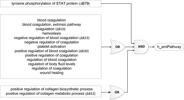
Logical circuit of h_amiPathway. Logical circuits described the relationships between GO biological processes and the MI pathway. We used multiple input single output logical gates AND and OR, where the GOBP were the inputs and h_amiPathway were the outputs. The extracted network of MI identified five major GOBP terms, including tyrosine phosphorylation of STAT protein (ΔB79), coagulation (ΔB10), negative and positive regulation of blood coagulation (ΔB23 & ΔB30), and positive regulation of collagen metabolic process (ΔB32), required to activate the MI pathway. The labels next to the name of the GOBP terms corresponded to the legend in Figure 6.
Figure 6.
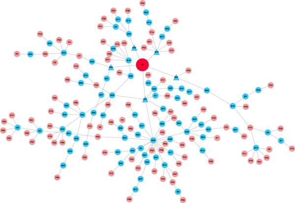
The extracted MI network. The acute MI pathway was colored in red while other pathways were colored in light red. Biological processes were represented in blue circles. GOBPs having direct impact on h_amiPathways were represented as blue triangles. A small branch of the network inside the blue rectangle involving coagulation was zoomed out for demonstration. Below are legends for selected pathways and processes (for the complete list of pathways and processes, see Supplemental Table 1). P3: h_amiPathway. P40: h_tgfbPathway. P58: hsa04350:TGF-betaSignalingPathway. P92: REACT_6844:Signaling by TGF beta. B10: coagulation. B30: positive regulation of blood coagulation. B32: positive regulation of collagen metabolic process. B44: positive regulation of protein kinase B signaling cascade. B58: regulation of kinase activity. B49: protein kinase cascade. B79: tyrosine phosphorylation of STAT protein.
Critical routes of the extracted MI network
The complete network of pathways and GOBP contains a huge amount of information although it could be overwhelming. We extracted the MI network and only retained the backbone to explore additional features that might not have been covered. Figure 6 showed the routes from MI-related pathways, represented as light red circles, to the h_amiPathway, whose color was in red, through biological processes in as blue circles. The graph was undirected, meaning some routes could be bidirectional. A small branch of the network inside the blue rectangle was zoomed out for illustration purpose. The complete list of pathways and GOBP can be viewed in Supplemental Table 1. We observed that all 7 pathways in those 6 branches needed to pass through coagulation to be connected to h_amiPathway. We found the cell cycle pathway, hsa04110:CellCycle, particularly interesting since the pathway was linked to h_fibrinolysisPathway, through cell growth. Heissig et. al (2007) showed that by deleting plasminogen, a classical fibrinolytic factor that controls hematopoietic stress response, in mice, hematopoietic stem cells were prevented from entering the cell cycle and undergoing multilineage differentiation after myelosuppression, leading to the death of the mice [24]. In other words, the plasminogen fibrinolytic pathway is crucial for hematopoietic regeneration. In another study, Heidt et al. (2014) showed that hematopoetic stem cells in the bone marrow could be activated by chronic stress, and further differentiated into increasing number of leukocytes. These leukocytes travel into the blood circulation and participate in the development of cardiovascular diseases [25]. Incidentally, fibrinolytic therapies have been used to enhance restoration of myocardial flow in the epcicardial infarct-related coronary artery [26]. Thus, it will be interesting to investigate the role of fibrinolysis and the increasing number of leukocytes in the cardiac remodelling post-MI and heart failures.
Discussion
In this study, we established a network by integrating GO biological processes and pathways from BioCarta, KEGG, and REACTOME enriched for MI-specific proteins using statistical measures and hierarchical structures. We examined the similarities between pathways and biological processes, and derived Boolean models of pathways in terms of standardized vocabulary with GOBP terms. This network can be used to explore critical routes that connect pathways and biological processes to the development of diseases or conditions. To demonstrate a functional interaction network, we started from the proteins in an MI-specific protein-protein interaction network we had previously constructed, acquired the enriched GO biological processes and pathways, constructed the GOBP graph and the functional pathway-process network, and determined the logical circuitry representing the involvement of GOBPs in pathways. The approach could be used with any set of genes or proteins, specific to any conditions or diseases, to develop additional features and visualizations.
This study presented three important results. First, we established a MI-specific functional biological pathway-process network, with demonstrated sub-networks shown in Figures 2 and 3. We standardized pathway descriptions by their connected GOBP terms, making it easier to compare differences and similarities between pathways, especially those with similar descriptions from different databases. We provided an example in section 3.2 with TGF-beta signaling pathways and pointed out the common and exclusive biological properties from BioCarta, KEGG and REACTOME. Second, we derived the relationships between GOBP terms based on the hierarchical structure defined in the GO Consortium and organized these terms into functional groups that could contribute differently to the pathways. For each pathway, GOBP terms that belonged to different functional groups should act simultaneously to activate the pathway, whereas only one process in a functional group was needed initiate the function. We used multiple input single output logical gates AND and OR, where the GOBP were the inputs and pathways were the outputs. We built two logic circuits corresponding to the MI and fibrinolysis pathways. It was shown that tyrosine phosphorylation of STAT protein, coagulation and regulation of collagen process were required to activate the MI pathway. We also provided experimental and clinical evidence for the association between the MI pathway and biological processes. Third, we illustrated a centralized version of the complete network of pathways and GOBP, providing insights of critical routes from and to the main pathway, h_amiPathway. Because MI was the major theme of this study, this extracted network allowed us to quickly visualize the connection between pathways before and after MI and their involvement in the changes in the post-MI myocardium.
Our results illustrated that using the functional biological pathway-process network is a promising method to identify biological properties of pathways under specific conditions. Pathways having similar descriptions encompassed both similar and diverse biological processes, indicating variation in their ability to share similar functional characteristics. The coverages of biological pathways can be increased with the incorporation of more biological processes and protein members, promoting more comprehensive pathways. As we discover and understand more about genes and proteins, the network helps to expand the participating genes or proteins in the pathways through the introduction of related genes in the GOBP. Pathways will be more comprehensive, leading to better knowledge of diseases. However, functional groups of GOBP terms based on hierarchical structures might need to be further evaluated for coherence. Moreover, GOBP functional groups might not have the same amount of contribution to the corresponding pathways; probabilistic Boolean models would allow more robustness in the face of uncertainty. In conclusion, we report here the establishment of the network of pathways and biological processes that can be used as a foundation to identify biological properties of pathways, providing interaction and visualization of biological systems at pathway level.
List of abbreviations used
AVRC: Arrhythmogenic Right Ventricular Cardiomyopathy
DCM: Dilated Cardiomyopathy
GO: Gene Ontology
GOBP: Gene Ontology Biological Process
HCM: Hypertrophic Cardiomyopathy
KEGG: Kyoto Encyclopaedia of Genes and Genomes
MI: Myocardial Infarction
MIPIN: Myocardial Infarction-Specific Protein-Protein Interaction Network
PANTHER: Protein Analysis Through Evolutionary Relationships
STAT: Signal Transducer and Activator of Transcription
TGF-beta: Transforming Growth Factor Beta
Competing interests
The authors declare that they have no competing interests.
Authors' contributions
Conceived and designed the experiments: NTN, MLL, and YFJ. Analyzed or reviewed the data: NTN, MLL, and YFJ. Contributed reagents/materials/analysis tools: NTN and YFJ. Wrote or edited the paper: NTN, MLL, and YFJ.
Supplementary Material
Pathways and GOBPs of MI network. This file contains names of pathways and GOBPs in the extracted network of MI with labels as displayed in Figure 6. Pathways were ordered alphabetically with prefix "P". GOBPs were ordered alphabetically with prefix "B".
Contributor Information
Nguyen T Nguyen, Email: wfw252@my.utsa.edu.
Merry L Lindsey, Email: mllindsey@umc.edu.
Yu-Fang Jin, Email: yufang.jin@utsa.edu.
Acknowledgements
We acknowledge funding support from the National Institutes of Health NHLBI from HHSN 268201000036C (N01-HV-00244) for the San Antonio Cardiovascular Proteomics Center and R01HL075360, and from the Biomedical Laboratory Research and Development Service of the Veterans Affairs Office of Research and Development Award 5I01BX000505 to MLL. The funders had no role in study design, data collection and analysis, decision to publish, or preparation of the manuscript.
Declarations
The publication costs for this article was funded by the corresponding author.
This article has been published as part of BMC Genomics Volume 16 Supplement 7, 2015: Selected articles from The International Conference on Intelligent Biology and Medicine (ICIBM) 2014: Genomics. The full contents of the supplement are available online at http://www.biomedcentral.com/bmcgenomics/supplements/16/S7.
References
- Kanehisa M, Goto S. KEGG: kyoto encyclopedia of genes and genomes. Nucleic Acids Res. 2000;28(1):27–30. doi: 10.1093/nar/28.1.27. [DOI] [PMC free article] [PubMed] [Google Scholar]
- Nishimura D. BioCarta. Biotech Software & Internet Report. 2001;2(3):117–120. doi: 10.1089/152791601750294344. [DOI] [Google Scholar]
- Croft D, Mundo AF, Haw R, Milacic M, Weiser J, Wu G, Caudy M, Garapati P, Gillespie M, Kamdar MR. et al. The Reactome pathway knowledgebase. Nucleic Acids Res. 2014;42(Database):D472–477. doi: 10.1093/nar/gkt1102. [DOI] [PMC free article] [PubMed] [Google Scholar]
- Thomas PD, Campbell MJ, Kejariwal A, Mi H, Karlak B, Daverman R, Diemer K, Muruganujan A, Narechania A. PANTHER: a library of protein families and subfamilies indexed by function. Genome Res. 2003;13(9):2129–2141. doi: 10.1101/gr.772403. [DOI] [PMC free article] [PubMed] [Google Scholar]
- Caspi R, Altman T, Billington R, Dreher K, Foerster H, Fulcher CA, Holland TA, Keseler IM, Kothari A, Kubo A. et al. The MetaCyc database of metabolic pathways and enzymes and the BioCyc collection of Pathway/Genome Databases. Nucleic Acids Res. 2014;42(Database):D459–471. doi: 10.1093/nar/gkt1103. [DOI] [PMC free article] [PubMed] [Google Scholar]
- Eichner J, Rosenbaum L, Wrzodek C, Haring HU, Zell A, Lehmann R. Integrated enrichment analysis and pathway-centered visualization of metabolomics, proteomics, transcriptomics, and genomics data by using the InCroMAP software. J Chromatogr B Analyt Technol Biomed Life Sci. 2014;966:77–82. doi: 10.1016/j.jchromb.2014.04.030. [DOI] [PubMed] [Google Scholar]
- Gruden K, Hren M, Herman A, Blejec A, Albrecht T, Selbig J, Bauer C, Schuchardt J, Or-Guil M, Zupancic K. et al. A "crossomics" study analysing variability of different components in peripheral blood of healthy caucasoid individuals. PLoS One. 2012;7(1):e28761. doi: 10.1371/journal.pone.0028761. [DOI] [PMC free article] [PubMed] [Google Scholar]
- Amiour N, Imbaud S, Clement G, Agier N, Zivy M, Valot B, Balliau T, Armengaud P, Quillere I, Canas R. et al. The use of metabolomics integrated with transcriptomic and proteomic studies for identifying key steps involved in the control of nitrogen metabolism in crops such as maize. Journal of Experimental Botany. 2012;63(14):5017–5033. doi: 10.1093/jxb/ers186. [DOI] [PubMed] [Google Scholar]
- Babur O, Dogrusoz U, Cakir M, Aksoy BA, Schultz N, Sander C, Demir E. Integrating biological pathways and genomic profiles with ChiBE 2. BMC Genomics. 2014;15:642. doi: 10.1186/1471-2164-15-642. [DOI] [PMC free article] [PubMed] [Google Scholar]
- Ashburner M, Ball CA, Blake JA, Botstein D, Butler H, Cherry JM, Davis AP, Dolinski K, Dwight SS, Eppig JT. et al. Gene ontology: tool for the unification of biology. The Gene Ontology Consortium. Nat Genet. 2000;25(1):25–29. doi: 10.1038/75556. [DOI] [PMC free article] [PubMed] [Google Scholar]
- Nguyen NT, Zhang X, Wu C, Lange RA, Chilton RJ, Lindsey ML, Jin YF. Integrative computational and experimental approaches to establish a post-myocardial infarction knowledge map. PLoS Comput Biol. 2014;10(3):e1003472. doi: 10.1371/journal.pcbi.1003472. [DOI] [PMC free article] [PubMed] [Google Scholar]
- Huang dW, Sherman BT, Lempicki RA. Systematic and integrative analysis of large gene lists using DAVID bioinformatics resources. Nat Protoc. 2009;4(1):44–57. doi: 10.1038/nprot.2008.211. [DOI] [PubMed] [Google Scholar]
- Cohen J. A Coefficient of Agreement for Nominal Scales. Educ Psychol Meas. 1960;20(1):37–46. doi: 10.1177/001316446002000104. [DOI] [Google Scholar]
- Carlson M. GO.db: A set of annotation maps describing the entire Gene Ontology. 2013.
- Csardi G NT. InterJournal, Complex Systems(1695); 2006. The igraph software package for complex network research. [Google Scholar]
- Gould L, Reddy CV, Swamy CR, Oh KC, Kim SG. Decline of serum phosphorus in acute myocardial infarction. Angiology. 1979;30(4):219–222. doi: 10.1177/000331977903000401. [DOI] [PubMed] [Google Scholar]
- Aronson D, Kapeliovich M, Hammerman H, Dragu R. The Relation between Serum Phosphorus Levels and Clinical Outcomes after Acute Myocardial Infarction. PLoS One. 2013;8(3) doi: 10.1371/journal.pone.0058348. [DOI] [PMC free article] [PubMed] [Google Scholar]
- Vaidyanathan D, Venkatesan S, Ramadesikan VK. Serum phosphate in acute myocardial infarction. Indian J Physiol Pharmacol. 2000;44(2):225–228. [PubMed] [Google Scholar]
- Knapp M, Baranowski M, Czarnowski D, Lisowska A, Zabielski P, Gorski J, Musial W. Plasma sphingosine-1-phosphate concentration is reduced in patients with myocardial infarction. Med Sci Monitor. 2009;15(9):Cr490–Cr493. [PubMed] [Google Scholar]
- Darsee JR, Heymsfield SB, Nutter DO. Hypertrophic cardiomyopathy and human leukocyte antigen linkage: differentiation of two forms of hypertrophic cardiomyopathy. N Engl J Med. 1979;300(16):877–882. doi: 10.1056/NEJM197904193001602. [DOI] [PubMed] [Google Scholar]
- Nutter DO, Heymsfield SB, Glenn JF. Retraction. Darsee JR, Heymsfield SB, Nutter DO. Hypertrophic cardiomyopathy and human leukocyte antigen linkage: differentiation of two forms of hypertrophic cardiomyopathy. N Engl J Med 1979;300:877-82. N Engl J Med. 1983;308(23):1400. doi: 10.1056/nejm198306093082307. [DOI] [PubMed] [Google Scholar]
- Lozano MD, Rubocki RJ, Wilson JE, McManus BM, Wisecarver JL. Human leukocyte antigen class II associations in patients with idiopathic dilated cardiomyopathy. Myocarditis Treatment Trial Investigators. J Card Fail. 1997;3(2):97–103. doi: 10.1016/S1071-9164(97)90041-5. [DOI] [PubMed] [Google Scholar]
- Li X, Luo R, Jiang R, Chen R, Hua W. Human leukocyte antigen-DQ beta 1 chain (DQB1) gene polymorphisms are associated with dilated cardiomyopathy: a systematic review and meta-analysis. Heart Lung. 2012;41(4):360–367. doi: 10.1016/j.hrtlng.2012.01.005. [DOI] [PubMed] [Google Scholar]
- Heissig B, Lund LR, Akiyama H, Ohki M, Morita Y, Romer J, Nakauchi H, Okumura K, Ogawa H, Werb Z. et al. The plasminogen fibrinolytic pathway is required for hematopoietic regeneration. Cell Stem Cell. 2007;1(6):658–670. doi: 10.1016/j.stem.2007.10.012. [DOI] [PMC free article] [PubMed] [Google Scholar]
- Heidt T, Sager HB, Courties G, Dutta P, Iwamoto Y, Zaltsman A, von Zur Muhlen C, Bode C, Fricchione GL, Denninger J. et al. Chronic variable stress activates hematopoietic stem cells. Nat Med. 2014;20(7):754–758. doi: 10.1038/nm.3589. [DOI] [PMC free article] [PubMed] [Google Scholar]
- Armstrong PW, Collen D, Antman E. Fibrinolysis for acute myocardial infarction: the future is here and now. Circulation. 2003;107(20):2533–2537. doi: 10.1161/01.CIR.0000072930.64775.DC. [DOI] [PubMed] [Google Scholar]
- Gilutz H, Siegel Y, Paran E, Cristal N, Quastel MR. Alpha 1-antitrypsin in acute myocardial infarction. Br Heart J. 1983;49(1):26–29. doi: 10.1136/hrt.49.1.26. [DOI] [PMC free article] [PubMed] [Google Scholar]
- Toldo S, Seropian IM, Mezzaroma E, Van Tassell BW, Salloum FN, Lewis EC, Voelkel N, Dinarello CA, Abbate A. Alpha-1 antitrypsin inhibits caspase-1 and protects from acute myocardial ischemia-reperfusion injury. J Mol Cell Cardiol. 2011;51(2):244–251. doi: 10.1016/j.yjmcc.2011.05.003. [DOI] [PubMed] [Google Scholar]
- Gonzalez-Nunez M, Munoz-Felix JM, Lopez-Novoa JM. The ALK-1/Smad1 pathway in cardiovascular physiopathology. A new target for therapy? Biochim Biophys Acta. 2013;1832(10):1492–1510. doi: 10.1016/j.bbadis.2013.05.016. [DOI] [PubMed] [Google Scholar]
- Tian F, Zhou AX, Smits AM, Larsson E, Goumans MJ, Heldin CH, Boren J, Akyurek LM. Endothelial cells are activated during hypoxia via endoglin/ALK-1/SMAD1/5 signaling in vivo and in vitro. Biochem Biophys Res Commun. 2010;392(3):283–288. doi: 10.1016/j.bbrc.2009.12.170. [DOI] [PubMed] [Google Scholar]
- Shimoda Y, Satoh M, Nakamura M, Akatsu T, Hiramori K. Activated tumour necrosis factor-alpha shedding process is associated with in-hospital complication in patients with acute myocardial infarction. Clinical science. 2005;108(4):339–347. doi: 10.1042/CS20040229. [DOI] [PubMed] [Google Scholar]
- Satoh M, Ishikawa Y, Itoh T, Minami Y, Takahashi Y, Nakamura M. The expression of TNF-alpha converting enzyme at the site of ruptured plaques in patients with acute myocardial infarction. Eur J Clin Invest. 2008;38(2):97–105. doi: 10.1111/j.1365-2362.2007.01912.x. [DOI] [PubMed] [Google Scholar]
- Nakayama T, Soma M, Saito S, Honye J, Sato M, Aoi N, Kosuge K, Haketa A, Kanmatsuse K, Kokubun S. Missense mutation of exon 3 in the type A human natriuretic peptide receptor gene is associated with myocardial infarction. Medical science monitor : international medical journal of experimental and clinical research. 2003;16(12):CR505–510. [PubMed] [Google Scholar]
- Nakanishi M, Saito Y, Kishimoto I, Harada M, Kuwahara K, Takahashi N, Kawakami R, Nakagawa Y, Tanimoto K, Yasuno S. et al. Role of natriuretic peptide receptor guanylyl cyclase-A in myocardial infarction evaluated using genetically engineered mice. Hypertension. 2005;46(2):441–447. doi: 10.1161/01.HYP.0000173420.31354.ef. [DOI] [PubMed] [Google Scholar]
- McQueen MJ, Hawken S, Wang X, Ounpuu S, Sniderman A, Probstfield J, Steyn K, Sanderson JE, Hasani M, Volkova E. et al. Lipids, lipoproteins, and apolipoproteins as risk markers of myocardial infarction in 52 countries (the INTERHEART study): a case-control study. Lancet. 2008;372(9634):224–233. doi: 10.1016/S0140-6736(08)61076-4. [DOI] [PubMed] [Google Scholar]
- Kamstrup PR, Tybjaerg-Hansen A, Nordestgaard BG. Lipoprotein(a) and risk of myocardial infarction--genetic epidemiologic evidence of causality. Scand J Clin Lab Invest. 2011;71(2):87–93. doi: 10.3109/00365513.2010.550311. [DOI] [PubMed] [Google Scholar]
- Jasmin JF, Rengo G, Lymperopoulos A, Gupta R, Eaton GJ, Quann K, Gonzales DM, Mercier I, Koch WJ, Lisanti MP. Caveolin-1 deficiency exacerbates cardiac dysfunction and reduces survival in mice with myocardial infarction. Am J Physiol Heart Circ Physiol. 2011;300(41):H1274–1281. doi: 10.1152/ajpheart.01173.2010. [DOI] [PMC free article] [PubMed] [Google Scholar]
- Shivshankar P, Halade GV, Calhoun C, Escobar GP, Mehr AJ, Jimenez F, Martinez C, Bhatnagar H, Mjaatvedt CH, Lindsey ML, Caveolin-1 deletion exacerbates cardiac interstitial fibrosis by promoting M2 macrophage activation in mice after myocardial infarction. J Mol Cell Cardiol. 2014. pp. 84–93. [DOI] [PMC free article] [PubMed]
- Juhan-Vague I, Morange PE, Aubert H, Henry M, Aillaud MF, Alessi MC, Samnegard A, Hawe E, Yudkin J, Margaglione M. et al. Plasma thrombin-activatable fibrinolysis inhibitor antigen concentration and genotype in relation to myocardial infarction in the north and south of Europe. Arterioscler Thromb Vasc Biol. 2002;22(5):867–873. doi: 10.1161/01.ATV.0000015445.22243.F4. [DOI] [PubMed] [Google Scholar]
- Kraft P, Schwarz T, Meijers JC, Stoll G, Kleinschnitz C. Thrombin-activatable fibrinolysis inhibitor (TAFI) deficient mice are susceptible to intracerebral thrombosis and ischemic stroke. PLoS One. 2010;5(7):e11658. doi: 10.1371/journal.pone.0011658. [DOI] [PMC free article] [PubMed] [Google Scholar]
- Pellikka M, Narhi L, Perola M, Penttila A, Karhunen PJ, Mikkelsson J. Platelet GPIbalpha, GPIV and vWF polymorphisms and fatal pre-hospital MI among middle-aged men. Journal of thrombosis and thrombolysis. 2008;26(2):91–96. doi: 10.1007/s11239-007-0072-2. [DOI] [PubMed] [Google Scholar]
- Knowles JW, Wang H, Itakura H, Southwick A, Myers RM, Iribarren C, Fortmann SP, Go AS, Quertermous T, Hlatky MA. Association of polymorphisms in platelet and hemostasis system genes with acute myocardial infarction. Am Heart J. 2007;154(6):1052–1058. doi: 10.1016/j.ahj.2007.05.021. [DOI] [PMC free article] [PubMed] [Google Scholar]
- Siegerink B, Algra A, Rosendaal FR. Genetic variants of coagulation factor XIII and the risk of myocardial infarction in young women. Br J Haematol. 2009;146(4):459–461. doi: 10.1111/j.1365-2141.2009.07805.x. [DOI] [PubMed] [Google Scholar]
- Rallidis LS, Politou M, Komporozos C, Panagiotakos DB, Belessi CI, Travlou A, Lekakis J, Kremastinos DT. Factor XIII Val34Leu polymorphism and the risk of myocardial infarction under the age of 36 years. Thromb Haemost. 2008;99(6):1085–1089. doi: 10.1160/TH07-12-0755. [DOI] [PubMed] [Google Scholar]
- Doggen CJ, Rosendaal FR, Meijers JC. Levels of intrinsic coagulation factors and the risk of myocardial infarction among men: Opposite and synergistic effects of factors XI and XII. Blood. 2006;108(13):4045–4051. doi: 10.1182/blood-2005-12-023697. [DOI] [PubMed] [Google Scholar]
- Minnema MC, Peters RJ, de Winter R, Lubbers YP, Barzegar S, Bauer KA, Rosenberg RD, Hack CE, ten Cate H. Activation of clotting factors XI and IX in patients with acute myocardial infarction. Arterioscler Thromb Vasc Biol. 2000;20(11):2489–2493. doi: 10.1161/01.ATV.20.11.2489. [DOI] [PubMed] [Google Scholar]
- Mannucci PM, Asselta R, Duga S, Guella I, Spreafico M, Lotta L, Merlini PA, Peyvandi F, Kathiresan S, Ardissino D. The association of factor V Leiden with myocardial infarction is replicated in 1880 patients with premature disease. J Thromb Haemost. 2010;8(10):2116–2121. doi: 10.1111/j.1538-7836.2010.03982.x. [DOI] [PubMed] [Google Scholar]
- Dowaidar M, Settin A. Risk of myocardial infarction related to factor V Leiden mutation: a meta-analysis. Genet Test Mol Biomarkers. 2010;14(4):493–498. doi: 10.1089/gtmb.2010.0017. [DOI] [PubMed] [Google Scholar]
- Mannila MN, Lovely RS, Kazmierczak SC, Eriksson P, Samnegard A, Farrell DH, Hamsten A, Silveira A. Elevated plasma fibrinogen gamma' concentration is associated with myocardial infarction: effects of variation in fibrinogen genes and environmental factors. J Thromb Haemost. 2007;5(4):766–773. doi: 10.1111/j.1538-7836.2007.02406.x. [DOI] [PubMed] [Google Scholar]
- Jacquemin B, Antoniades C, Nyberg F, Plana E, Muller M, Greven S, Salomaa V, Sunyer J, Bellander T, Chalamandaris AG. et al. Common genetic polymorphisms and haplotypes of fibrinogen alpha, beta, and gamma chains affect fibrinogen levels and the response to proinflammatory stimulation in myocardial infarction survivors: the AIRGENE study. J Am Coll Cardiol. 2008;52(11):941–952. doi: 10.1016/j.jacc.2008.06.016. [DOI] [PubMed] [Google Scholar]
- Ulrich MM, Janssen AM, Daemen MJ, Rappaport L, Samuel JL, Contard F, Smits JF, Cleutjens JP. Increased expression of fibronectin isoforms after myocardial infarction in rats. J Mol Cell Cardiol. 1997;29(9):2533–2543. doi: 10.1006/jmcc.1997.0486. [DOI] [PubMed] [Google Scholar]
- van Dijk A, Niessen HW, Ursem W, Twisk JW, Visser FC, van Milligen FJ. Accumulation of fibronectin in the heart after myocardial infarction: a putative stimulator of adhesion and proliferation of adipose-derived stem cells. Cell Tissue Res. 2008;332(2):289–298. doi: 10.1007/s00441-008-0573-0. [DOI] [PMC free article] [PubMed] [Google Scholar]
- Aleil B, Mossard JM, Wiesel ML, Lanza F, Cazenave JP. Increased plasma levels of soluble platelet glycoprotein V in patients with acute myocardial infarction. J Thromb Haemost. 2003;1(8):1846–1847. doi: 10.1046/j.1538-7836.2003.00319.x. [DOI] [PubMed] [Google Scholar]
- Morel O, Hugel B, Jesel L, Lanza F, Douchet MP, Zupan M, Chauvin M, Cazenave JP, Freyssinet JM, Toti F. Sustained elevated amounts of circulating procoagulant membrane microparticles and soluble GPV after acute myocardial infarction in diabetes mellitus. Thromb Haemost. 2004;91(2):345–353. doi: 10.1160/TH03-05-0294. [DOI] [PubMed] [Google Scholar]
- Tekin D, Dursun AD, Xi L. Hypoxia inducible factor 1 (HIF-1) and cardioprotection. Acta pharmacologica Sinica. 2010;31(9):1085–1094. doi: 10.1038/aps.2010.132. [DOI] [PMC free article] [PubMed] [Google Scholar]
- Kido M, Du L, Sullivan CC, Li X, Deutsch R, Jamieson SW, Thistlethwaite PA. Hypoxia-inducible factor 1-alpha reduces infarction and attenuates progression of cardiac dysfunction after myocardial infarction in the mouse. J Am Coll Cardiol. 2005;46(11):2116–2124. doi: 10.1016/j.jacc.2005.08.045. [DOI] [PubMed] [Google Scholar]
- Wouters D, Wagenaar-Bos I, van Ham M, Zeerleder S. C1 inhibitor: just a serine protease inhibitor? New and old considerations on therapeutic applications of C1 inhibitor. Expert opinion on biological therapy. 2008;8(8):1225–1240. doi: 10.1517/14712598.8.8.1225. [DOI] [PubMed] [Google Scholar]
- Rennie JA, Crawford GP, Ogston D. Changes in protease inhibitors after acute myocardial infarction. J Clin Pathol. 1976;29(7):639–641. doi: 10.1136/jcp.29.7.639. [DOI] [PMC free article] [PubMed] [Google Scholar]
- Szkodzinski J, Hudzik B, Osuch M, Romanowski W, Szygula-Jurkiewicz B, Polonski L, Zubelewicz-Szkodzinska B. Serum concentrations of interleukin-4 and interferon-gamma in relation to severe left ventricular dysfunction in patients with acute myocardial infarction undergoing percutaneous coronary intervention. Heart and vessels. 2011;26(4):399–407. doi: 10.1007/s00380-010-0076-2. [DOI] [PubMed] [Google Scholar]
- Yang ZQ, Xu YQ, Linden J, Kron IL, French BA. Reduced Myocardial Infarct Size in Interferon-Gamma Knock-out Mice Implicates CD4+T Cells in Reperfusion Injury. Circulation. 2009;120(18):S1165–S1166. [Google Scholar]
- Nawata J, Ohno I, Isoyama S, Suzuki J, Miura S, Ikeda J, Shirato K. Differential expression of alpha 1, alpha 3 and alpha 5 integrin subunits in acute and chronic stages of myocardial infarction in rats. Cardiovascular Research. 1999;43(2):371–381. doi: 10.1016/S0008-6363(99)00117-0. [DOI] [PubMed] [Google Scholar]
- Sahul Z, Dione DP, Dobrucki L, Kalinowski L, Brennan M, Mekkaoui C, Cavaliere P, Hawley C, Hu X, Haramis H. et al. Targeted alpha-v integrin imaging defines spatial and temporal changes in the angiogenic process post myocardial infarction. Circulation. 2006;114(18):499–499. [Google Scholar]
- Siegerink B, Rosendaal FR, Algra A. High-molecular-weight kininogen and the risk of a myocardial infarction and ischemic stroke in young women: the RATIO case-control study. Journal of Thrombosis and Haemostasis. 2012;10(11):2409–2412. doi: 10.1111/j.1538-7836.2012.04927.x. [DOI] [PubMed] [Google Scholar]
- Ito H, Hayashi I, Izumi T, Majima M. Bradykinin inhibits development of myocardial infarction through B2 receptor signalling by increment of regional blood flow around the ischaemic lesions in rats. Br J Pharmacol. 2003;138(1):225–233. doi: 10.1038/sj.bjp.0705013. [DOI] [PMC free article] [PubMed] [Google Scholar]
- Xie Y, Chen J, Han P, Yang P, Hou J, Kang YJ. Immunohistochemical detection of differentially localized up-regulation of lysyl oxidase and down-regulation of matrix metalloproteinase-1 in rhesus monkey model of chronic myocardial infarction. Exp Biol Med (Maywood) 2012;237(7):853–859. doi: 10.1258/ebm.2012.012070. [DOI] [PubMed] [Google Scholar]
- Lerman RH, Apstein CS, Kagan HM, Osmers EL, Chichester CO, Vogel WM, Connelly CM, Steffee WP. Myocardial healing and repair after experimental infarction in the rabbit. Circ Res. 1983;53(3):378–388. doi: 10.1161/01.RES.53.3.378. [DOI] [PubMed] [Google Scholar]
- Antoniak S, Pawlinski R, Mackman N. Protease-activated receptors and myocardial infarction. IUBMB life. 2011;63(6):383–389. doi: 10.1002/iub.441. [DOI] [PMC free article] [PubMed] [Google Scholar]
- Zhong B, Wang DH. Protease-activated receptor 2-mediated protection of myocardial ischemia-reperfusion injury: role of transient receptor potential vanilloid receptors. Am J Physiol Regul Integr Comp Physiol. 2009;297(6):R1681–1690. doi: 10.1152/ajpregu.90746.2008. [DOI] [PMC free article] [PubMed] [Google Scholar]
- Seqqat R, Rafiq K, Hanscom M, Kunapuli SP, Steinberg SF, Houser SR, Sabri A. Protease activated receptor-4 regulates post-infarction ventricular remodeling and cardiac function. Circulation. 2007;116(16):45–45. [Google Scholar]
- Zymek P, Bujak M, Chatila K, Cieslak A, Thakker G, Entman ML, Frangogiannis NG. The role of platelet-derived growth factor signaling in healing myocardial infarcts. J Am Coll Cardiol. 2006;48(11):2315–2323. doi: 10.1016/j.jacc.2006.07.060. [DOI] [PubMed] [Google Scholar]
- Edelberg JM, Lee SH, Kaur M, Tang L, Feirt NM, McCabe S, Bramwell O, Wong SC, Hong MK. Platelet-derived growth factor-AB limits the extent of myocardial infarction in a rat model: feasibility of restoring impaired angiogenic capacity in the aging heart. Circulation. 2002;105(5):608–613. doi: 10.1161/hc0502.103672. [DOI] [PubMed] [Google Scholar]
- Dymicka-Piekarska V, Kemona H, Mantur M, Stogowski A, Kemona-Chetnik I, Bychowski J. Platelet factor 4 as a marker of platelet activation in patients with acute myocardial infarction. Roczniki Akademii Medycznej w Bialymstoku. 2000;45:96–103. [PubMed] [Google Scholar]
- Kuijpers PM, Hamulyak K, Strik JJ, Wellens HJ, Honig A. Beta-thromboglobulin and platelet factor 4 levels in post-myocardial infarction patients with major depression. Psychiatry research. 2002;109(2):207–210. doi: 10.1016/S0165-1781(02)00017-3. [DOI] [PubMed] [Google Scholar]
- Fedi S, Sofi F, Brogi D, Tellini I, Cesari F, Sestini I, Gazzini A, Comeglio M, Abbate R, Gensini GF. Low protein Z plasma levels are independently associated with acute coronary syndromes. Thromb Haemost. 2003;90(6):1173–1178. doi: 10.1160/TH03-04-0237. [DOI] [PubMed] [Google Scholar]
- Le Cam-Duchez V, Soria C, Sollier CBD, Borg JY, Coudert M, Montalescot G, Esposito G, Drouet L, Collet JP. Rare genotypes of protein Z gene are a risk factor for premature myocardial infarction but not protein Z plasma level. Thromb Haemostasis. 2009;102(1):131–136. doi: 10.1160/TH09-01-0007. [DOI] [PubMed] [Google Scholar]
- Dobaczewski M, Bujak M, Li N, Gonzalez-Quesada C, Mendoza LH, Wang XF, Frangogiannis NG. Smad3 Signaling Critically Regulates Fibroblast Phenotype and Function in Healing Myocardial Infarction. Circulation Research. 2010;107(3):418–U176. doi: 10.1161/CIRCRESAHA.109.216101. [DOI] [PMC free article] [PubMed] [Google Scholar]
- Deten A, Holzl A, Leicht M, Barth W, Zimmer HG. Changes in extracellular matrix and in transforming growth factor beta isoforms after coronary artery ligation in rats. Journal of Molecular and Cellular Cardiology. 2001;33(6):1191–1207. doi: 10.1006/jmcc.2001.1383. [DOI] [PubMed] [Google Scholar]
- Singla DK, Singla RD, Lamm S, Glass C. TGF-beta 2 treatment enhances cytoprotective factors released from embryonic stem cells and inhibits apoptosis in infarcted myocardium. Am J Physiol-Heart C. 2011;300(4):H1442–H1450. doi: 10.1152/ajpheart.00917.2010. [DOI] [PMC free article] [PubMed] [Google Scholar]
- Bujak M, Frangogiannis NG. The role of TGF-beta signaling in myocardial infarction and cardiac remodeling. Cardiovasc Res. 2007;74(2):184–195. doi: 10.1016/j.cardiores.2006.10.002. [DOI] [PMC free article] [PubMed] [Google Scholar]
- Okada H, Takemura G, Kosai KI, Li YW, Takahashi T, Esaki M, Yuge K, Miyata S, Maruyama R, Mikami A. et al. Postinfarction gene therapy against transforming growth factor-beta signal modulates infarct tissue dynamics and attenuates left ventricular remodeling and heart failure. Circulation. 2005;111(19):2430–2437. doi: 10.1161/01.CIR.0000165066.71481.8E. [DOI] [PubMed] [Google Scholar]
- Ireland H, Kunz G, Kyriakoulis K, Stubbs PJ, Lane DA. Thrombomodulin gene mutations associated with myocardial infarction. Circulation. 1997;96(1):15–18. doi: 10.1161/01.CIR.96.1.15. [DOI] [PubMed] [Google Scholar]
- Chao TH, Li YH, Chen JH, Wu HL, Shi GY, Tsai WC, Chen PS, Liu PY. Relation of thrombomodulin gene polymorphisms to acute myocardial infarction in patients <= 50 years of age. American Journal of Cardiology. 2004;93(2):204–207. doi: 10.1016/j.amjcard.2003.09.039. [DOI] [PubMed] [Google Scholar]
- Sezaki S, Hirohata S, Iwabu A, Nakamura K, Toeda K, Miyoshi T, Yamawaki H, Demircan K, Kusachi S, Shiratori Y. et al. Thrombospondin-1 is induced in rat myocardial infarction and its induction is accelerated by ischemia/reperfusion. Exp Biol Med. 2005;230(9):621–630. doi: 10.1177/153537020523000904. [DOI] [PubMed] [Google Scholar]
- Zwicker JI, Peyvandi F, Palla R, Lombardi R, Canciani MT, Cairo A, Ardissino D, Bernardinelli L, Bauer KA, Lawler J. et al. The thrombospondin-1 N700S polymorphism is associated with early myocardial infarction without altering von Willebrand factor multimer size. Blood. 2006;108(4):1280–1283. doi: 10.1182/blood-2006-04-015701. [DOI] [PMC free article] [PubMed] [Google Scholar]
- Stavropoulou A, Philippou A, Halapas A, Sourla A, Pissimissis N, Koutsilieris M. uPA, uPAR and TGFbeta(1) expression during early and late post myocardial infarction period in rat myocardium. In vivo. 2010;24(5):647–652. [PubMed] [Google Scholar]
- Minami E, Castellani C, Malchodi L, Deem J, Bertko K, Meznarich J, Dishmon M, Murry CE, Stempien-Otero A. The role of macrophage-derived urokinase plasminogen activator in myocardial infarct repair: urokinase attenuates ventricular remodeling. J Mol Cell Cardiol. 2010;49(3):516–524. doi: 10.1016/j.yjmcc.2010.03.022. [DOI] [PMC free article] [PubMed] [Google Scholar]
- Huebener P, Abou-Khamis T, Zymek P, Bujak M, Ying X, Chatila K, Haudek S, Thakker G, Frangogiannis NG. CD44 is critically involved in infarct healing by regulating the inflammatory and fibrotic response. J Immunol. 2008;180(4):2625–2633. doi: 10.4049/jimmunol.180.4.2625. [DOI] [PubMed] [Google Scholar]
- Huang SS, Huang PH, Chen YH, Sung SH, Chiang KH, Chen JW, Lin SJ. Plasma heparin cofactor II activity is an independent predictor of future cardiovascular events in patients after acute myocardial infarction. Coron Artery Dis. 2008;19(8):597–602. doi: 10.1097/MCA.0b013e3283155579. [DOI] [PubMed] [Google Scholar]
- Voight BF, Peloso GM, Orho-Melander M, Frikke-Schmidt R, Barbalic M, Jensen MK, Hindy G, Holm H, Ding EL, Johnson T. et al. Plasma HDL cholesterol and risk of myocardial infarction: a mendelian randomisation study. Lancet. 2012;380(9841):572–580. doi: 10.1016/S0140-6736(12)60312-2. [DOI] [PMC free article] [PubMed] [Google Scholar]
- Camors E, Monceau V, Charlemagne D. Annexins and Ca2+ handling in the heart. Cardiovasc Res. 2005;65(4):793–802. doi: 10.1016/j.cardiores.2004.11.010. [DOI] [PubMed] [Google Scholar]
- Spencer JA, Hacker SL, Davis EC, Mecham RP, Knutsen RH, Li DY, Gerard RD, Richardson JA, Olson EN, Yanagisawa H. Altered vascular remodeling in fibulin-5-deficient mice reveals a role of fibulin-5 in smooth muscle cell proliferation and migration. Proc Natl Acad Sci USA. 2005;102(8):2946–2951. doi: 10.1073/pnas.0500058102. [DOI] [PMC free article] [PubMed] [Google Scholar]
- Galli D, Innocenzi A, Staszewsky L, Zanetta L, Sampaolesi M, Bai A, Martinoli E, Carlo E, Balconi G, Fiordaliso F. et al. Mesoangioblasts, vessel-associated multipotent stem cells, repair the infarcted heart by multiple cellular mechanisms: a comparison with bone marrow progenitors, fibroblasts, and endothelial cells. Arterioscler Thromb Vasc Biol. 2005;25(4):692–697. doi: 10.1161/01.ATV.0000156402.52029.ce. [DOI] [PubMed] [Google Scholar]
- Sawaki D, Suzuki T, Aizawa K, Matsumura T, Munemasa Y, Ishida J, Fridman SL, Nagai R. KLF6 Modulates Recruitment and Polarization of Inflammatory Cells Through Cardiomyocytes in Initiation of Cardiac Fibrosis. Circulation. 2011;124(21) [Google Scholar]
- Cornelissen I, Palmer D, David T, Wilsbacher L, Concengco C, Conley P, Pandey A, Coughlin SR. Roles and interactions among protease-activated receptors and P2ry12 in hemostasis and thrombosis. Proc Natl Acad Sci USA. 2010;107(43):18605–18610. doi: 10.1073/pnas.1013309107. [DOI] [PMC free article] [PubMed] [Google Scholar]
- Brenner B, Sanchez-Vega B, Wu SM, Lanir N, Stafford DV, Solera J. A missense mutation in gamma-glutamyl carboxylase gene causes combined deficiency of all vitamin K-dependent blood coagulation factors. Blood. 1998;92(12):4554–4559. [PubMed] [Google Scholar]
Associated Data
This section collects any data citations, data availability statements, or supplementary materials included in this article.
Supplementary Materials
Pathways and GOBPs of MI network. This file contains names of pathways and GOBPs in the extracted network of MI with labels as displayed in Figure 6. Pathways were ordered alphabetically with prefix "P". GOBPs were ordered alphabetically with prefix "B".