Abstract
The minimal invasiveness of endoscopic submucosal dissection (ESD) prompted us to apply this technique to large-size early esophageal squamous cell carcinoma and Barrett’s adenocarcinoma, despite the limitations in the study population and surveillance duration. A post-ESD ulceration of greater than three-fourths of esophageal circumference was advocated as an important risk factor for refractory strictures that require several sessions of dilation therapy. Most of the preoperative conditions are asymptomatic, but dilatation treatment for dysphagia associated with the stricture has potential risks of severe complications and a worsening of quality of life. Possible mechanisms of dysphasia were demonstrated based on dysmotility and pathological abnormalities at the site: (1) delayed mucosal healing; (2) severe inflammation and disorganized fibrosis with abundant extracellular matrices in the submucosa; and (3) atrophy in the muscularis proper. However, reports on the administration of anti-scarring agents, preventive dilation therapies, and regenerative medicine demonstrated limited success in stricture prevention, and there were discrepancies in the study designs and protocols of these reports. The development and consequent long-term assessments of new prophylactic technologies on the promotion of wound healing and control of the inflammatory/tumor microenvironment will require collaboration among various research fields because of the limited accuracy of preoperative staging and high-risk of local recurrence.
Keywords: Esophageal stricture, Dysphasia, Endoscopic submucosal dissection
Core tip: The number of cases of refractory post-endoscopic submucosal dissection (ESD) strictures will increase as the indications for ESD expand. Dysphagia related to the stricture is primarily treated using repeated dilatation treatments, which risk complications and a diminished quality of life. Dysmotility and inflammation-associated disorders at the site may reflect the mechanisms of dysphasia. However, anti-scarring agent administration, endoscopic modalities, and regenerative medicine have limited effects. The development and subsequent long-term assessment of new technologies for the prevention and control of carcinogenesis will be required based on the limited accuracy of preoperative staging and the risk of local recurrence.
INTRODUCTION
The management of superficial esophageal neoplasms (SENs) has shifted from surgery to endoscopic treatment with recent developments in endoscopic technologies because of minimal invasiveness and positive outcomes[1,2]. Notably, endoscopic submucosal dissection (ESD) achieves optimal histopathological staging using en bloc resection compared to conventional endoscopic mucosal resection (EMR)[3-5]. Recently, ESD was successfully applied worldwide in large early esophageal squamous cell carcinoma (ESCC) and Barrett’s adenocarcinoma[6-12].
However, there are no established criteria for curability of ESD for larger-sized ESCC and Barrett’s adenocarcinoma. Existing treatment guidelines state that ESCC that are limited to intraepithelial or lamina propria mucosae (LPM) with nominal lymph node metastasis are candidates for endoscopic treatment[13], but the rates of lymph node metastases of ESCC invading the muscularis mucosae (MM) or the upper third of the submucosa (SM1) were 9% or 19%, respectively[14]. Previous retrospective studies have demonstrated that the curability of ESD for large-sized high-grade intraepithelial neoplasms of the esophagus or superficial ESCC were equivalent to surgical resection[7,15], which expanded the indication for endoscopic treatment of SENs[16,17]. However, successful resection of these lesions using ESD was reported in relatively small numbers of subjects with limited-term surveillance. EMR followed by radiofrequency ablation therapy is mainly performed in patients with early stage Barrett’s-associated adenocarcinoma, as an alternative to esophagectomy in Western countries with promising results[18,19]. EMR has limitations in resectable size, which results in a higher risk of local recurrence[13], and ESD has been used to remove large carcinomatous lesions concomitant with background Barrett’s mucosa with malignant potential[9,10,20]. However, whether it is reasonable to evaluate the curability for Barrett’s adenocarcinoma that invades SM1 using the same criteria for gastric cancer or ESCC remains controversial[13]. Therefore, further prospective studies using large numbers of subjects and long-term surveillance are required to confirm the usefulness and safety of ESD.
Many studies have suggested that strictures after endoscopic treatment for large-sized SENs might be refractory to dilation therapy (Figure 1). The occurrence of refractory strictures after non-surgical treatments was significantly higher than the occurrence after surgery, but the overall rates of strictures after esophagogastrectomy, chemo-radiation therapy (CRT), and endoscopic resection were 1.6%-25%, 3.3%-40%, and 6.0%-18%, respectively[1,7,15,21-23]. Most strictures that occurred after endoscopic treatment were classified as complex strictures, e.g., (1) a simple stricture that is a short, focal, and symmetric stenosis with a luminal diameter that allows the passage of a standard endoscope; and (2) complex strictures that are defined as having one or more of the following features: a severely narrowed luminal diameter ≤ 12 mm, length ≥ 20 mm, angulation, or asymmetry[24,25]. Therefore, it is important to prevent severe stricture of the esophagus from occurring after endoscopic treatment. Several retrospective single-center studies identified more than three-fourths of the circumferential extent of a mucosal defect following endoscopic treatment using “(sub)circumferential resection” or pathological tumor staging deeper than the LPM as important predictors of severe stricture (Table 1). The stricture rate after esophageal (sub)circumferential ESD was reported to be 88% to 100%, which was higher than the 86% and 66% reported using CRT and EMR[1,7,22,27-29]. The length of the stricture may also be related to its severity[25,30,31]. Takahashi et al[25] revealed that the average length of strictures that needed more than 10 sessions of dilation was greater than strictures that required less than 10 sessions (53 mm vs 39 mm, respectively). A study at Nagasaki University Hospital showed that patients with at least two of the following risk factors might be at a higher risk to develop stricture: mucosal defect of more than three-fourths of the circumference, a longitudinal tumor diameter greater than 40 mm, or cervical location[31]. Consequently, the length, circumference, location, and pathological depth of SENs should be taken into account when considering ESD as a treatment strategy.
Figure 1.
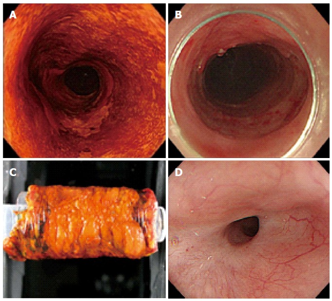
Representative endoscopic imaging of a stricture after circumferential endoscopic submucosal dissection for a large-sized superficial esophageal squamous cell carcinoma. A: Lugor chromoendoscopy imaging; B: Circumferential post-ESD ulceration; C: ESD specimen; D: Endoscopic imaging of post-ESD stricture 3 mo after ESD. ESD: Endoscopic submucosal dissection.
Table 1.
Risk factors of esophageal stricture formation after endo-therapy
| Ref. | Setting | Study design | Patients (n) | Mechanical stenosis and/or symptom(dysphasia) | Risk factor | Additional therapy of chemo/radiation | Follow-up period (mo) |
| Ono et al[15] | Single center | Retrospective | 65 | Necessity of EBD | More than three-fourths of circumferential extension of mucosal defect (OR = 44.2; 95%CI: 4.4-443.6) histologic depth to the LPM cancer (OR = 14.2; 95%CI: 2.7-74.2) | Excluded | Unknown |
| Shi et al[26] | Single center | Retrospective | 362 | Failure to pass a standard endoscope (11 mm-diameter) | More than three-fourths of circumferential extension of mucosal defect (OR = 44.2, 95%CI: 4.4-443.6) depth of invasion above the LPM cancer (OR = 14.2, 95%CI: 2.7-74.2) | Excluded | 41 (16-77) |
| Takahashi et al[25] | Single center | Retrospective | 76 | Failure of both symptomatic relief of dysphagia and the passage of a standard endoscope (9.2 mm or 9.0 mm-diameter) without any resistance 4-wk after the last session | More than three-fourths of the circumferential extent of mucosal defect (OR = 305.9; 95%CI: 89.387-1046.8) | Excluded | Stricture group: 30.0 (5-142) nonstricture group: 45.0 (6-174) |
EBD: Endoscopic balloon dilation.
(Sub)circumferential ESD was performed in the clinical setting in patients who were unwilling to undergo esophagectomy or patients with severe comorbidities[13,14,31]. Thereafter, most of these patients required several sessions of dilation because of severe/refractory dysphasia, although their preoperative conditions were asymptomatic. Instead, repeated dilatation has the potential risk of tears/perforation, which results in potentially lethal complications and a worsening of quality of life (QOL)[32,33]. The relatively high risk of recurrence after endoscopic treatment for SENs may occur because severe strictures after ESD may mask recurrent lesions that are hidden in the blind spots of the surveillance endoscopic inspection and disturb their endoscopic treatment procedure[20]. Therefore, the control of these refractory strictures with symptomatic dysphagia after (sub)circumferential ESD is an urgent task for broader acceptance of ESD as a treatment strategy for SEN. We review recent achievements in the preventive and treatment strategies of esophageal strictures after endoscopic resection and the mechanism of their development.
MECHANISM OF THE SYMPTOMATIC/MECHANICAL STRICTURE AFTER ESD
We include mechanical stricture and esophageal dysmotility as major causes of post-ESD dysphasia.
First, we discuss the mechanisms of mechanical stricture development. In general, wound healing occurs by way of the following series of events: (1) induction of inflammatory response and organization of granulation tissue; (2) proliferation of epithelial cells in the border of the wound and the covering of the wound with granulation; (3) growth of subepithelial granulation; and (4) scar formation and contraction after remodeling, such as dedifferentiation from fibroblasts into fiber cells or contraction of collagen fibers[34]. A cascade of inflammatory cytokines and reactive oxygen species is activated immediately after injury, and inflammation continues until the process is complete[35,36]. The following three pathological characteristics that are unique to the process of stricture formation after esophageal EMR/ESD were demonstrated: (1) delayed mucosal healing; (2) severe inflammation, which results in deeper ulceration and extensive fibrosis, in the submucosa; and (3) atrophy in the muscularis proper[37-40]. These factors may be induced by deep thermal injury because of the electric current effect of endoscopic therapy or the prolonged loss of the esophageal epithelium as a barrier against the external environment, including exogenous material, saliva, food, microorganisms or refluxate[29,41-43]. Our immunohistochemical study of esophageal specimens surgically obtained from patients with post-ESD strictures revealed rich collagen fibers with inflammatory cells in the submucosa and atrophic changes in the muscularis proper (Figure 2). Collagen fibers are the major components of connective tissue, which provide structural support in scars, and their disordered orientation may be associated with the elasticity and strength of esophageal tissue[38,44]. Previous studies suggested that the atrophy of muscle fibers might originate from passive fiber atrophy or a dynamic alteration of the muscle fibers into myofibroblasts, activated fibroblasts, which are deeply associated with inflammation, and carcinogenesis. The disorganized fibrosis and the abundant production of extracellular matrices in the submucosa and atrophy in the muscularis propria are key factors in post-ESD strictures.
Figure 2.
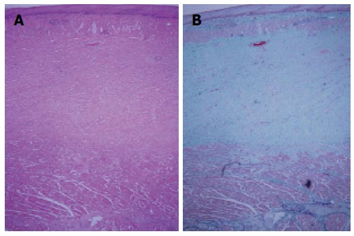
Representative histological findings of specimens surgically obtained from patients with post-endoscopic submucosal dissection strictures of the esophagus (magnification × 40). A: Hematoxylin-Eosin staining; B: Elastica-Masson staining. These results indicated rich collagen fibers with inflammatory cells in the submucosa and atrophic changes in muscularis proper at the stricture site.
Second, we should consider the possible roles of esophageal dysmotility in the occurrence of symptomatic dysphagia. Previous studies also suggested that dysphagia associated with gastroesophageal reflux disease, caustic esophageal burns and mucosal damage by endoscopic photodynamic therapy may be caused by the underlying esophageal dysmotility[45-48]. Practically, some post-ESD patients complain of sporadic dysphagia without mechanical strictures on endoscopic findings, especially when swallowing a large mass of food. Bu et al[45] demonstrated that ineffective esophageal motility after esophageal ESD was related to dysphasia symptoms using a high-resolution manometry system and a published symptomatic scoring system. These results suggested that the ESD procedure itself can induce a dysmotility related to irreversible symptomatic dysphagia without mechanical stricture. However, no study revealed the relationship between symptomatic dysphasia and esophageal dysmotility in pre- or post-ESD patients.
THERAPEUTIC STRATEGY FOR POST ESD-STRICTURES OF THE ESOPHAGUS
Endoscopic dilation therapy
Currently, endoscopic balloon dilation (EBD) is the preferred method to achieve mechanical dilation of the strictures. EBD includes possible complications of perforation, massive bleeding, and bacteremia[24,25,32,49]. Notably, the potential risk of perforation from dilation therapies for post-EMR/ESD strictures of the esophagus is slightly higher than peptic or anastomotic strictures[24,25,50]. We demonstrated in detail that the perforation rate in total EBD procedures for post-ESD strictures was 1.02%, which was slightly greater than the 0.4% rate that was reported in a retrospective survey for peptic strictures[29]. Takahashi et al[25] identified two independent risk factors for perforation using Maloney and Savary wire-guided bougienage, i.e., multiple dilations (OR = 1.185; 95%CI: 1.038-1.353; P = 0.012), and location in the lower esophagus, which is an area with a relative lack of muscle and/or surrounding supportive tissues (OR = 12.763; 95%CI: 1.089-149.563; P = 0.043)[25]. Moreover, the dilation effect of endoscopic dilation therapies is uncertain, and suboptimal improvements in symptoms may occur. Repeated dilations can worsen scarring, which results in the recurrence of more refractory strictures[25,51]. Therefore, a carefully planned strategy for dilation treatment is also needed for patients with strictures.
Endoscopic radial incision and cutting method
Recently, the endoscopic radial incision and cutting method (ERIC) method was developed to directly remove/slice the fibrotic tissue of a stricture using ESD-oriented devices. Minamino et al[52] demonstrated the usefulness and safety of ERIC in two patients with refractory strictures of the esophagus after subcircumferential ESD followed by repeated EBD concomitant with locoregional steroid injections. However, the indication for and possible complications of this technique are uncertain because of the lack of studies using a large number of subjects and long-term surveillance. Actually, we experienced one case of stricture after a 10-cm long circumferential ESD that was too severe to be cured using repeated EBD with steroid injections followed by ERIC. It was very hard to perform the incision and slice during the procedure because the stricture was too tight to enable manipulation of the devices (Figure 3). Therefore, further studies will be required to determine the appropriate indications for ERIC and overcome these limitations.
Figure 3.
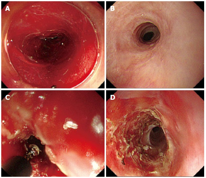
Representative endoscopic images of refractory strictures of the esophagus. A: Endoscopic images immediately after 10-cm long circumferential ESD; B: Images 3 mo after ESD followed by repeated EBD with steroid injection; C: ERIC procedure with IT-2 knife (Olympus, Tokyo, Japan); D: Severe stricture 1 mo after ERIC. ESD: Endoscopic submucosal dissection; ERIC: Endoscopic radial incision and cutting; EBD: Endoscopic balloon dilation.
Stent use
Metallic esophageal stents were initially used for minimally invasive and permanent treatment of fistulas and unresectable malignant stenosis. The use of stents resulted in complications, such as granulation tissue hyperplasia, pain, stent displacement, and esophageal ulceration[30,53,54]. Improvements in removable stents, such as temporary self-expandable metallic stents and biodegradable stents, gradually increased the use of stent implantation as a new treatment option for refractory benign esophageal strictures[55-57].
Some studies that reported the usefulness of temporary self-expandable metallic stents for the treatment of esophageal benign stenosis noted their major advantages, including a sustained dilatation effect and ease of removal[55,58,59]. Matsumoto et al[60] reported that a patient with refractory stenosis 1 mo after ESD was cured by a 7-d implantation of a stent that caused no complications and no recurrence during the 1-mo follow-up. Wen et al[61] performed a single-center randomized controlled trial (RCT) and showed that the stricture rate and number of additional dilatations during a short-term follow-up period were significantly lower in subjects treated by stent placement for 8-wk immediately after ESD than patients who were not treated with stents. However, other studies noted that the long-term treatment effect and complication rates were not as satisfactory as expected[55,62].
Biodegradable stents were developed to overcome the drawbacks of metallic/plastic stents for benign strictures. Two case reports with short-term (up to 6 mo) surveillance demonstrated the usefulness and safety of polylactide biodegradable stents for stenosis after esophageal ESD that were greater than seven-eighth of the circumference[63,64]. Further studies will be needed to clarify the long-term outcome, but hyperplastic regenerative changes might make it difficult to distinguish between inflammatory changes and neoplastic residues during the surveillance.
PREVENTIVE STRATEGIES FOR POST-ESD STRICTURES
No strategy for the prevention of post-ESD strictures has been established. Previous studies have reported the usefulness of the local/systemic administration of anti-scarring agents, preventive EBD, and regenerative medicine in the prevention of strictures, albeit with limited success (Table 2). A relatively small number of subjects can result in a type II error. Moreover, there are differences in study designs (basic or clinical), definitions of esophageal stricture and symptomatic dysphasia after endoscopic resection, the indication/method/devices of EBD, follow-up period after the treatment, the treatment history of additional therapies before/after endoscopic treatment, and pathological tumor staging of SENs. Notably, the circumferential extent of post-ESD ulceration can be easily affected by the extent of esophageal wall expansion due to the inflated air amount on endoscopic examination or by shrinkage of the residual mucosa after ESD. Therefore, it is difficult to make direct comparisons to clarify the best choice of prophylactic strategies for strictures. Further prospective, randomized, double-blinded, controlled investigations of a larger number of subjects are required.
Table 2.
Results of previous studies on preventive strategies for esophageal stricture after endo-therapy
| Ref. |
Subjects |
Stenosis |
Dilation therapy |
Outcome |
||||||||||
| Preventive strategy | Study design | n | Tumor stage | Circumference of the mucosal defect | Additional therapy of chemo/radiation | Difinition of stenosis | Dysphasia (symptom) | Dysphasia score | Indication | Device | Follow-up period | Stenosis rate | Therapeutic ED number | |
| Ezoe et al[41] | Preventive EBD | Case series, retrospective | 41 | T1a-T1b | Subcircumference (> 3/4) -circumference | Excluded | No passage of an endoscope (Φ11 mm) | Complaint of dysphagia | - | Mechanical/symptomatic | CRE balloon (four size) | Median 84 mo | 59% | 4.5 (0-35) |
| Uno et al[29] | Preventive EBD + tranilast | Case-control, prospective | 31 | T1a-T1b (SM1) | Subcircumference (> 3/4) -circumference of the lesion | Excluded | Vomiting at least once a week + no passage of an endoscope (Φ10.8 mm) | Dysphagia to regular solid meal | + | Mechanical + symptomatic | CRE balloon (15-18 mm) | 24.3 ± 7.4 mo | 33.30% | 0.0 (0.0-1.75) |
| Wen et al[61] | Fully covered metalic stent | Cohort study, prospective | 22 | T1a-T1b (SM1) | Subcircumference (> 3/4) -circumference | Excluded | No passage of an endoscope (Φ9.8 mm) | Persistent dysphagia to regular solid meal | - | Mechanical | Savary-Gilliard dilator | 12 wk | 18.20% | 0.45 (0-3) |
| Hashimoto et al[71] | Local injection of steroid | Case-control, retrospective | 41 | T1a-T1b (SM3) | Subcircumference (> 3/4) | Included | Complaint of dysphasia/no passage of an endoscope (Φ11.4 mm) | Complaint of dysphagia | - | Mechanical | CRE balloon (unclear) | Unclear | 19.00% | 1.7 (0-15) |
| Hanaoka et al[72] | Local injection of steroid | Cohort study, prospective | 59 | T1a-T1b | Subcircumference (> 3/4) | Included (chemotherapy) | Dysphagia to some solids/no passage of an endoscope (Φ9.2 mm) | Dysphagia to some solids | + | Symptomatic | Unclear | 8 wk | 10% | 0 (0-2) |
| Yamaguchi et al[65] | Systemic administration of steroid | Cohort study, retrospective | 41 | T1a-T1b (SM1) | Subcircumference (> 3/4) -circumference | Excluded | Unclear | Complaint of dysphagia | - | Symptomatic | CRE balloon (15-18 mm) | 3 mo | 5.30% | |
| 6-12 mo | 1.7 (0-7) | |||||||||||||
| Sato et al[77] | Systemic administration of steroid + EBD | Cohort study, retrospective | 23 | T1a-T1b (SM3) | Circumference | Included | Resistence/failure to pass an endoscope (Φ9.9 mm) | - | - | Mechanical | CRE baloon (15-18 mm) | No fininition | - | 13.8 ± 6.9 |
| Ohki et al[98] | Autologous oral mucosal epithelial cell sheets | Single-arm | 9 | Unknown | Subcircumference (> 2/3) | Unknown | No definition | - | + | Mechanical | Unclear | - | - | 2.33 (0-21) |
| Sakaguchi et al[106] | PGA | Case series, prospective | 8 | T1a-T1b (SM2) | Subcircumference (> 3/4) | Unknown | Failure to pass a endoscope (Φ9.8 mm) | Symptoms of dysphasia | - | Mechanical | CRE balloon (12-18 mm) | No definition | 37.5 % (about 28 POD) | 0.8 ± 1.2 |
| Iizuka et al[107] | PGA | Case series, prospective | 15 | T1a-T1b (SM2) | Subcircumference (> 1/2) | Excluded | Unclear | Unclear | - | Unclear | EBD (unclear) | 6 wk | 7.70% | 5 |
PGA: Polyglycolic acid; ESD: Endoscopic submucosal dissection; EBD: Endoscopic balloon dilation.
Preventive EBD
Previous studies demonstrated that preventive EBD was scheduled immediately after endoscopic resection, which may be useful for the prevention of post-EMR/ESD strictures[41]. However, patients with large-sized lesions underwent > 13 sessions of preventive EBD after sub-circumferential ESD or > 30 sessions after circumferential ESD, which worsens their QOL and increases the severity of the stricture and the occurrence of EBD-associated complications[5,29,41,65]. Therefore, the development of new strategies to reduce the numbers of EBD sessions is needed.
Anti-inflammatory agents
The earlier administration of anti-inflammatory agents might have an impact on the initial inflammation, subsequent hyperplasia of granulation and fibrosis in the submucosa. However, the efficacy of anti-inflammatory approaches in stricture prevention is expected[66]. Corticosteroids possess anti-inflammatory activity[67], inhibit collagen synthesis and enhance the breakdown of collagen fibers, thereby preventing the cross-linking of collagen fibers in the stricture formation[68]. Nonaka et al[37] used a pig model and demonstrated that steroid injection after circumferential ESD might modify the arrangement and proliferation of spindle-shaped myofibroblasts during the healing process in a normal or near-normal way. Recently, endoscopic injection or systemic administration of steroids was primarily used in clinical studies.
Endoscopic intralesional injections of steroids: The use of triamcinolone acetonide (TA) in the endoscopic field was proposed previously[68-70]. Hashimoto et al[71] used TA injection into the site on the 3rd/7th/10th days after ESD at a dose of 18 to 62 mg for each session in a single-center retrospective study and demonstrated that the stricture rate in patients with TA was significantly lower than in patients without TA (19% vs 75%, respectively). In another single-center prospective study, Hanaoka et al[72] used a single local injection of 100 mg of TA immediately after ESD, which resulted in a 10% stricture rate and 0 in the median number of EBD sessions in TA subjects compared with 66% and 2%, respectively, in their historical control without TA. However, some patients who were treated using (sub)circumferential ESD followed by steroid injection with EBD still suffered from refractory strictures[31,52,73].
The results of previous studies exhibit discrepancies[37,38]. Some studies noted that the use of local steroid injections did not prevent strictures, but it induced serious complications, such as periesophageal abscess/perforation[28,74,75]. Several studies suggested the following causal factors of these discrepancies. First, the delay in wound healing processes by steroid treatment exhibits bifacial effects. Instead of delayed scar formation, delayed epithelialization might facilitate bacterial infection and the subsequent extension of inflammation, which results in a worsening of the healing process and deepening of fibrotic changes[38,49]. Second, local steroid administration may induce an excessive inhibition of fibrosis during an early phase of the healing process, which results in a weakening of the wall[49]. Eventually, a deep ulceration and subsequent perforation can be induced by TA injection, especially when the muscle fibers are missing (Figure 4)[75]. These changes are associated with a relatively higher risk of perforation from EBD in post-ESD patients who received steroid therapy than in patients who underwent EBD alone[75]. Third, this method has critical/technical limitations in the certainty of drug delivery[76]. Actually, we often encountered situations in which many of submucosal TA injections leaked out during/immediately after the injection session, which means that the injected agent may not be as evenly distributed as we intended. Therefore, it can be difficult to measure/establish the precise/optimal amount of the drug that was actually delivered because no studies used tracing modalities for the injected drugs. Further studies are needed to establish a stable drug delivery system and define the optimal dose, duration, and timing of steroid administration for stricture prevention.
Figure 4.
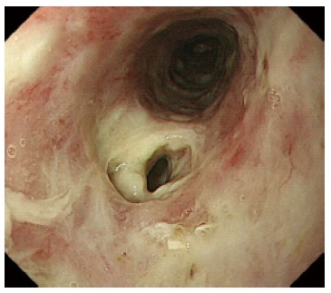
Representative image of esophageal perforation 2 d after local injection of triamcinolone acetonide for endoscopic submucosal dissection ulceration.
Systemic steroid therapy: Yamaguchi et al[65] demonstrated the efficacy of systemic steroid therapy for stricture prevention. Oral prednisolone was initiated 2 d after ESD at a dose of 30 mg/d, and the dose was gradually tapered in decrements of 5 mg/d every 2 wk for 1 mo followed by decrements of 5 mg/d every week for the next 4 wk for 8 wk after ESD. A comparison of the stricture rate of 19 patients with systemic steroid therapy with 22 patients with preventive EBD alone revealed that the rate in the former group was significantly lower than that in the latter group (5.3% vs 31.8%, respectively). Sato et al[77] also reported that early, rather than late, administration of systemic steroid therapy concomitant with therapeutic EBD was more effective than therapeutic EBD alone in the prevention of esophageal strictures after circumferential ESD. However, we should consider that there were only limited effects of steroids on stricture prevention, especially after long-length (sub)circumferential ESD or circumferential ESD, before extrapolating these results from study settings to practical clinical settings[73,78]. We should also pay careful attention to the adverse effects of systemic administration, such as immune suppression, psychiatric disturbances, diabetes, and peptic ulceration[65]. One case report described that systemic steroid therapy after esophageal ESD may have caused disseminated nocardiosis[79].
Potential drugs to target fibrotic formation: Machida et al[80] demonstrated that local injection of mitomycin C (MMC) into the site improved recurrent dysphagia or re-stenosis without serious complications in 5 patients. MMC has an anti-proliferative effect on fibroblasts in strictures, but the reproducibility of these results was poor[76,81]. In contrast, MMC may cause critical adverse events, such as delayed mucosal healing, ulcer formation, perforation, and secondary malignancy[82,83].
N-acetylcysteine (NAC) is also expected to prevent esophageal strictures because of the efficacy of NAC against pulmonary fibrosis and restructuring after EBD for intestinal strictures of Crohn’s disease or coronary angioplasty[84-86]. NAC is an antioxidant compound with antifibrotic effects, which primarily occur through the inhibition of transforming growth factor-β signaling, and anti-inflammatory effects that occur via the down-regulation of tumor necrosis factor-α, interleukin-6 and interleukin-8 synthesis[87-90]. Barret et al[91] demonstrated that the systemic administration of NAC after circumferential ESD did not exhibit a significant benefit on the occurrence of esophageal fibrosis and subsequent esophageal stricture one month after ESD in a pig model, which had the special condition of continuous acid aggression in the esophagus, compared to other models[91]. We performed a prospective, stratified-randomized, open-label trial in humans and demonstrated the efficacy and safety of preventive EBD combined with the commercially available oral agent NAC Tranilast for the prevention of esophageal strictures after 5 circumferential or 26 near-circumferential ESD cases (Figure 5). We demonstrated that the percentage of post-ESD strictures and the median numbers of additional EBD sessions and published dysphagia score 16 and 24 wk after ESD in subjects with preventive EBD concomitant with Tranilast intake were significantly lower than patients without Tranilast intake (33.3%, 0.0%, 0.0%, 0.0%, vs 68.8%, 4.0%, 5.0%, 3.0%, respectively)[29]. A previous study suggested that combination therapy involving NAC and other treatments may exhibit synergy effects[92], and we are currently performing a randomized controlled trial to elucidate the synergistic effect of NAC on the preventive effect of oral steroid administration.
Figure 5.
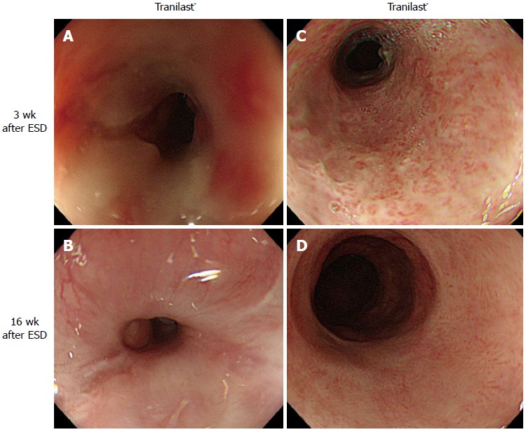
Representative image of the ulcer healing process after circumferential endoscopic submucosal dissection of the esophagus. A: 3 wk after ESD of patient without Tranilast intake; B: 16 wk after ESD of patient without Tranilast intake; C: 3 wk after ESD of patient with Tranilast intake; D: 16 wk after ESD of patient with Tranilast intake. ESD: Endoscopic submucosal dissection.
Regenerative medicine approaches
Tissue engineering approaches and scaffold-based therapy/cell-based therapy are based on the concept that transplanted tissue with/without supportive materials can induce early repair and replace damaged tissues.
Cell-based therapies: Transplanted cells are expected to induce an early repair of wounds through their interaction with the host tissue, but their efficacies were demonstrated in only some animal studies. Sakurai et al[93] demonstrated the effect of autologous keratinocyte implantation in the prevention of scar formation 2 wk after EMR, but there was no significant difference in inflammatory cell infiltration[93]. Zuercher et al[33] demonstrated that stricture formation with circumferential transmural fibrosis 6 mo after a 6-cm long circumferential EMR of sheep esophagus was inhibited by the injection of autologous keratinocytes into the sites. The further development of regenerative medicine will be achieved by the use of adipose-derived stem cells (ADSCs), which are easily obtained from adipose tissue, for the targeted prevention of esophageal strictures. ADSCs exhibit biological features that are similar to those of bone marrow-derived stem cells, which enhance early wound healing through humoral factor secretion, differentiation into multiple cell-types, suppression of inflammatory cells, and the promotion of angiogenesis[94-96]. Honda et al[39] demonstrated that local injection of autologous ADSCs after post-EMR ulceration prevented esophageal strictures in a canine model, but no investigations of surface markers, multi-potentiality, or cell proliferation during the healing process were performed.
However, other studies noted possible difficulties in the engraftment of transplanted cells at the site because of their low viability and difficulty in confirming their injection into the targeted site. Whether the injected cells can really form a regenerating epithelial layer or facilitate epithelial cells of the ulcer edge to cover the site remains unclear because of the difficulty in tracing the cells.
Technology of autologous oral mucosal epithelial cell sheets: A breakthrough system for epithelial cell sheets on temperature-responsive culture inserts was developed at Tokyo Women’s Medical University[97], and the cell membrane proteins and cell-to-cell junctions in these sheets can be directly grafted to the site without suturing or the use of any adhesive materials. Sheet transplantation to mucosal defects immediately after ESD promoted re-epithelization in an animal/clinical model[40,43,98,99]. Transplantation of cell sheets prevented esophageal strictures after hemi-circumferential to sub-circumferential ESD in 8 of 9 cases in a clinical study, and mucosal healing was rapidly completed within 3 to 5 wk. Only one case required 21 sessions of EBD[98]. These results suggested that cell sheets promote epithelial healing and prevent stricture formation. However, the transplantation of these sheets may be disturbed by esophageal wall motion, the passage of intakes, or gravity, depending on the location of the site. Therefore, further studies of the development of devices that facilitate stable sheet transplantation are warranted to confirm the usefulness and safety of this therapy.
Scaffold-based therapies: Temporary scaffolds developed from biodegradable materials may provide key proteins to establish a microenvironment that is suitable to facilitate cell adhesion and migration/proliferation, which results in early wound repair[100].
Previous studies have suggested that the extracellular matrix plays an important role in the prevention of strictures. Badylak et al[101] found that stents combined with extracellular matrix and autologous muscle tissues enabled the reconstruction of esophageal structures and the recovery of function without stricture formation in a canine model. Nieponice et al[102] demonstrated that implantation of a temporary stent wrapped with an extracellular matrix biological scaffold composed of porcine-derived small intestinal submucosa macroscopically/microscopically prevented esophageal stenosis even after a long-length circumferential EMR of the esophagus in another canine model.
Commercially available biological scaffold sheets composed of polyglycolic acid (PGA), poly-L-lactic acid (PLLA), or human amniotic membrane (AM) are expected to minimize scar contracture in various clinical fields[64,103,104]. PGA/PLLA are widely used as biodegradable suture materials because they are synthetic compounds that are completely degraded into nontoxic products in physiological conditions[104]. A recent case-series/report applied PGA sheets for the prevention of post-ESD stricture[105-107]. Sakaguchi et al[106] demonstrated that the use of sheets after ESD with mucosal defects of more than three-quarters of the esophageal circumference resulted in a stricture rate of 37.5% 4 wk after ESD. Iizuka et al[107] demonstrated a stricture rate of 7.7% 6 wk after ESD with more than half of the mucosal defects. Saito et al[64] demonstrated the usefulness of biological scaffolds composed of PLLA as temporary stent supports for stricture prevention in 2 cases. AM grafts, which consist of an avascular stroma and a monostratified cylinder cell epithelium with very few antigens[108], are used clinically in the field of ophthalmology. However, Barret et al[108-110] demonstrated that AM grafts had only limited benefit in preventing strictures in a swine model. More recently, Schomisch et al[111] revealed that no commercially available biological mesh with stent prevented esophageal stricture after 5-10-cm long circumferential ESD in a swine model. Therefore, the usefulness of ‘clinically available’ biological scaffold sheets in stricture prevention remains controversial.
Technical difficulties in the delivery of cell sheets to the esophageal wall also remain. Delivery using over-the-scope applications using clips or through-the-scope applications using dilation balloons or temporary self-expandable stents are time-consuming, and hole defects after ESD remain uncovered by the sheets in some cases. Instead, the patched sheets easily drop off, probably due to peristaltic esophageal wall motions or gravity depending on the anatomical location. Uno et al[29] and Iizuka et al[107] demonstrated that the sheets remained in place 1 wk after ESD in 86.7% of the patients and 2 wk after ESD in 40% of the patients, although a much longer time was needed for complete healing. Moreover, these sheets exhibit potential adverse events, such as local recurrences and infections, because the scaffold itself provides a good environment for the engraftment of malignant cells or microorganisms in the esophageal lumen and oral cavity.
Therefore, these biological scaffold-based approaches that target the control of the tissue microenvironment will shed new light on the development of new technologies for the prevention of strictures. However, sufficiently stable and firm covering technologies without adverse effects have not been developed.
PERSPECTIVE
Previous studies have suggested that each of these preventive strategies might reduce the risk of esophageal stricture after (sub)circumferential ESD from 88%-100% to 5.3%-59%. However, these strategies have a reduced or no effect on prevention in some cases, such as circumferential mucosal defects after ESD and sub-circumferential mucosal defects of a longitudinal length longer than 50 mm (Table 2).
Further studies using a combination of the published strategies, rather than a single strategy, are warranted as the first step to overcome this issue. The establishment of accurate, consistent, and safe methods to deliver agents through an endoscope is one of the most important topics in this field. Recent developments in stent technologies, such as recyclable covered stents, drug-eluting stents, anti-displacement stents, and biodegradable stents, promote future improvements in stents with an adherent substance that contains release agents/nanoparticles, which may enable a more reliable and feasible drug delivery system.
Another approach is the development of transplanting engineered tissues. The rationale for this idea is that tissue engineering approaches have the ability to create a temporary mechanical barrier and promote epithelial re-growth because of their anti-inflammatory and anti-fibrotic properties. Future prospects in tissue engineering approaches have great potential to cause damaged tissues to exhibit (near-) normal physiological functions. However, there remain concerns about possible complications from the existing technologies, such as local infection, immunological responses, and tumorigenesis, remain because some previous studies included patients with large-sized SENs in which the pathological assessment of the ESD specimens revealed submucosal invasion or positive vertical margins. Myofibroblasts are a major component of the microenvironment, and these cells might play a central role in the wound healing process and the development of carcinogenesis. Therefore, further works that focus on the control of fibroblast activity, such as stable wound-covering stent/cell sheets with biological agents/nanoparticles with anti-fibrotic activity, could provide a big step in the development of new modalities.
CONCLUSION
We reviewed the recent achievements in preventive strategies for post-ESD strictures of the esophagus, which demonstrated only limited success. The limitations of preoperative staging accuracy and the relatively high risk of local recurrence during the surveillance support the establishment of preventive strategies as an urgent task to maintain prognosis and improve QOL. Therefore, the strengthening of the unity of knowledge of the biological/physiological mechanisms of post-ESD strictures in various research fields, such as basic/clinical medical research, nanotechnology, and medical engineering, is required to establish new strategies to promote wound healing and control the tumor/inflammatory microenvironment in carcinogenesis. After the efficacy and safety of these new strategies are confirmed in long-term careful assessments, (sub)circumferential ESD of the esophagus will be accepted more broadly as the major treatment strategy for SENs.
Footnotes
Conflict-of-interest: The authors declare no conflict of interests.
Open-Access: This article is an open-access article which was selected by an in-house editor and fully peer-reviewed by external reviewers. It is distributed in accordance with the Creative Commons Attribution Non Commercial (CC BY-NC 4.0) license, which permits others to distribute, remix, adapt, build upon this work non-commercially, and license their derivative works on different terms, provided the original work is properly cited and the use is non-commercial. See: http://creativecommons.org/licenses/by-nc/4.0/
Peer-review started: February 11, 2015
First decision: March 10, 2015
Article in press: April 28, 2015
P- Reviewer: Cremers MI, Kalaitzakis E S- Editor: Ma YJ L- Editor: A E- Editor: Liu XM
References
- 1.Katada C, Muto M, Manabe T, Boku N, Ohtsu A, Yoshida S. Esophageal stenosis after endoscopic mucosal resection of superficial esophageal lesions. Gastrointest Endosc. 2003;57:165–169. doi: 10.1067/mge.2003.73. [DOI] [PubMed] [Google Scholar]
- 2.Chennat J, Konda VJ, Ross AS, de Tejada AH, Noffsinger A, Hart J, Lin S, Ferguson MK, Posner MC, Waxman I. Complete Barrett’s eradication endoscopic mucosal resection: an effective treatment modality for high-grade dysplasia and intramucosal carcinoma--an American single-center experience. Am J Gastroenterol. 2009;104:2684–2692. doi: 10.1038/ajg.2009.465. [DOI] [PubMed] [Google Scholar]
- 3.Gotoda T, Yamamoto H, Soetikno RM. Endoscopic submucosal dissection of early gastric cancer. J Gastroenterol. 2006;41:929–942. doi: 10.1007/s00535-006-1954-3. [DOI] [PubMed] [Google Scholar]
- 4.Ishihara R, Iishi H, Uedo N, Takeuchi Y, Yamamoto S, Yamada T, Masuda E, Higashino K, Kato M, Narahara H, et al. Comparison of EMR and endoscopic submucosal dissection for en bloc resection of early esophageal cancers in Japan. Gastrointest Endosc. 2008;68:1066–1072. doi: 10.1016/j.gie.2008.03.1114. [DOI] [PubMed] [Google Scholar]
- 5.Oyama T, Tomori A, Hotta K, Morita S, Kominato K, Tanaka M, Miyata Y. Endoscopic submucosal dissection of early esophageal cancer. Clin Gastroenterol Hepatol. 2005;3:S67–S70. doi: 10.1016/s1542-3565(05)00291-0. [DOI] [PubMed] [Google Scholar]
- 6.Takahashi H, Arimura Y, Masao H, Okahara S, Tanuma T, Kodaira J, Kagaya H, Shimizu Y, Hokari K, Tsukagoshi H, et al. Endoscopic submucosal dissection is superior to conventional endoscopic resection as a curative treatment for early squamous cell carcinoma of the esophagus (with video) Gastrointest Endosc. 2010;72:255–264, 264.e1-e2. doi: 10.1016/j.gie.2010.02.040. [DOI] [PubMed] [Google Scholar]
- 7.Ono S, Fujishiro M, Niimi K, Goto O, Kodashima S, Yamamichi N, Omata M. Long-term outcomes of endoscopic submucosal dissection for superficial esophageal squamous cell neoplasms. Gastrointest Endosc. 2009;70:860–866. doi: 10.1016/j.gie.2009.04.044. [DOI] [PubMed] [Google Scholar]
- 8.Hirasawa K, Kokawa A, Oka H, Yahara S, Sasaki T, Nozawa A, Tanaka K. Superficial adenocarcinoma of the esophagogastric junction: long-term results of endoscopic submucosal dissection. Gastrointest Endosc. 2010;72:960–966. doi: 10.1016/j.gie.2010.07.030. [DOI] [PubMed] [Google Scholar]
- 9.Kakushima N, Yahagi N, Fujishiro M, Kodashima S, Nakamura M, Omata M. Efficacy and safety of endoscopic submucosal dissection for tumors of the esophagogastric junction. Endoscopy. 2006;38:170–174. doi: 10.1055/s-2005-921039. [DOI] [PubMed] [Google Scholar]
- 10.Yoshinaga S, Gotoda T, Kusano C, Oda I, Nakamura K, Takayanagi R. Clinical impact of endoscopic submucosal dissection for superficial adenocarcinoma located at the esophagogastric junction. Gastrointest Endosc. 2008;67:202–209. doi: 10.1016/j.gie.2007.09.054. [DOI] [PubMed] [Google Scholar]
- 11.Repici A, Hassan C, Carlino A, Pagano N, Zullo A, Rando G, Strangio G, Romeo F, Nicita R, Rosati R, et al. Endoscopic submucosal dissection in patients with early esophageal squamous cell carcinoma: results from a prospective Western series. Gastrointest Endosc. 2010;71:715–721. doi: 10.1016/j.gie.2009.11.020. [DOI] [PubMed] [Google Scholar]
- 12.Höbel S, Dautel P, Baumbach R, Oldhafer KJ, Stang A, Feyerabend B, Yahagi N, Schrader C, Faiss S. Single center experience of endoscopic submucosal dissection (ESD) in early Barrett´s adenocarcinoma. Surg Endosc. 2015;29:1591–1597. doi: 10.1007/s00464-014-3847-5. [DOI] [PubMed] [Google Scholar]
- 13.Fujishiro M. Perspective on the practical indications of endoscopic submucosal dissection of gastrointestinal neoplasms. World J Gastroenterol. 2008;14:4289–4295. doi: 10.3748/wjg.14.4289. [DOI] [PMC free article] [PubMed] [Google Scholar]
- 14.Oyama T, Miyata Y, Shimaya S. Lymph nodal metastasis of m3, sm1 esophageal cancer [in Japanese] Stom and Intest. 2002;37:71–74. [Google Scholar]
- 15.Ono S, Fujishiro M, Niimi K, Goto O, Kodashima S, Yamamichi N, Omata M. Predictors of postoperative stricture after esophageal endoscopic submucosal dissection for superficial squamous cell neoplasms. Endoscopy. 2009;41:661–665. doi: 10.1055/s-0029-1214867. [DOI] [PubMed] [Google Scholar]
- 16.Safranek PM, Cubitt J, Booth MI, Dehn TC. Review of open and minimal access approaches to oesophagectomy for cancer. Br J Surg. 2010;97:1845–1853. doi: 10.1002/bjs.7231. [DOI] [PubMed] [Google Scholar]
- 17.Ishihara R, Yamamoto S, Iishi H, Takeuchi Y, Sugimoto N, Higashino K, Uedo N, Tatsuta M, Yano M, Imai A, et al. Factors predictive of tumor recurrence and survival after initial complete response of esophageal squamous cell carcinoma to definitive chemoradiotherapy. Int J Radiat Oncol Biol Phys. 2010;76:123–129. doi: 10.1016/j.ijrobp.2009.01.038. [DOI] [PubMed] [Google Scholar]
- 18.Phoa KN, Pouw RE, van Vilsteren FG, Sondermeijer CM, Ten Kate FJ, Visser M, Meijer SL, van Berge Henegouwen MI, Weusten BL, Schoon EJ, et al. Remission of Barrett’s esophagus with early neoplasia 5 years after radiofrequency ablation with endoscopic resection: a Netherlands cohort study. Gastroenterology. 2013;145:96–104. doi: 10.1053/j.gastro.2013.03.046. [DOI] [PubMed] [Google Scholar]
- 19.Orman ES, Li N, Shaheen NJ. Efficacy and durability of radiofrequency ablation for Barrett‘s Esophagus: systematic review and meta-analysis. Clin Gastroenterol Hepatol. 2013;11:1245–1255. doi: 10.1016/j.cgh.2013.03.039. [DOI] [PMC free article] [PubMed] [Google Scholar]
- 20.Nakagawa K, Koike T, Iijima K, Shinkai H, Hatta W, Endo H, Ara N, Uno K, Asano N, Imatani A, et al. Comparison of the long-term outcomes of endoscopic resection for superficial squamous cell carcinoma and adenocarcinoma of the esophagus in Japan. Am J Gastroenterol. 2014;109:348–356. doi: 10.1038/ajg.2013.450. [DOI] [PubMed] [Google Scholar]
- 21.Newaishy GA, Read GA, Duncan W, Kerr GR. Results of radical radiotherapy of squamous cell carcinoma of the oesophagus. Clin Radiol. 1982;33:347–352. doi: 10.1016/s0009-9260(82)80288-2. [DOI] [PubMed] [Google Scholar]
- 22.Yoda Y, Yano T, Kaneko K, Tsuruta S, Oono Y, Kojima T, Minashi K, Ikematsu H, Ohtsu A. Endoscopic balloon dilatation for benign fibrotic strictures after curative nonsurgical treatment for esophageal cancer. Surg Endosc. 2012;26:2877–2883. doi: 10.1007/s00464-012-2273-9. [DOI] [PubMed] [Google Scholar]
- 23.McManus KG, Ritchie AJ, McGuigan J, Stevenson HM, Gibbons JR. Sutures, staplers, leaks and strictures. A review of anastomoses in oesophageal resection at Royal Victoria Hospital, Belfast 1977-1986. Eur J Cardiothorac Surg. 1990;4:97–100. doi: 10.1016/1010-7940(90)90222-l. [DOI] [PubMed] [Google Scholar]
- 24.Silvis SE, Nebel O, Rogers G, Sugawa C, Mandelstam P. Endoscopic complications. Results of the 1974 American Society for Gastrointestinal Endoscopy Survey. JAMA. 1976;235:928–930. doi: 10.1001/jama.235.9.928. [DOI] [PubMed] [Google Scholar]
- 25.Takahashi H, Arimura Y, Okahara S, Uchida S, Ishigaki S, Tsukagoshi H, Shinomura Y, Hosokawa M. Risk of perforation during dilation for esophageal strictures after endoscopic resection in patients with early squamous cell carcinoma. Endoscopy. 2011;43:184–189. doi: 10.1055/s-0030-1256109. [DOI] [PubMed] [Google Scholar]
- 26.Shi Q, Ju H, Yao LQ, Zhou PH, Xu MD, Chen T, Zhou JM, Chen TY, Zhong YS. Risk factors for postoperative stricture after endoscopic submucosal dissection for superficial esophageal carcinoma. Endoscopy. 2014;46:640–644. doi: 10.1055/s-0034-1365648. [DOI] [PubMed] [Google Scholar]
- 27.Isomoto H, Yamaguchi N, Nakayama T, Hayashi T, Nishiyama H, Ohnita K, Takeshima F, Shikuwa S, Kohno S, Nakao K. Management of esophageal stricture after complete circular endoscopic submucosal dissection for superficial esophageal squamous cell carcinoma. BMC Gastroenterol. 2011;11:46. doi: 10.1186/1471-230X-11-46. [DOI] [PMC free article] [PubMed] [Google Scholar]
- 28.van Vilsteren FG, Pouw RE, Seewald S, Alvarez Herrero L, Sondermeijer CM, Visser M, Ten Kate FJ, Yu Kim Teng KC, Soehendra N, Rösch T, et al. Stepwise radical endoscopic resection versus radiofrequency ablation for Barrett’s oesophagus with high-grade dysplasia or early cancer: a multicentre randomised trial. Gut. 2011;60:765–773. doi: 10.1136/gut.2010.229310. [DOI] [PubMed] [Google Scholar]
- 29.Uno K, Iijima K, Koike T, Abe Y, Asano N, Ara N, Shimosegawa T. A pilot study of scheduled endoscopic balloon dilation with oral agent tranilast to improve the efficacy of stricture dilation after endoscopic submucosal dissection of the esophagus. J Clin Gastroenterol. 2012;46:e76–e82. doi: 10.1097/MCG.0b013e31824fff76. [DOI] [PubMed] [Google Scholar]
- 30.Chiu YC, Hsu CC, Chiu KW, Chuah SK, Changchien CS, Wu KL, Chou YP. Factors influencing clinical applications of endoscopic balloon dilation for benign esophageal strictures. Endoscopy. 2004;36:595–600. doi: 10.1055/s-2004-814520. [DOI] [PubMed] [Google Scholar]
- 31.Isomoto H, Yamaguchi N, Minami H, Nakao K. Management of complications associated with endoscopic submucosal dissection/endoscopic mucosal resection for esophageal cancer. Dig Endosc. 2013;25 Suppl 1:29–38. doi: 10.1111/j.1443-1661.2012.01388.x. [DOI] [PubMed] [Google Scholar]
- 32.Tsunada S, Ogata S, Mannen K, Arima S, Sakata Y, Shiraishi R, Shimoda R, Ootani H, Yamaguchi K, Fujise T, et al. Case series of endoscopic balloon dilation to treat a stricture caused by circumferential resection of the gastric antrum by endoscopic submucosal dissection. Gastrointest Endosc. 2008;67:979–983. doi: 10.1016/j.gie.2007.12.023. [DOI] [PubMed] [Google Scholar]
- 33.Zuercher BF, George M, Escher A, Piotet E, Ikonomidis C, Andrejevic SB, Monnier P. Stricture prevention after extended circumferential endoscopic mucosal resection by injecting autologous keratinocytes in the sheep esophagus. Surg Endosc. 2013;27:1022–1028. doi: 10.1007/s00464-012-2509-8. [DOI] [PubMed] [Google Scholar]
- 34.Kumar V, Abbas AK, Fausto N, Aster J. Pathologic basis of disease. 8th ed. Philadelphia (PA): Sanders; 2010. [Google Scholar]
- 35.Adzick NS, Lorenz HP. Cells, matrix, growth factors, and the surgeon. The biology of scarless fetal wound repair. Ann Surg. 1994;220:10–18. doi: 10.1097/00000658-199407000-00003. [DOI] [PMC free article] [PubMed] [Google Scholar]
- 36.Ferguson MW, Duncan J, Bond J, Bush J, Durani P, So K, Taylor L, Chantrey J, Mason T, James G, et al. Prophylactic administration of avotermin for improvement of skin scarring: three double-blind, placebo-controlled, phase I/II studies. Lancet. 2009;373:1264–1274. doi: 10.1016/S0140-6736(09)60322-6. [DOI] [PubMed] [Google Scholar]
- 37.Nonaka K, Miyazawa M, Ban S, Aikawa M, Akimoto N, Koyama I, Kita H. Different healing process of esophageal large mucosal defects by endoscopic mucosal dissection between with and without steroid injection in an animal model. BMC Gastroenterol. 2013;13:72. doi: 10.1186/1471-230X-13-72. [DOI] [PMC free article] [PubMed] [Google Scholar]
- 38.Honda M, Nakamura T, Hori Y, Shionoya Y, Yamamoto K, Nishizawa Y, Kojima F, Shigeno K. Feasibility study of corticosteroid treatment for esophageal ulcer after EMR in a canine model. J Gastroenterol. 2011;46:866–872. doi: 10.1007/s00535-011-0400-3. [DOI] [PubMed] [Google Scholar]
- 39.Honda M, Hori Y, Nakada A, Uji M, Nishizawa Y, Yamamoto K, Kobayashi T, Shimada H, Kida N, Sato T, et al. Use of adipose tissue-derived stromal cells for prevention of esophageal stricture after circumferential EMR in a canine model. Gastrointest Endosc. 2011;73:777–784. doi: 10.1016/j.gie.2010.11.008. [DOI] [PubMed] [Google Scholar]
- 40.Kanai N, Yamato M, Ohki T, Yamamoto M, Okano T. Fabricated autologous epidermal cell sheets for the prevention of esophageal stricture after circumferential ESD in a porcine model. Gastrointest Endosc. 2012;76:873–881. doi: 10.1016/j.gie.2012.06.017. [DOI] [PubMed] [Google Scholar]
- 41.Ezoe Y, Muto M, Horimatsu T, Morita S, Miyamoto S, Mochizuki S, Minashi K, Yano T, Ohtsu A, Chiba T. Efficacy of preventive endoscopic balloon dilation for esophageal stricture after endoscopic resection. J Clin Gastroenterol. 2011;45:222–227. doi: 10.1097/MCG.0b013e3181f39f4e. [DOI] [PubMed] [Google Scholar]
- 42.Conio M, Sorbi D, Batts KP, Gostout CJ. Endoscopic circumferential esophageal mucosectomy in a porcine model: an assessment of technical feasibility, safety, and outcome. Endoscopy. 2001;33:791–794. doi: 10.1055/s-2001-16516. [DOI] [PubMed] [Google Scholar]
- 43.Ohki T, Yamato M, Murakami D, Takagi R, Yang J, Namiki H, Okano T, Takasaki K. Treatment of oesophageal ulcerations using endoscopic transplantation of tissue-engineered autologous oral mucosal epithelial cell sheets in a canine model. Gut. 2006;55:1704–1710. doi: 10.1136/gut.2005.088518. [DOI] [PMC free article] [PubMed] [Google Scholar]
- 44.Ramage JI, Rumalla A, Baron TH, Pochron NL, Zinsmeister AR, Murray JA, Norton ID, Diehl N, Romero Y. A prospective, randomized, double-blind, placebo-controlled trial of endoscopic steroid injection therapy for recalcitrant esophageal peptic strictures. Am J Gastroenterol. 2005;100:2419–2425. doi: 10.1111/j.1572-0241.2005.00331.x. [DOI] [PubMed] [Google Scholar]
- 45.Bu BG, Linghu EQ, Li HK, Wang XX, Guo RB, Peng LH. Influence of endoscopic submucosal dissection on esophageal motility. World J Gastroenterol. 2013;19:4781–4785. doi: 10.3748/wjg.v19.i29.4781. [DOI] [PMC free article] [PubMed] [Google Scholar]
- 46.Bautista A, Varela R, Villanueva A, Estevez E, Tojo R, Cadranel S. Motor function of the esophagus after caustic burn. Eur J Pediatr Surg. 1996;6:204–207. doi: 10.1055/s-2008-1066508. [DOI] [PubMed] [Google Scholar]
- 47.Malhi-Chowla N, Wolfsen HC, DeVault KR. Esophageal dysmotility in patients undergoing photodynamic therapy. Mayo Clin Proc. 2001;76:987–989. doi: 10.4065/76.10.987. [DOI] [PubMed] [Google Scholar]
- 48.Hemmink GJ, Alvarez Herrero L, Bogte A, Bredenoord AJ, Bergman JJ, Smout AJ, Weusten BL. Esophageal motility and impedance characteristics in patients with Barrett’s esophagus before and after radiofrequency ablation. Eur J Gastroenterol Hepatol. 2013;25:1024–1032. doi: 10.1097/MEG.0b013e32836283dc. [DOI] [PubMed] [Google Scholar]
- 49.Egan JV, Baron TH, Adler DG, Davila R, Faigel DO, Gan SL, Hirota WK, Leighton JA, Lichtenstein D, Qureshi WA, et al. Esophageal dilation. Gastrointest Endosc. 2006;63:755–760. doi: 10.1016/j.gie.2006.02.031. [DOI] [PubMed] [Google Scholar]
- 50.Riley SA, Attwood SE. Guidelines on the use of oesophageal dilatation in clinical practice. Gut. 2004;53 Suppl 1:i1–i6. doi: 10.1136/gut.53.suppl_1.i1. [DOI] [PMC free article] [PubMed] [Google Scholar]
- 51.Cheng YS, Li MH, Yang RJ, Zhang HZ, Ding ZX, Zhuang QX, Jiang ZM, Shang KZ. Restenosis following balloon dilation of benign esophageal stenosis. World J Gastroenterol. 2003;9:2605–2608. doi: 10.3748/wjg.v9.i11.2605. [DOI] [PMC free article] [PubMed] [Google Scholar]
- 52.Minamino H, Machida H, Tominaga K, Sugimori S, Okazaki H, Tanigawa T, Yamagami H, Watanabe K, Watanabe T, Fujiwara Y, et al. Endoscopic radial incision and cutting method for refractory esophageal stricture after endoscopic submucosal dissection of superficial esophageal carcinoma. Dig Endosc. 2013;25:200–203. doi: 10.1111/j.1443-1661.2012.01348.x. [DOI] [PubMed] [Google Scholar]
- 53.Shin JH, Song HY, Ko GY, Lim JO, Yoon HK, Sung KB. Esophagorespiratory fistula: long-term results of palliative treatment with covered expandable metallic stents in 61 patients. Radiology. 2004;232:252–259. doi: 10.1148/radiol.2321030733. [DOI] [PubMed] [Google Scholar]
- 54.Kim JH, Song HY, Shin JH, Kim TW, Kim KR, Kim SB, Park SI, Kim JH, Choi E. Palliative treatment of unresectable esophagogastric junction tumors: balloon dilation combined with chemotherapy and/or radiation therapy and metallic stent placement. J Vasc Interv Radiol. 2008;19:912–917. doi: 10.1016/j.jvir.2008.02.020. [DOI] [PubMed] [Google Scholar]
- 55.Kim JH, Song HY, Choi EK, Kim KR, Shin JH, Lim JO. Temporary metallic stent placement in the treatment of refractory benign esophageal strictures: results and factors associated with outcome in 55 patients. Eur Radiol. 2009;19:384–390. doi: 10.1007/s00330-008-1151-2. [DOI] [PubMed] [Google Scholar]
- 56.Holm AN, de la Mora Levy JG, Gostout CJ, Topazian MD, Baron TH. Self-expanding plastic stents in treatment of benign esophageal conditions. Gastrointest Endosc. 2008;67:20–25. doi: 10.1016/j.gie.2007.04.031. [DOI] [PubMed] [Google Scholar]
- 57.Dua KS, Vleggaar FP, Santharam R, Siersema PD. Removable self-expanding plastic esophageal stent as a continuous, non-permanent dilator in treating refractory benign esophageal strictures: a prospective two-center study. Am J Gastroenterol. 2008;103:2988–2994. doi: 10.1111/j.1572-0241.2008.02177.x. [DOI] [PubMed] [Google Scholar]
- 58.Wadhwa RP, Kozarek RA, France RE, Brandabur JJ, Gluck M, Low DE, Traverso LW, Moonka R. Use of self-expandable metallic stents in benign GI diseases. Gastrointest Endosc. 2003;58:207–212. doi: 10.1067/mge.2003.343. [DOI] [PubMed] [Google Scholar]
- 59.Cheng YS, Li MH, Chen WX, Chen NW, Zhuang QX, Shang KZ. Temporary partially-covered metal stent insertion in benign esophageal stricture. World J Gastroenterol. 2003;9:2359–2361. doi: 10.3748/wjg.v9.i10.2359. [DOI] [PMC free article] [PubMed] [Google Scholar]
- 60.Matsumoto S, Miyatani H, Yoshida Y, Nokubi M. Cicatricial stenosis after endoscopic submucosal dissection of esophageal cancer effectively treated with a temporary self-expandable metal stent. Gastrointest Endosc. 2011;73:1309–1312. doi: 10.1016/j.gie.2010.11.007. [DOI] [PubMed] [Google Scholar]
- 61.Wen J, Lu Z, Yang Y, Liu Q, Yang J, Wang S, Wang X, Du H, Meng J, Wang H, et al. Preventing stricture formation by covered esophageal stent placement after endoscopic submucosal dissection for early esophageal cancer. Dig Dis Sci. 2014;59:658–663. doi: 10.1007/s10620-013-2958-5. [DOI] [PubMed] [Google Scholar]
- 62.Kim JH, Song HY, Park SW, Yoon CJ, Shin JH, Yook JH, Kim BS. Early symptomatic strictures after gastric surgery: palliation with balloon dilation and stent placement. J Vasc Interv Radiol. 2008;19:565–570. doi: 10.1016/j.jvir.2007.11.015. [DOI] [PubMed] [Google Scholar]
- 63.Tanaka T, Takahashi M, Nitta N, Furukawa A, Andoh A, Saito Y, Fujiyama Y, Murata K. Newly developed biodegradable stents for benign gastrointestinal tract stenoses: a preliminary clinical trial. Digestion. 2006;74:199–205. doi: 10.1159/000100504. [DOI] [PubMed] [Google Scholar]
- 64.Saito Y, Tanaka T, Andoh A, Minematsu H, Hata K, Tsujikawa T, Nitta N, Murata K, Fujiyama Y. Novel biodegradable stents for benign esophageal strictures following endoscopic submucosal dissection. Dig Dis Sci. 2008;53:330–333. doi: 10.1007/s10620-007-9873-6. [DOI] [PubMed] [Google Scholar]
- 65.Yamaguchi N, Isomoto H, Nakayama T, Hayashi T, Nishiyama H, Ohnita K, Takeshima F, Shikuwa S, Kohno S, Nakao K. Usefulness of oral prednisolone in the treatment of esophageal stricture after endoscopic submucosal dissection for superficial esophageal squamous cell carcinoma. Gastrointest Endosc. 2011;73:1115–1121. doi: 10.1016/j.gie.2011.02.005. [DOI] [PubMed] [Google Scholar]
- 66.Miyashita M, Onda M, Okawa K, Matsutani T, Yoshiyuki T, Sasajima K, Kyono S, Yamashita K. Endoscopic dexamethasone injection following balloon dilatation of anastomotic stricture after esophagogastrostomy. Am J Surg. 1997;174:442–444. doi: 10.1016/s0002-9610(97)00116-5. [DOI] [PubMed] [Google Scholar]
- 67.Carrico TJ, Mehrhof AI, Cohen IK. Biology of wound healing. Surg Clin North Am. 1984;64:721–733. doi: 10.1016/s0039-6109(16)43388-8. [DOI] [PubMed] [Google Scholar]
- 68.Kochhar R, Makharia GK. Usefulness of intralesional triamcinolone in treatment of benign esophageal strictures. Gastrointest Endosc. 2002;56:829–834. doi: 10.1067/mge.2002.129871. [DOI] [PubMed] [Google Scholar]
- 69.Russell SB, Trupin JS, Myers JC, Broquist AH, Smith JC, Myles ME, Russell JD. Differential glucocorticoid regulation of collagen mRNAs in human dermal fibroblasts. Keloid-derived and fetal fibroblasts are refractory to down-regulation. J Biol Chem. 1989;264:13730–13735. [PubMed] [Google Scholar]
- 70.Kochhar R, Ray JD, Sriram PV, Kumar S, Singh K. Intralesional steroids augment the effects of endoscopic dilation in corrosive esophageal strictures. Gastrointest Endosc. 1999;49:509–513. doi: 10.1016/s0016-5107(99)70052-0. [DOI] [PubMed] [Google Scholar]
- 71.Hashimoto S, Kobayashi M, Takeuchi M, Sato Y, Narisawa R, Aoyagi Y. The efficacy of endoscopic triamcinolone injection for the prevention of esophageal stricture after endoscopic submucosal dissection. Gastrointest Endosc. 2011;74:1389–1393. doi: 10.1016/j.gie.2011.07.070. [DOI] [PubMed] [Google Scholar]
- 72.Hanaoka N, Ishihara R, Takeuchi Y, Uedo N, Higashino K, Ohta T, Kanzaki H, Hanafusa M, Nagai K, Matsui F, et al. Intralesional steroid injection to prevent stricture after endoscopic submucosal dissection for esophageal cancer: a controlled prospective study. Endoscopy. 2012;44:1007–1011. doi: 10.1055/s-0032-1310107. [DOI] [PubMed] [Google Scholar]
- 73.Kobayashi S, Kanai N, Ohki T, Takagi R, Yamaguchi N, Isomoto H, Kasai Y, Hosoi T, Nakao K, Eguchi S, et al. Prevention of esophageal strictures after endoscopic submucosal dissection. World J Gastroenterol. 2014;20:15098–15109. doi: 10.3748/wjg.v20.i41.15098. [DOI] [PMC free article] [PubMed] [Google Scholar]
- 74.Rajan E, Gostout C, Feitoza A, Herman L, Knipschield M, Burgart L, Chung S, Cotton P, Hawes R, Kalloo A, et al. Widespread endoscopic mucosal resection of the esophagus with strategies for stricture prevention: a preclinical study. Endoscopy. 2005;37:1111–1115. doi: 10.1055/s-2005-870531. [DOI] [PubMed] [Google Scholar]
- 75.Yamashina T, Uedo N, Fujii M, Ishihara R, Mikamori M, Motoori M, Yano M, Iishi H. Delayed perforation after intralesional triamcinolone injection for esophageal stricture following endoscopic submucosal dissection. Endoscopy. 2013;45 Suppl 2 UCTN:E92. doi: 10.1055/s-0032-1326253. [DOI] [PubMed] [Google Scholar]
- 76.Wu Y, Schomisch SJ, Cipriano C, Chak A, Lash RH, Ponsky JL, Marks JM. Preliminary results of antiscarring therapy in the prevention of postendoscopic esophageal mucosectomy strictures. Surg Endosc. 2014;28:447–455. doi: 10.1007/s00464-013-3210-2. [DOI] [PMC free article] [PubMed] [Google Scholar]
- 77.Sato H, Inoue H, Kobayashi Y, Maselli R, Santi EG, Hayee B, Igarashi K, Yoshida A, Ikeda H, Onimaru M, et al. Control of severe strictures after circumferential endoscopic submucosal dissection for esophageal carcinoma: oral steroid therapy with balloon dilation or balloon dilation alone. Gastrointest Endosc. 2013;78:250–257. doi: 10.1016/j.gie.2013.01.008. [DOI] [PubMed] [Google Scholar]
- 78.Pelclová D, Navrátil T. Do corticosteroids prevent oesophageal stricture after corrosive ingestion? Toxicol Rev. 2005;24:125–129. doi: 10.2165/00139709-200524020-00006. [DOI] [PubMed] [Google Scholar]
- 79.Ishida T, Morita Y, Hoshi N, Yoshizaki T, Ohara Y, Kawara F, Tanaka S, Yamamoto Y, Matsuo H, Iwata K, et al. Disseminated nocardiosis during systemic steroid therapy for the prevention of esophageal stricture after endoscopic submucosal dissection. Dig Endosc. 2015;27:388–391. doi: 10.1111/den.12317. [DOI] [PubMed] [Google Scholar]
- 80.Machida H, Tominaga K, Minamino H, Sugimori S, Okazaki H, Yamagami H, Tanigawa T, Watanabe K, Watanabe T, Fujiwara Y, et al. Locoregional mitomycin C injection for esophageal stricture after endoscopic submucosal dissection. Endoscopy. 2012;44:622–625. doi: 10.1055/s-0032-1306775. [DOI] [PubMed] [Google Scholar]
- 81.Berger M, Ure B, Lacher M. Mitomycin C in the therapy of recurrent esophageal strictures: hype or hope? Eur J Pediatr Surg. 2012;22:109–116. doi: 10.1055/s-0032-1311695. [DOI] [PubMed] [Google Scholar]
- 82.Bakshi SR, Patel RK, Roy SK, Alladi P, Trivedi AH, Bhatavdekar JM, Patel DD, Shah PM, Rawal UM. Mitomycin C induced chromosomal aberrations in young cancer patients. Mutat Res. 1998;422:223–228. doi: 10.1016/s0027-5107(98)00197-3. [DOI] [PubMed] [Google Scholar]
- 83.Spiecker M, Lorenz I, Marx N, Darius H. Tranilast inhibits cytokine-induced nuclear factor kappaB activation in vascular endothelial cells. Mol Pharmacol. 2002;62:856–863. doi: 10.1124/mol.62.4.856. [DOI] [PubMed] [Google Scholar]
- 84.Demedts M, Behr J, Buhl R, Costabel U, Dekhuijzen R, Jansen HM, MacNee W, Thomeer M, Wallaert B, Laurent F, et al. High-dose acetylcysteine in idiopathic pulmonary fibrosis. N Engl J Med. 2005;353:2229–2242. doi: 10.1056/NEJMoa042976. [DOI] [PubMed] [Google Scholar]
- 85.Tamai H, Katoh K, Yamaguchi T, Hayakawa H, Kanmatsuse K, Haze K, Aizawa T, Nakanishi S, Suzuki S, Suzuki T, et al. The impact of tranilast on restenosis after coronary angioplasty: the Second Tranilast Restenosis Following Angioplasty Trial (TREAT-2) Am Heart J. 2002;143:506–513. doi: 10.1067/mhj.2002.120770. [DOI] [PubMed] [Google Scholar]
- 86.Oshitani N, Yamagami H, Watanabe K, Higuchi K, Arakawa T. Long-term prospective pilot study with tranilast for the prevention of stricture progression in patients with Crohn’s disease. Gut. 2007;56:599–600. doi: 10.1136/gut.2006.115469. [DOI] [PMC free article] [PubMed] [Google Scholar]
- 87.Azuma H, Banno K, Yoshimura T. Pharmacological properties of N-(3’,4’-dimethoxycinnamoyl) anthranilic acid (N-5’), a new anti-atopic agent. Br J Pharmacol. 1976;58:483–488. doi: 10.1111/j.1476-5381.1976.tb08614.x. [DOI] [PMC free article] [PubMed] [Google Scholar]
- 88.Shimizu T, Kanai K, Kyo Y, Asano K, Hisamitsu T, Suzaki H. Effect of tranilast on matrix metalloproteinase production from neutrophils in-vitro. J Pharm Pharmacol. 2006;58:91–99. doi: 10.1211/jpp.58.1.0011. [DOI] [PubMed] [Google Scholar]
- 89.Yamada H, Tajima S, Nishikawa T, Murad S, Pinnell SR. Tranilast, a selective inhibitor of collagen synthesis in human skin fibroblasts. J Biochem. 1994;116:892–897. doi: 10.1093/oxfordjournals.jbchem.a124612. [DOI] [PubMed] [Google Scholar]
- 90.Miyazawa K, Kikuchi S, Fukuyama J, Hamano S, Ujiie A. Inhibition of PDGF- and TGF-beta 1-induced collagen synthesis, migration and proliferation by tranilast in vascular smooth muscle cells from spontaneously hypertensive rats. Atherosclerosis. 1995;118:213–221. doi: 10.1016/0021-9150(95)05607-6. [DOI] [PubMed] [Google Scholar]
- 91.Barret M, Batteux F, Beuvon F, Mangialavori L, Chryssostalis A, Pratico C, Chaussade S, Prat F. N-acetylcysteine for the prevention of stricture after circumferential endoscopic submucosal dissection of the esophagus: a randomized trial in a porcine model. Fibrogenesis Tissue Repair. 2012;5:8. doi: 10.1186/1755-1536-5-8. [DOI] [PMC free article] [PubMed] [Google Scholar]
- 92.Sarchahi AA, Maimandi A, Tafti AK, Amani M. Effects of acetylcysteine and dexamethasone on experimental corneal wounds in rabbits. Ophthalmic Res. 2008;40:41–48. doi: 10.1159/000111158. [DOI] [PubMed] [Google Scholar]
- 93.Sakurai T, Miyazaki S, Miyata G, Satomi S, Hori Y. Autologous buccal keratinocyte implantation for the prevention of stenosis after EMR of the esophagus. Gastrointest Endosc. 2007;66:167–173. doi: 10.1016/j.gie.2006.12.062. [DOI] [PubMed] [Google Scholar]
- 94.Huang SP, Hsu CC, Chang SC, Wang CH, Deng SC, Dai NT, Chen TM, Chan JY, Chen SG, Huang SM. Adipose-derived stem cells seeded on acellular dermal matrix grafts enhance wound healing in a murine model of a full-thickness defect. Ann Plast Surg. 2012;69:656–662. doi: 10.1097/SAP.0b013e318273f909. [DOI] [PubMed] [Google Scholar]
- 95.Komatsu I, Yang J, Zhang Y, Levin LS, Erdmann D, Klitzman B, Hollenbeck ST. Interstitial engraftment of adipose-derived stem cells into an acellular dermal matrix results in improved inward angiogenesis and tissue incorporation. J Biomed Mater Res A. 2013;101:2939–2947. doi: 10.1002/jbm.a.34582. [DOI] [PubMed] [Google Scholar]
- 96.Guadalajara H, Herreros D, De-La-Quintana P, Trebol J, Garcia-Arranz M, Garcia-Olmo D. Long-term follow-up of patients undergoing adipose-derived adult stem cell administration to treat complex perianal fistulas. Int J Colorectal Dis. 2012;27:595–600. doi: 10.1007/s00384-011-1350-1. [DOI] [PubMed] [Google Scholar]
- 97.Yamato M, Utsumi M, Kushida A, Konno C, Kikuchi A, Okano T. Thermo-responsive culture dishes allow the intact harvest of multilayered keratinocyte sheets without dispase by reducing temperature. Tissue Eng. 2001;7:473–480. doi: 10.1089/10763270152436517. [DOI] [PubMed] [Google Scholar]
- 98.Ohki T, Yamato M, Ota M, Takagi R, Murakami D, Kondo M, Sasaki R, Namiki H, Okano T, Yamamoto M. Prevention of esophageal stricture after endoscopic submucosal dissection using tissue-engineered cell sheets. Gastroenterology. 2012;143:582–588.e1-e2. doi: 10.1053/j.gastro.2012.04.050. [DOI] [PubMed] [Google Scholar]
- 99.Ohki T, Yamato M, Ota M, Takagi R, Kondo M, Kanai N, Okano T, Yamamoto M. Application of regenerative medical technology using tissue-engineered cell sheets for endoscopic submucosal dissection of esophageal neoplasms. Dig Endosc. 2015;27:182–188. doi: 10.1111/den.12354. [DOI] [PubMed] [Google Scholar]
- 100.Keane TJ, Londono R, Carey RM, Carruthers CA, Reing JE, Dearth CL, D’Amore A, Medberry CJ, Badylak SF. Preparation and characterization of a biologic scaffold from esophageal mucosa. Biomaterials. 2013;34:6729–6737. doi: 10.1016/j.biomaterials.2013.05.052. [DOI] [PMC free article] [PubMed] [Google Scholar]
- 101.Badylak SF, Vorp DA, Spievack AR, Simmons-Byrd A, Hanke J, Freytes DO, Thapa A, Gilbert TW, Nieponice A. Esophageal reconstruction with ECM and muscle tissue in a dog model. J Surg Res. 2005;128:87–97. doi: 10.1016/j.jss.2005.03.002. [DOI] [PubMed] [Google Scholar]
- 102.Nieponice A, McGrath K, Qureshi I, Beckman EJ, Luketich JD, Gilbert TW, Badylak SF. An extracellular matrix scaffold for esophageal stricture prevention after circumferential EMR. Gastrointest Endosc. 2009;69:289–296. doi: 10.1016/j.gie.2008.04.022. [DOI] [PubMed] [Google Scholar]
- 103.Shinozaki T, Hayashi R, Ebihara M, Miyazaki M, Tomioka T. Mucosal defect repair with a polyglycolic acid sheet. Jpn J Clin Oncol. 2013;43:33–36. doi: 10.1093/jjco/hys186. [DOI] [PubMed] [Google Scholar]
- 104.Middleton JC, Tipton AJ. Synthetic biodegradable polymers as orthopedic devices. Biomaterials. 2000;21:2335–2346. doi: 10.1016/s0142-9612(00)00101-0. [DOI] [PubMed] [Google Scholar]
- 105.Tsuji Y, Ohata K, Gunji T, Shozushima M, Hamanaka J, Ohno A, Ito T, Yamamichi N, Fujishiro M, Matsuhashi N, et al. Endoscopic tissue shielding method with polyglycolic acid sheets and fibrin glue to cover wounds after colorectal endoscopic submucosal dissection (with video) Gastrointest Endosc. 2014;79:151–155. doi: 10.1016/j.gie.2013.08.041. [DOI] [PubMed] [Google Scholar]
- 106.Sakaguchi Y, Tsuji Y, Ono S, Saito I, Kataoka Y, Takahashi Y, Nakayama C, Shichijo S, Matsuda R, Minatsuki C, et al. Polyglycolic acid sheets with fibrin glue can prevent esophageal stricture after endoscopic submucosal dissection. Endoscopy. 2015;47:336–340. doi: 10.1055/s-0034-1390787. [DOI] [PubMed] [Google Scholar]
- 107.Iizuka T, Kikuchi D, Yamada A, Hoteya S, Kajiyama Y, Kaise M. Polyglycolic acid sheet application to prevent esophageal stricture after endoscopic submucosal dissection for esophageal squamous cell carcinoma. Endoscopy. 2015;47:341–344. doi: 10.1055/s-0034-1390770. [DOI] [PubMed] [Google Scholar]
- 108.Akle CA, Adinolfi M, Welsh KI, Leibowitz S, McColl I. Immunogenicity of human amniotic epithelial cells after transplantation into volunteers. Lancet. 1981;2:1003–1005. doi: 10.1016/s0140-6736(81)91212-5. [DOI] [PubMed] [Google Scholar]
- 109.Fraser JF, Cuttle L, Kempf M, Phillips GE, Hayes MT, Kimble RM. A randomised controlled trial of amniotic membrane in the treatment of a standardised burn injury in the merino lamb. Burns. 2009;35:998–1003. doi: 10.1016/j.burns.2009.01.003. [DOI] [PubMed] [Google Scholar]
- 110.Barret M, Pratico CA, Camus M, Beuvon F, Jarraya M, Nicco C, Mangialavori L, Chaussade S, Batteux F, Prat F. Amniotic membrane grafts for the prevention of esophageal stricture after circumferential endoscopic submucosal dissection. PLoS One. 2014;9:e100236. doi: 10.1371/journal.pone.0100236. [DOI] [PMC free article] [PubMed] [Google Scholar]
- 111.Schomisch SJ, Yu L, Wu Y, Pauli EM, Cipriano C, Chak A, Lash RH, Ponsky JL, Marks JM. Commercially available biological mesh does not prevent stricture after esophageal mucosectomy. Endoscopy. 2014;46:144–148. doi: 10.1055/s-0033-1344997. [DOI] [PMC free article] [PubMed] [Google Scholar]


